
EN

Home>
Resources>
Learning>
Articles>
TfR1 (CD71): From an Iron Transport Receptor to a Blood–Brain Barrier Delivery Target
TfR1 (CD71): From an Iron Transport Receptor to a Blood–Brain Barrier Delivery Target
Most people working on TfR1-mediated delivery have run into the same odd result. The antibody has subnanomolar affinity and gives a beautiful internalization curve in cells, yet brain exposure in mice stubbornly refuses to rise. Increase the dose and it actually drops.
The first time it happens, you wonder whether you got the experiment wrong. The antibody is simply binding too tightly.
TfR1 spends its days bringing iron into cells and returning to the membrane. Carrying cargo across an endothelial cell is a different job. Anything attached to the receptor faces the same sorting decision in the endosome: back to the luminal surface, across to the abluminal surface, or into a lysosome for degradation. Affinity and valency change the outcome of that decision. For CNS delivery, the value of TfR1 lies in rapid, controllable internalization. High expression is the rationale in oncology. Controllable internalization is the rationale in CNS delivery.
TfR1’s day job is moving iron. Drug delivery is outside its usual remit
The TFRC gene encodes a type II transmembrane glycoprotein with a single transmembrane helix and most of its sequence exposed extracellularly. At the membrane it forms a homodimer, with each monomer binding one molecule of iron-loaded transferrin (holo-Tf). The extracellular region has three domains: a protease-like domain, an apical domain, and a helical domain. The helical domain mediates dimerization. The apical domain faces outward, provides the binding site for most antibodies, and happens to play no part in transferrin binding.
The iron uptake cycle starts when holo-Tf binds the receptor at neutral pH. The complex is internalized via clathrin-coated pits, and the early endosome acidifies to pH 5.5–6.0. Iron dissociates and is transported into the cytosol by DMT1. Iron-free transferrin (apo-Tf) remains attached to the receptor and returns with it to the cell surface through recycling tubules. At neutral pH, apo-Tf comes off and the cycle starts again.
The early endosome is where the decision is made. It serves as a sorting station, routing incoming cargo to rapid recycling, slow recycling through Rab11-positive endosomes, or lysosomal degradation. Transcytosis essentially borrows the recycling pathway. In endothelial cells, some complexes are delivered to the abluminal surface for release instead of returning to the luminal surface.
The amount internalized therefore tells you little about how much can be delivered. Efficiency depends on which route the cargo takes out of the early endosome.
The higher the affinity, the less reaches the brain
This is the most counterintuitive lesson in the field, and it comes down to four observations that have held up repeatedly over the past decade.
Valency matters more than affinity. Bivalent binding cross-links multiple TfR1 molecules into clusters, which are more readily ubiquitinated and routed to lysosomes. Monovalent binding avoids cross-linking and is more likely to lead to recycling or transcytosis. Public data show that in mice, a monovalent module with low to moderate affinity can increase brain target engagement by about 55-fold compared with a high-avidity bivalent format.
pH-dependent dissociation. The final step in transcytosis is for the antibody to let go of the receptor at the neutral pH of the abluminal surface. Antibodies that still hold on tightly in acidic endosomes are carried along the degradation pathway. Antibodies with lower affinity at pH 5.5 that can still bind at pH 7.4 are therefore usually more efficient. That is why many shuttle platforms use histidine scanning on the binding arm.
Choose an epitope away from the transferrin-binding surface. An epitope that overlaps the holo-Tf binding site leads to competitive inhibition of iron uptake by brain endothelial cells, raising safety concerns with long-term dosing. The apical domain has become the usual choice precisely because it does not participate in Tf binding.
Silence the Fc. Reticulocyte precursors express high levels of TfR1. IgGs that retain ADCC or CDC may be cleared in the periphery before reaching the brain endothelium and may even cause erythroid toxicity. Most shuttle antibodies in development therefore carry mutations such as LALA or LALAPG.
Taken together, the four observations turn the intuitive answer on its head. Affinity, valency, pH dependence, and epitope all have to fall within a workable window. That is why in vitro evaluation needs to look beyond binding.
Lysosomal storage diseases got there first. Alzheimer’s is still waiting
Lysosomal storage diseases were the first indications in which CNS delivery paid off, ahead of Alzheimer’s disease. The mechanism is clear, there is a defined biomarker in cerebrospinal fluid heparan sulfate (CSF HS), and the regulatory path is more straightforward.
IZCARGO (pabinafusp alfa) is JCR’s fusion of an anti-TfR1 antibody with IDS. It was approved in Japan in 2021 and received authorization in the United Arab Emirates and Kuwait in 2026. Global phase III is still underway.
AVLAYAH (tividenofusp alfa, DNL310) comes from Denali and follows the same design, a TfR1 module plus IDS. On March 24, 2026, the FDA granted it accelerated approval for the CNS manifestations of Hunter syndrome. Phase I/II data from 47 patients showed a mean 91% reduction in CSF HS at week 24. The accelerated approval was based on a surrogate endpoint. The confirmatory trial is ongoing, and the prescribing information carries a boxed warning for hypersensitivity reactions.
Trontinemab is a TfR1 × Aβ bispecific antibody from Roche’s BrainShuttle platform. Its anti-Aβ arm is attached to a monovalent TfR1 module, putting the design principle from the previous section into practice. The phase Ib/IIa data stand out. At week 28 in the 3.6 mg/kg group, 91%–92% of participants had brain amyloid levels below 24 centiloids, and the incidence of ARIA-E was below 5%. These are early data from a limited sample without a control group, and clinical benefit has yet to be shown. Until TRONTIER 1 reads out, with about 800 participants and expected primary completion in June 2028, there is still a gap between clearing plaques quickly and slowing cognitive decline.
Oncology is a different story. Proliferating cells need more iron, upregulate TfR1, and internalize it rapidly, making TfR1 look like a good ADC target. CX-2029 is a conditionally activated anti-CD71 ADC designed with a masking peptide to limit peripheral toxicity. Phase II showed some activity, with an ORR of 21% in squamous esophageal cancer and 10% in squamous NSCLC. But anemia occurred in 82.6% of patients and reached grade 3 or higher in 76.1%, a classic case of on-target, off-tumor toxicity. AbbVie ended the collaboration in March 2023.
Table 1. Development progress of representative TfR1-targeted molecules
|
Molecule |
Latest progress |
Key point |
|
IZCARGO (pabinafusp alfa) |
Marketed in Japan in 2021. United Arab Emirates and Kuwait in 2026. Global phase III. |
The first TfR1 × IDS fusion protein to make it to market. |
|
AVLAYAH (tividenofusp alfa) |
FDA accelerated approval in 2026-03. |
47 patients, 91% reduction in CSF HS. Approved on a surrogate endpoint, with a boxed warning. |
|
Trontinemab |
Phase III, about 800 participants. |
Monovalent module. 92% fell below 24 CL, and ARIA-E was below 5%. |
|
CX-2029 |
Terminated in 2023. |
ORR of 21%/10%, but anemia in 82.6%. |
Put the two approaches side by side and the distinction is clear. TfR1 is valuable as a route for targeted delivery, while high expression alone makes a poor basis for targeted killing. The CNS approach works precisely because the BBB endothelium is one of the few relatively localized interfaces in the body with high TfR1 expression.
From binding to internalization to species choice
1. Is the receptor present, and at what density? Confirm surface expression by flow cytometry, quantify receptors per cell, and compare the human and monkey binding curves. This step is easy to underestimate. In TfR1 antibody screening, a strong result on the human receptor often drops by an order of magnitude on the monkey receptor. Cynomolgus monkeys are the only primate species regulators accept for pre-IND studies, and without parallel validation in monkey cells, it is hard to build a case for in vivo experiments.
2. Where does the antibody go after internalization? The usual approach is to let it bind at 4 °C, allow internalization at 37 °C, acid-wash away surface antibody, and measure the fraction internalized. That number is sufficient for an ADC but falls short for a shuttle antibody. It lumps early endosome entry and lysosome entry into a single value. Early endosome-specific uptake is a more discriminating readout. Molecules that reach this compartment can still be recycled or undergo transcytosis, while those that reach the lysosome are left to be degraded. Measure total internalization and early endosomal localization separately for the same antibody in the same cellular background, and you can screen out molecules that appear to internalize well but will actually be degraded.
3. Is the species appropriate? The human and cynomolgus TfR1 apical domains share more than 95% sequence homology, and human antibodies usually behave similarly on the monkey receptor. Homology between human and mouse is only about 73%, and many human antibodies do not bind the mouse receptor at all or recognize a different epitope. Conclusions from testing a shuttle mechanism in mice often cannot be extrapolated.
Three cell lines, each answering a different question
For those three levels of evaluation, we offer three complementary cell lines.
1. CHO-K1 Human TFRC Cell (RQP74327): Stably expresses full-length human TFRC and is maintained under puromycin selection. Surface expression was confirmed by flow cytometry. The line is used for antibody binding, FACS screening, and competition assays.

Figure 1. Recombinant TFRC CHO stably expressing TFRC.
2. CHO-K1 Cynomolgus TFRC Cell (RQP74657): Stably expresses cynomolgus TFRC and is maintained under hygromycin B selection. The line is used to compare binding across species and rank candidates.
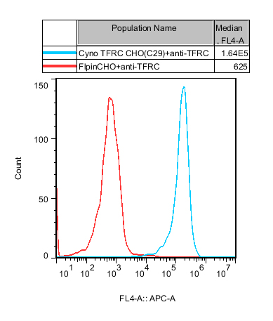
Figure 2. Recombinant CHO-K1 Cynomolgus TFRC Cell constitutively expressing Cyno TFRC .
3. TfR1 Internalization (Early-endosome specific) Cell (C15): Quantifies how efficiently antibodies enter early endosomes, distinguishing that measurement from total internalization.
Both overexpression lines are cultured as adherent cells in F12K + 10% FBS. Passage stability was established through passage 32, and both lines tested negative for mycoplasma.
We deliberately chose full-length protein over the extracellular domain (ECD) alone. Many TfR1 antibody epitopes depend on the dimer conformation, and only the full-length protein displayed at the membrane reproduces the actual binding geometry. This matters especially when judging whether binding is monovalent or bivalent, because valency is what determines where the cargo is sorted.
C15 has a specific use. When two candidates give nearly identical total internalization curves but differ severalfold in brain exposure in vivo, the difference often lies in what happens after entry into early endosomes. C15 ranks candidates more closely in line with in vivo results than total internalization does. In the validation data, trontinemab gives a clear dose-dependent response and can be used directly as a positive control.
There are three limits to keep in mind. Overexpression differs from physiological receptor density, so the results cannot be converted directly into in vivo flux. CHO cells lack the polarized architecture of the BBB. They can tell you which compartment the cargo reached, while crossing the barrier remains a separate question. The internalization readout also depends on the time window, so compare whole curves rather than a single time point.
Common pitfalls in experimental design
Using total internalization as a proxy for transport efficiency. High-affinity bivalent antibodies often give the best-looking curves and are also the most likely to end up in lysosomes.
Overlooking transferrin in the culture medium. Holo-Tf in medium containing 10% serum is sufficient to compete for some binding sites. When the epitope is close to the Tf binding surface, readouts will be systematically underestimated and can also drift between lots.
Relying on mice alone to test the mechanism. At 73% homology, many human antibodies fail to bind the mouse receptor. Using that result to rule out a candidate is risky.
Applying ADC data to shuttle antibodies. An ADC needs to get into a cell, be degraded, and release its payload. A shuttle antibody needs to get through the cell intact. The same experiment can lead to exactly opposite conclusions.
A model needs to show how cargo is sorted after internalization
TfR1 combines a well-understood biology with unusual scope for engineering. The mechanism is clear enough to tune the combination of affinity, valency, pH, and epitope. With so many parameters in play, though, a small oversight in vitro can produce a misleading ranking.
What makes a model useful is its ability to distinguish sorting outcomes after internalization, regardless of how high expression is. The two full-length cell lines, human and cynomolgus, address binding and species differences. The early endosome-specific model addresses where the cargo went. Getting both answers right is what makes the in vivo experiments meaningful.
Frequently Asked Questions
A:Do TfR1, CD71, and TFRC refer to the same thing?
Yes. The protein name is transferrin receptor 1, the gene symbol is TFRC, and the CD designation is CD71. They refer to the same molecule.
A:Why make a TfR1 antibody monovalent?
Bivalent binding cross-links receptors into clusters, which are more likely to be routed to lysosomes for degradation, reducing brain exposure. Monovalent binding avoids cross-linking and makes transcytosis more likely.
A:Can a total internalization assay identify good shuttle antibodies?
Total internalization alone falls short. It combines early endosome entry and lysosome entry in one number, even though these two routes have opposite implications for delivery. An early endosome-specific readout is needed to tell them apart.
Related Products: CHO-K1 Human TFRC Cell (RQP74327), CHO-K1 Cynomolgus TFRC Cell (RQP74657), TFRC target model


news recommendation
CaSR: The Calcium Homeostasis Sentinel and a Core GPCR Target for Metabolic and Kidney Disease Research
CD3E: The T-Cell Activation Switch and Cell Models for Targeted Therapeutics
NFAT Signaling Pathway: A Critical Target for Drug R&D and Cell Models
TSHR Cell Model for Thyroid Cancer Drug Screening | GPCR Assay Platform-Reqbio
Worldwide Commercial Licensing Rights Secured:Jurkat E6.1, CHO-K1, HEK293, THP-1, Raji Cell Lines-Expanding Our Capabilities in Cell-Based Research.
Explore


info@reqbio.com
