

Home>
Resources>
Learning>
Articles>
CD3E: The T-Cell Activation Switch and Cell Models for Targeted Therapeutics
CD3E: The T-Cell Activation Switch and Cell Models for Targeted Therapeutics
In the adaptive immune system, T cells are the core force in defending against pathogens and tumors. The activation of T cells relies on a sophisticated molecular switch—the TCR-CD3 complex. Among its components, CD3E (CD3ε), as one of the earliest emerging and functionally most central subunits in this complex, is responsible for converting the antigen-recognition signal of the TCR into an intracellular cascade, ultimately driving T cell proliferation, differentiation, and killing. A lack of CD3E leads to T cell developmental arrest, whereas bispecific T-cell engagers (BiTE/TCE) targeting CD3E have achieved breakthroughs in hematological malignancies. As the heat around bispecific antibodies, CAR-T, and autoimmune disease treatments continues to rise, CD3E has become a star target bridging fundamental immunology and translational medicine. Leveraging its gene-editing platform, ReqBio has successfully developed a CD3E knockout (KO) NFAT-reporter Jurkat cell line, providing a precise tool for the activity screening and specificity evaluation of CD3E-targeting drugs.
I. CD3E: The Signaling Hub within the TCR-CD3 Complex
CD3E is a single-pass transmembrane protein whose structure is divided into three parts:
Extracellular Domain: Contains an Ig-like domain, responsible for forming stable heterodimers with TCRα/β and CD3γ/δ.
Transmembrane Domain: Rich in charged amino acids, maintaining the assembly of the complex through electrostatic interactions.
Intracellular Domain: Contains an ITAM (Immunoreceptor Tyrosine-based Activation Motif) motif, which serves as the core site for recruiting ZAP70 and initiating downstream signaling.
Upon TCR recognition of the antigen peptide-MHC complex, the ITAM of CD3E is phosphorylated by Lck, recruiting and activating ZAP70. This in turn phosphorylates LAT and SLP76, initiating multiple signaling pathways such as Ras-MAPK and calcineurin-NFAT, which ultimately induces IL-2 expression and T cell activation. The functional integrity of CD3E is an absolute requirement for T cell development, maturation, and the initiation of effector functions.
II. Therapeutics R&D Targeting CD3E: Bispecifics, Monoclonals, and Novel Molecules
CD3E expression is highly restricted to T cells, and its activation can effectively trigger T cell-mediated killing. Consequently, it has become an ideal target in the fields of tumor immunotherapy, autoimmunity, and transplantation.
Primary R&D Directions:
|
Drug Type |
Representative Drug |
Mechanism |
Indication |
Stage |
|
Bispecific T-cell Engager (TCE) |
Alnuctamab(CD3×BCMA) |
Bridges CD3E and tumor-associated antigens |
Relapsed multiple myeloma, SLE |
Phase 3 |
|
Monoclonal Antibody |
Foralumab |
Regulatory T cell agonist |
Alzheimer's disease, multiple system atrophy |
Phase 2 |
|
Antibody-Drug Conjugate (ADC) |
Dafsolimab setaritox(CD3×CD7) |
Depletes double-positive T cells |
Diffuse scleroderma |
Phase 1/2 |
|
Antibody-Drug Conjugate (ADC) |
NILK-2301(CD3×CEACAM5) |
Targets solid tumor antigens |
Solid tumors |
Phase 1 |
III. Accelerating CD3E-Targeted Drug R&D: ReqBio's KO Reporter Cell Models
During the screening process for CD3E-targeted therapeutics, conventional wild-type Jurkat cells cannot distinguish whether a compound-induced signal is mediated by CD3E or by other off-target effects. CD3E knockout (KO) cell lines serve as ideal negative controls to validate the target specificity of drugs.
To address the CD3E target, ReqBio has developed two core cell models:
|
Cell Name |
Cat. No. |
Background Cell |
Features |
Core Applications |
|
Jurkat E6.1 Human CD3E KO NFAT-Luc Cell |
RQP74469 |
NFAT-Luc Jurkat |
Stably integrates the NFAT-Luciferase reporter gene, with the CD3E gene knocked out. |
Evaluating CD3E agonists (e.g., OKT3). KO cells show no response while wild-type cells respond, used to validate target specificity. |
|
Jurkat E6.1 Human CD3E KO Cell |
RQP74475 |
Jurkat(wild-type) |
Only the CD3E gene is knocked out; contains no reporter gene. |
Serving as a negative control for binding assays such as FACS and Western Blot to validate antibody binding specificity. |
Table 1: CD3E Target Cell Models
Validation Data and Advantage Analysis:
1.Jurkat E6.1 Human CD3E KO NFAT-Luc Cell(RQP74469)
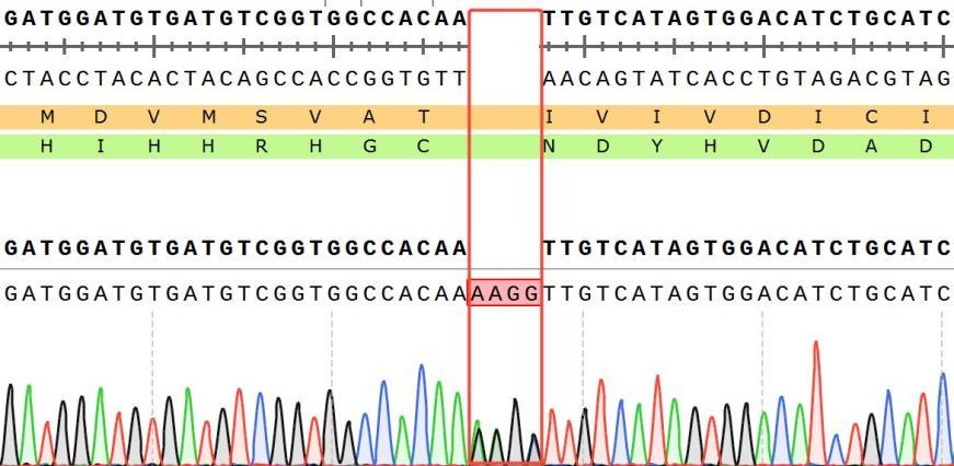
Figure 2 (Sequencing Validation): Displays the mutation sequence information of the CD3E gene [c.397_398ins4bp / p.I133Kfs], definitively confirming that the CD3E gene was successfully knocked out, resulting in a frameshift mutation that leads to protein truncation. Validation at the sequence level ensures a clear and traceable genetic background for the cell model.
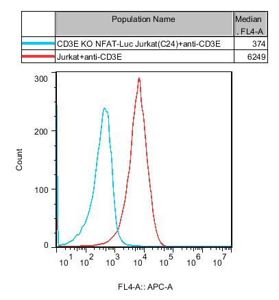
Figure 3 (Expression Validation): FACS or Western Blot analysis shows that compared to wild-type NFAT-Luc Jurkat, the CD3E protein is completely absent from the surface of the KO cells. This ensures that in subsequent functional assays, any residual signal is not mediated by CD3E.
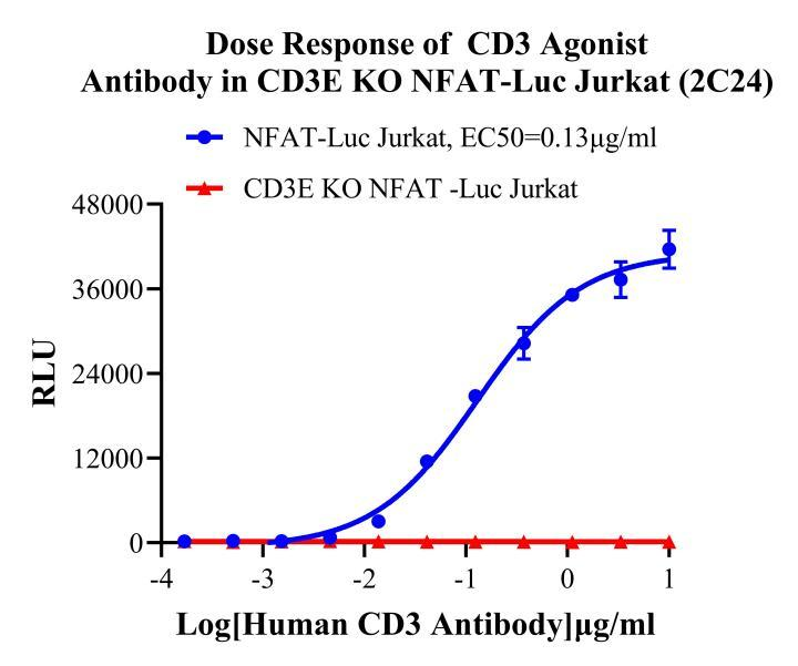
Figure 4 (Functional Validation – Core Highlight):
Assay Principle: In NFAT-Luc Jurkat cells, a CD3E agonist antibody (such as OKT3) activates the TCR signaling pathway. This triggers the activation of the NFAT transcription factor, driving the expression of luciferase (Luc).
Data Interpretation: In wild-type cells, the CD3E agonist antibody exhibits a typical dose-dependent activation profile characterized by a sharp sigmoidal curve and a wide assay window. Conversely, in the CD3E KO cells, there is absolutely no response across the identical concentration gradient, with the signal remaining strictly at background levels.
Advantage Analysis:
✅ The Gold Standard for Target Specificity Validation: Allows for the direct determination of whether a candidate molecule indeed exerts its effect through CD3E, effectively ruling out false positives.
✅ Highly Sensitive Reporter System: The NFAT-Luc signal amplification delivers a wide dynamic range, making it exceptionally well-suited for high-throughput screening.
✅ Ready-to-Use Control: Paired with wild-type NFAT-Luc cells, it enables the rapid calculation of a compound's selectivity index.
2.Jurkat E6.1 Human CD3E KO Cell(RQP74475)
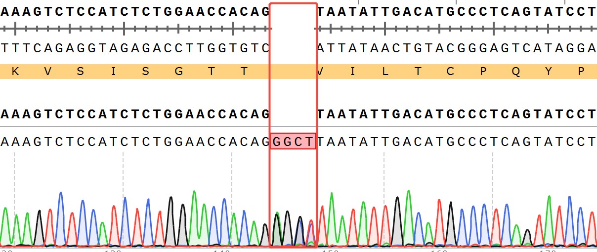
Figure 5 (Sequencing Validation): Displays the mutation information of another independent KO clone [c.133_134insGGCT / p.V45Gfs*13], which likewise represents a frameshift mutation, ensuring that no functional CD3E protein is produced.
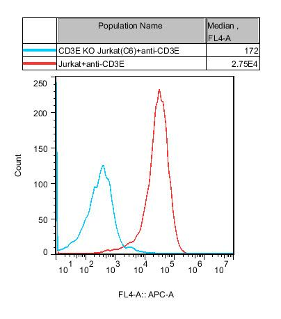
Figure 6 (Expression Validation): Confirms the absence of CD3E protein on the cell surface.
Core Applications: This model does not contain a reporter gene and is primarily utilized for binding assays (such as flow cytometry to evaluate the binding specificity of antibodies to CD3E), signaling pathway background subtraction, and off-target effect assessments.
Summary of Core Product Advantages:
|
Advantage Dimension |
Detailed Description |
|
High Target Relevance |
CD3E is the core target for TCE bispecifics, CAR-T, and immunomodulatory monoclonal antibodies, attracting high market interest and a broad base of R&D clients. |
|
Dual Validation: KO + Reporter |
Provides KO cell lines backed by rigorous, triple validation across sequencing, protein expression, and functional activity, ensuring highly reliable and publication-ready data. |
|
Powerful Specificity Assessment |
When paired with wild-type reporter cells, it allows researchers to rapidly differentiate between CD3E-mediated specific signals and non-specific background noise. |
|
Highly Sensitive NFAT-Luc System |
The luciferase reporter assay is simple, sensitive, and features a wide dynamic range, making it exceptionally well-suited for medium-to-high-throughput screening. |
|
Flexible Choice of Two KO Models |
Features a reporter-equipped model for functional activity assays and a non-reporter model for binding specificity validation, seamlessly adapting to diverse experimental requirements. |
|
Ready-to-Use Products |
These cell models undergo strict cryopreservation quality control, allowing them to be deployed in experiments immediately upon arrival, significantly shortening R&D timelines. |
IV. Application Scenarios: From Bispecific Screening to CAR-T Optimization
Early Screening of Bispecific Antibodies (TCEs): Evaluates whether candidate molecules rely on CD3E to activate T cells; a negative result in the KO cells definitively confirms target specificity.
Functional Characterization of CD3E Monoclonal Antibodies: Differentiates between agonist and antagonist antibodies, explicitly clarifying whether their mechanism of action is strictly dependent on CD3E.
Functional Controls for CAR-T Therapy: Serves as ideal negative target cells to evaluate whether CAR-T cells are being non-specifically activated through pathways outside of CD3E.
Off-Target Assessments for Autoimmune Disease Therapeutics: Ensures that candidate molecules do not unintentionally trigger non-specific activation of the CD3E signaling pathway, minimizing safety risks.
V. Conclusion
As the core switch for T-cell activation, CD3E is not only a fundamental key to understanding adaptive immunity, but also a crucial target in the development of immuno-oncology and autoimmune disease therapeutics. With the continuous iteration of bispecific antibody and CAR-T technologies, the demand for precise CD3E target-specificity evaluation tools has become increasingly urgent.
ReqBio's newly launched CD3E KO NFAT-Luc Jurkat and CD3E KO Jurkat cell models—backed by well-defined genetic backgrounds and comprehensive functional validation—provide global drug developers with a highly accurate and efficient suite of assessment tools to accelerate next-generation therapies.


news recommendation
CaSR: The Calcium Homeostasis Sentinel and a Core GPCR Target for Metabolic and Kidney Disease Research
NFAT Signaling Pathway: A Critical Target for Drug R&D and Cell Models
TSHR Cell Model for Thyroid Cancer Drug Screening | GPCR Assay Platform-Reqbio
We Are Pleased to Announce: Global Commercial Licensing Rights for Jurkat E6.1, CHO-K1, HEK293, THP-1 and RAJI Cell Lines Officially Secured.
Explore