

Home>
Resources>
Learning>
Articles>
CaSR: The Calcium Homeostasis Sentinel and a Core GPCR Target for Metabolic and Kidney Disease Research
CaSR: The Calcium Homeostasis Sentinel and a Core GPCR Target for Metabolic and Kidney Disease Research
Within the Class C G protein-coupled receptor (GPCR) family, the calcium-sensing receptor (CaSR) serves as a central molecular sentinel for systemic calcium and phosphate homeostasis. It precisely detects subtle changes in extracellular Ca²⁺ concentrations and coordinates responses across the parathyroid glands, kidneys, bones, and other organs to regulate parathyroid hormone (PTH) secretion and ion balance. CaSR dysfunction directly contributes to chronic kidney disease–secondary hyperparathyroidism (CKD-SHPT), osteoporosis, and disorders of calcium metabolism. With a well-defined function and established druggability, CaSR has become one of the most successful GPCR targets in CKD-SHPT, with multiple drugs approved worldwide. Its potential roles in inflammation, energy metabolism, and cancer are also being explored. Leveraging its established GPCR drug screening platform, Reqbio has developed a series of CaSR cell models covering Gq pathway detection, providing researchers worldwide with reliable and efficient cell-based tools for target-based drug screening.
I. CaSR: A Precision Sensor of Calcium Homeostasis
CaSR is encoded by the human CASR gene and is a representative member of the Class C GPCR family. Its principal function is to continuously monitor small changes in extracellular Ca²⁺ concentration, with sensitivity at the 0.1 mM level, and regulate PTH secretion through a negative-feedback mechanism. When blood calcium levels rise, CaSR is activated, suppressing PTH release and reducing calcium release from bone and renal calcium reabsorption. When blood calcium levels fall, CaSR activity decreases and PTH secretion increases, mobilizing calcium from bone and promoting renal calcium reabsorption. This tightly controlled negative-feedback loop provides the physiological basis for systemic calcium homeostasis.
CaSR is highly expressed not only in parathyroid chief cells but also in the kidneys, bones, intestine, thyroid C cells, vascular smooth muscle, adipose tissue, immune cells, and tumor cells. Its functions extend beyond calcium homeostasis to the regulation of inflammation, energy metabolism, cardiovascular protection, and tumor progression.
II. CaSR Structure and Signal Transduction
CaSR functions as a homodimer, a characteristic feature of Class C GPCRs. Each monomer contains three principal domains:
|
Domain |
Components |
Function |
|
Extracellular N-terminal domain (ECD) |
Venus flytrap domain (VFT) + cysteine-rich domain (CRD) |
The VFT contains five Ca²⁺-binding sites and serves as the primary ligand-sensing region; the CRD mediates dimerization and conformational transmission |
|
Seven-transmembrane domain (7TMD) |
Seven transmembrane helices |
Allosteric drug-binding region responsible for transmitting extracellular signals to the intracellular side |
|
Intracellular C-terminal domain (ICD) |
G protein-binding sites + β-arrestin recruitment region |
Regulates downstream signaling pathway selection, receptor desensitization, and internalization |
CaSR follows a cooperative dimer activation model. Extracellular Ca²⁺ and aromatic amino acids cooperatively bind to the VFT, inducing VFT closure → a conformational change in the CRD → 7TMD helix rearrangement → closer apposition of the dimeric transmembrane domains → exposure of G protein-binding sites → initiation of downstream signaling cascades.
Following activation, CaSR can couple to multiple G proteins, forming a multidimensional regulatory network:
|
Signaling Pathway |
G Protein |
Core Molecular Events |
Primary Biological Functions |
|
Gq/11–PLC–IP3/DAG |
Gq/11 |
PLC activation → IP3/DAG → Ca²⁺ release and PKC activation |
Principal pathway: suppresses PTH secretion, regulates renal calcium reabsorption, and inhibits osteoclast activity |
|
Gi/o–cAMP–PKA |
Gi/o |
AC inhibition → decreased cAMP → PKA inhibition |
Cooperates in suppressing PTH synthesis and secretion and regulates GLP-1 secretion and insulin release |
|
β-arrestin–ERK/Akt |
β-arrestin |
ERK1/2 and Akt activation |
Cell proliferation, apoptosis, and migration in cancer; inflammatory cytokine release; and bone cell differentiation |
III. CaSR-Targeted Drug Development: Continued Expansion of an Established Target
CaSR is one of the most successful GPCR targets in CKD-SHPT and disorders of calcium metabolism. Multiple drugs, including cinacalcet and etelcalcetide, have been approved worldwide, while development continues in osteoporosis, cancer, and inflammation.
Representative Clinical-Stage Programs:
|
Name |
Developer |
Stage |
Type |
Target |
Indication |
|
Strontium Ranelate |
Les Laboratoires Servier |
Phase 3 |
Small molecule |
CaSR |
Postmenopausal osteoporosis |
|
MT 1013 |
Shaanxi Micot Technology |
Phase 3 |
Synthetic peptide |
CaSR & OGP |
Secondary hyperparathyroidism |
|
DS-9194b |
Daiichi Sankyo |
Phase 1 |
Small molecule |
CaSR |
Osteoporosis |
IV. Accelerating CaSR-Targeted Drug Development: Reqbio Cell Models and Data Advantages
In drug screening for Class C GPCRs such as CaSR, conventional cAMP or β-arrestin assays are often not the most direct readouts because the receptor has relatively high constitutive activity and its ligand is an ion (Ca²⁺). The Gq/11–PLC–IP3/DAG–Ca²⁺ pathway is the principal CaSR signaling axis, and measurement of IP1 accumulation, a stable metabolite of IP3, is considered a gold-standard approach for evaluating CaSR activity. Reqbio has developed three complementary CaSR cell models that support different assay platforms and application scenarios.
Reqbio CaSR Cell Models:
|
Cell Line |
Catalog No. |
Host Cell |
Assay Format |
Signaling Pathway |
Primary Application |
|
HEK293 Human CaSR NFAT-Luc Cell Line |
RQP71562 |
HEK293 |
NFAT luciferase reporter |
Gq–PLC–Ca²⁺–NFAT |
Screening of CaSR agonists and allosteric modulators (Figures 1, 2, and 3) |
|
HEK293 Human CaSR Gα15 Cell Line |
RQP71568 |
HEK293 |
HTRF IP-One |
Gq–PLC–IP1 |
Screening of CaSR agonists and antagonists; quantitative activity evaluation (Figures 4 and 5) |
Validation Data and Advantages:
1. HEK293 Human CaSR NFAT-Luc Cell Line (RQP71562)
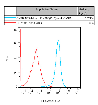
Figure 1. Stable expression of CaSR in HEK293 cells was confirmed by flow cytometry or Western blotting.
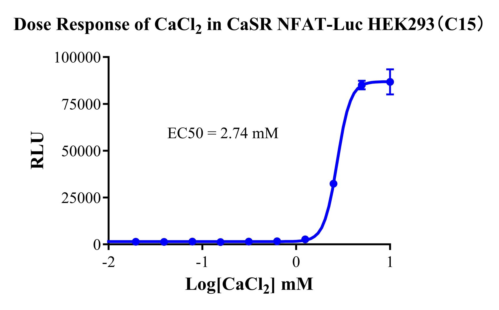
Figure 2. Dose response of CaCl₂ in CaSR NFAT-Luc HEK293 cells (C15).
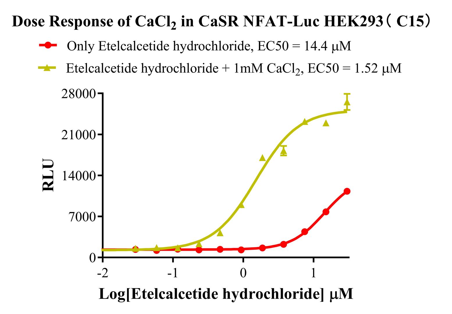
Figure 3. Dose response of CaCl₂ in CaSR NFAT-Luc HEK293 cells (C15).
Figures 2 and 3 (functional validation): CaCl₂ produced a characteristic dose-dependent sigmoidal activation curve. The response of CaSR to Ca²⁺ is naturally sigmoidal, consistent with its physiological role as a concentration sensor. The EC₅₀ was within the 2–5 mM range, corresponding to the physiological blood calcium concentration window, with a broad signal window.
Advantages:
NFAT reporter system: Ca²⁺ influx activates calcineurin, leading to NFAT dephosphorylation and nuclear translocation, which drives luciferase expression. This amplifies the Gq–Ca²⁺ signal and provides greater sensitivity than direct IP1 measurement.
Suitable for high-throughput screening: The luciferase assay is straightforward to perform, compatible with 384-well plates, and suitable for compound library screening.
Broad dynamic range: Figures 2 and 3 show consistent dose responses with broad assay windows, demonstrating model robustness and reproducibility.
Compatible with allosteric modulators: CaSR allosteric agonists, or calcimimetics such as cinacalcet, can shift the response curve to the left by increasing Ca²⁺ sensitivity. This model can be used to screen novel allosteric modulators.
2. HEK293 Human CaSR Gα15 Cell Line (RQP71568)
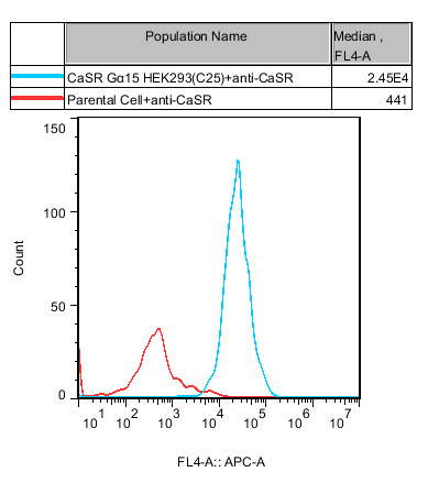
Figure 4. Flow cytometry confirmed stable CaSR expression in HEK293 cells together with Gα15, a G protein α subunit that can efficiently couple receptors to the Gq pathway in HEK293 cells and increase the sensitivity of CaSR Gq signaling assays.
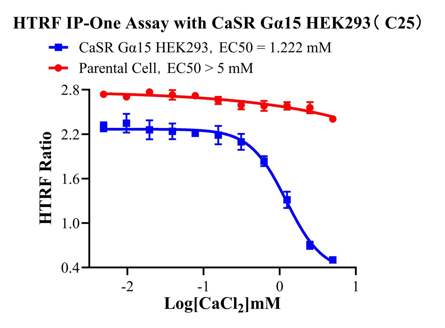
Figure 5 (functional validation). Using the HTRF IP-One assay platform, CaCl₂ induced IP1 accumulation in a dose-dependent manner, with a stable EC₅₀.
Advantages:
HTRF IP-One is a gold-standard assay for studying Gq signaling. It directly quantifies IP1 levels, providing a more direct and quantitative readout suitable for precise potency comparisons.
Gα15-enhanced design: Endogenous Gq expression is limited in wild-type HEK293 cells. Co-expression of Gα15 enhances the coupling efficiency of CaSR to Gq signaling, producing a broader assay window and a more stable EC₅₀.
Compatible with antagonist screening: At a fixed Ca²⁺ concentration, the model can be used to evaluate the inhibition of IP1 signaling by CaSR antagonists, or calcilytics such as DS-9194b.
Summary of Core Product Advantages:
|
Advantage |
Description |
|
Established target |
CaSR is a validated GPCR target in CKD-SHPT and calcium metabolism, with approved drugs and a defined market. |
|
Pathway-matched models |
All two models are based on the native Gq–PLC–IP3–Ca²⁺ signaling axis and therefore align with the physiological mechanism of CaSR. |
|
Two assay platforms |
NFAT luciferase reporter and HTRF IP-One formats support different instrumentation and throughput requirements. |
|
Gα15-enhanced design |
Co-expression of Gα15 enhances Gq signaling coupling, broadening the assay window and improving data stability. |
|
Compatible with allosteric modulators |
The models clearly reflect shifts in Ca²⁺ response curves induced by CaSR allosteric modulators and are suitable for screening next-generation allosteric compounds. |
|
Ready-to-use products |
Each model has undergone monoclonal screening and functional validation and can be used after recovery. |
V. Representative Applications
Screening CaSR agonists (calcimimetics) for CKD-SHPT: CaSR NFAT-Luc HEK293 or CaSR Gα15 HEK293 cells can be used to screen small molecules that increase CaSR sensitivity to Ca²⁺ for the treatment of CKD-SHPT, including compounds with mechanisms similar to cinacalcet.
Screening CaSR antagonists (calcilytics) for osteoporosis: Against a fixed background concentration of Ca²⁺, HTRF IP-One models can be used to evaluate the inhibitory effects of compounds on CaSR signaling for osteoporosis research, including analogs of DS-9194b.
Characterization of allosteric modulators: All three models can be used to evaluate the effects of allosteric modulators on CaSR dose-response curves, including left or right shifts and changes in maximal response, enabling comprehensive characterization of compound pharmacology.
Biological activity testing: Standardized CaSR agonists, such as Ca²⁺ or cinacalcet, can be used as reference materials for quality control of lot-to-lot biological activity of CaSR-targeted drugs.
VI. Conclusion
As a molecular sentinel of calcium homeostasis, CaSR has become an essential drug target in CKD-SHPT, osteoporosis, and disorders of calcium metabolism because of its precise Ca²⁺-sensing capability and multi-organ regulatory network. The success of approved drugs has established its druggability, while its potential roles in inflammation, energy metabolism, and cancer provide additional opportunities for next-generation drug development. Reqbio offers three CaSR cell models spanning NFAT luciferase reporter and HTRF IP-One assay platforms, with both the broadly used HEK293 host. Together, these models provide drug discovery researchers with a precise, flexible, and cross-validatable set of evaluation tools.
Through its cell engineering capabilities and high-precision gene-editing platform, Reqbio continues to provide drug screening cell models covering GPCRs, immunotherapy targets, kinases, and other target classes, together with high-quality cell-based bioassay services.


news recommendation
CD3E: The T-Cell Activation Switch and Cell Models for Targeted Therapeutics
NFAT Signaling Pathway: A Critical Target for Drug R&D and Cell Models
TSHR Cell Model for Thyroid Cancer Drug Screening | GPCR Assay Platform-Reqbio
We Are Pleased to Announce: Global Commercial Licensing Rights for Jurkat E6.1, CHO-K1, HEK293, THP-1 and RAJI Cell Lines Officially Secured.
Explore