
HEK293 Human ACVR2A Effector Reporter Cell
Cat. No: RQP74277
Size: 1 vial of frozen cells (>1E6 per vial in 1 mL)
Unit Price: Contact For Pricing
Product Info
Description
Biological Information
Assay Data
Cell Culture
| Cat. No | RQP74277 |
| Product Name | HEK293 Human ACVR2A Effector Reporter Cell |
| Product Type | Reporter Cell |
| Culture Properties | Adherent |
| Stability | 32passages (in-house test, that not means the cell line will be instable beyond the passages we tested.) |
| Mycoplasma Status | Negative |
| Culture Medium | DMEM+10%FBS+ 200 μg/ml Hygromycin B |
| Freeze Medium | 90% FBS+10% DMSO |
| Storage Conditions | Liquid nitrogen immediately upon delivery |
| Application | Functional(Report Gene) Assay |
For research use only. Not intended for human or animal clinical trials, therapeutic or diagnostic use.
Activin A is a key member of the transforming growth factor-beta (TGF-β) superfamily and a pleiotropic factor involved in the pathogenesis of immune-related disorders, such as allergies, autoimmune diseases, and cancer.
Activin A signals through two types of type I receptors and two types of type II receptors, which assemble into a functional receptor complex upon ligand binding. The type I receptors include activin receptor type 1A (or activin receptor-like kinase 2, ALK2), activin receptor type 1B (or ALK4), and activin receptor type 1C (or ALK7). Activin A preferentially binds to ALK4, while exhibiting lower affinity for ALK2 and ALK7. The type II receptors are activin receptor type IIA (ActRIIA) and activin receptor type IIB (ActRIIB), both characterized by constitutive serine/threonine kinase activity. Upon binding of Activin A to its type II receptors, two type I receptors are recruited and phosphorylated by the type II receptors, an event that activates their kinase activity. While most TGF-β superfamily members bind to ActRIIB or ActRIIA, only a few ligands bind to ALK4 with high affinity. In addition to the canonical receptor serine/threonine kinase-SMAD axis, Activin A has been shown to participate in signaling pathways such as p38, ERK, PI3K, and Wnt.
The HEK293 Human ACVR2A Effector Reporter Cell model effectively simulates the in vivo ACVR2A signal transduction process, the principle is illustrated in the figure below.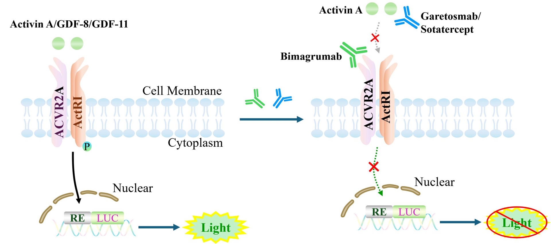
Figure 1. Schematic diagram of the HEK293 Human ACVR2A Effector Reporter Cell model
| Classification | Cytokine&Growth Factor |
| Family | Transforming growth factor-β (TGF-β) receptor family |
| Gene Name | ACVR2A |
| Gene Aliases | ACVR2;ACTRII;activin A receptor, type II;activin A receptor, type IIA |
| Gene ID | 92 |
| Accession Number | NM_001616.5 |
| UniProt Number | P27037 |
| Protein Name | Activin receptor type-2A |
| Protein Aliases | Activin receptor type IIA (ACTR-IIA; ACTRIIA) |
| Target Species | Human |
| Host cell | HEK293 |
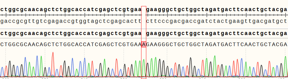
Figure 2. Sanger of ACVR2A Effector Reporter Cell
ACVR2B(NM_001106.4): c.221_222insA/ACVR2B:p.K74Kfs*7
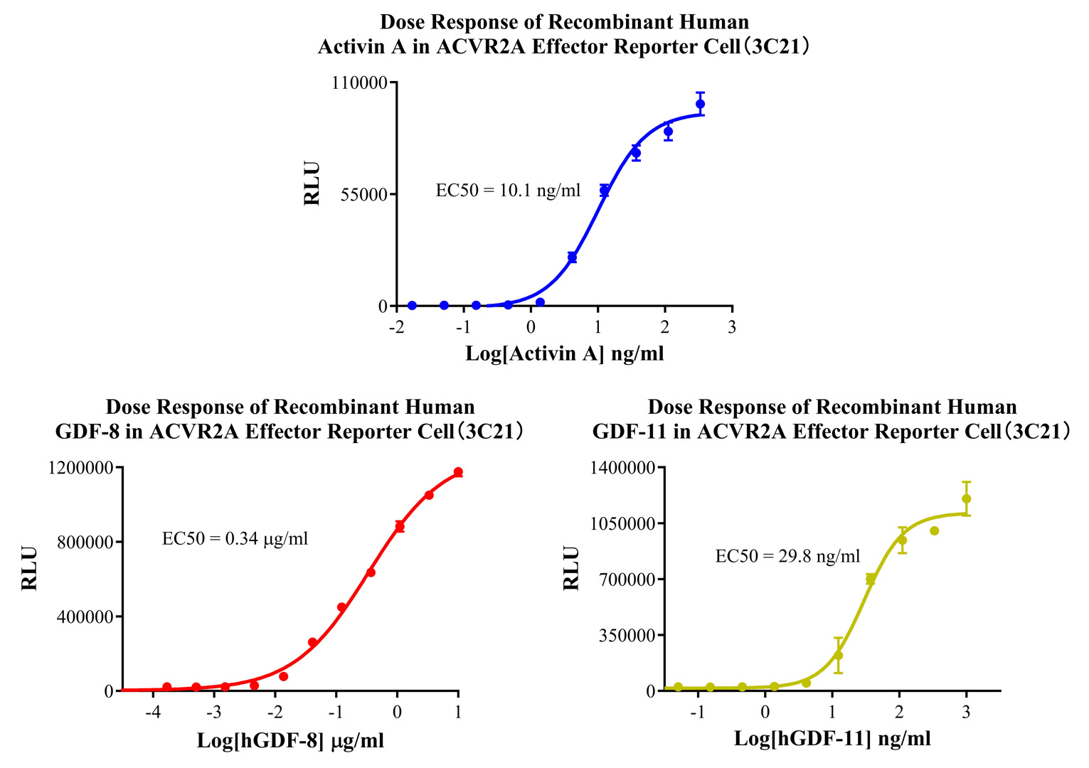
Figure 3. Dose Response of Recombinant Human Activin A in ACVR2A Effector Reporter Cell(3C21).Dose Response of Recombinant Human GDF-8 in ACVR2A Effector Reporter Cell (3C21). Dose Response of Recombinant Human GDF-11 in ACVR2A Effector Reporter Cell (3C21).
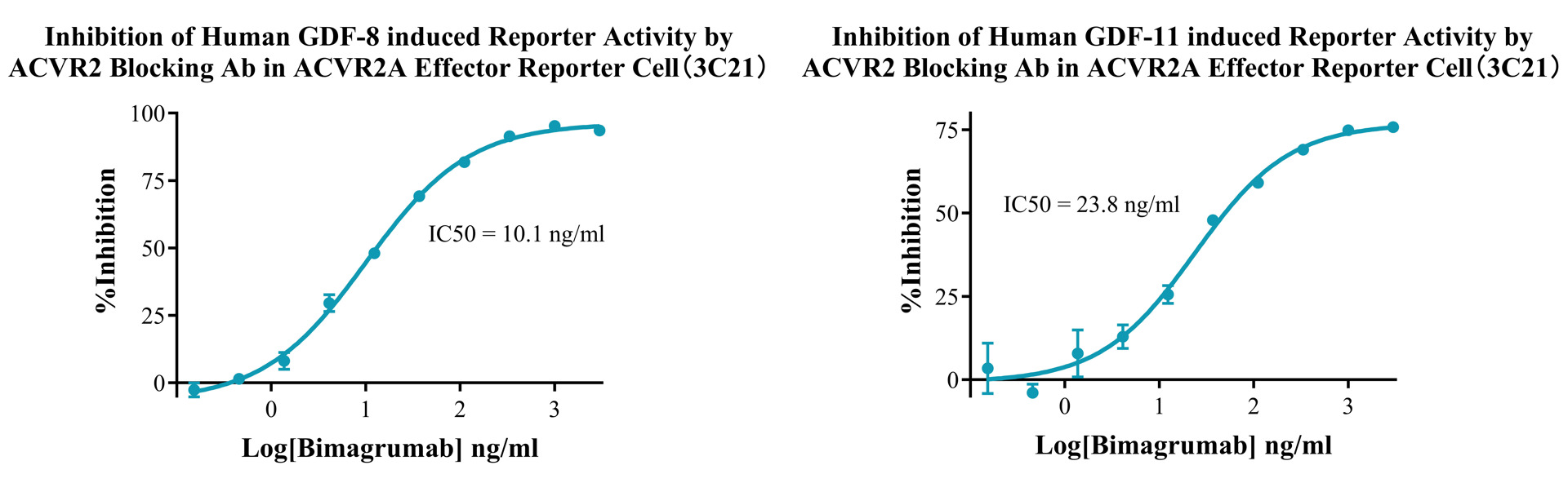
Figure 4. Inhibition of Human GDF-8 induced Reporter Activity by ACVR2 Blocking Ab in ACVR2A Effector Reporter Cell (3C21).Inhibition of Human GDF-11 induced Reporter Activity by ACVR2 Blocking Ab in ACVR2A Effector Reporter Cell (3C21).
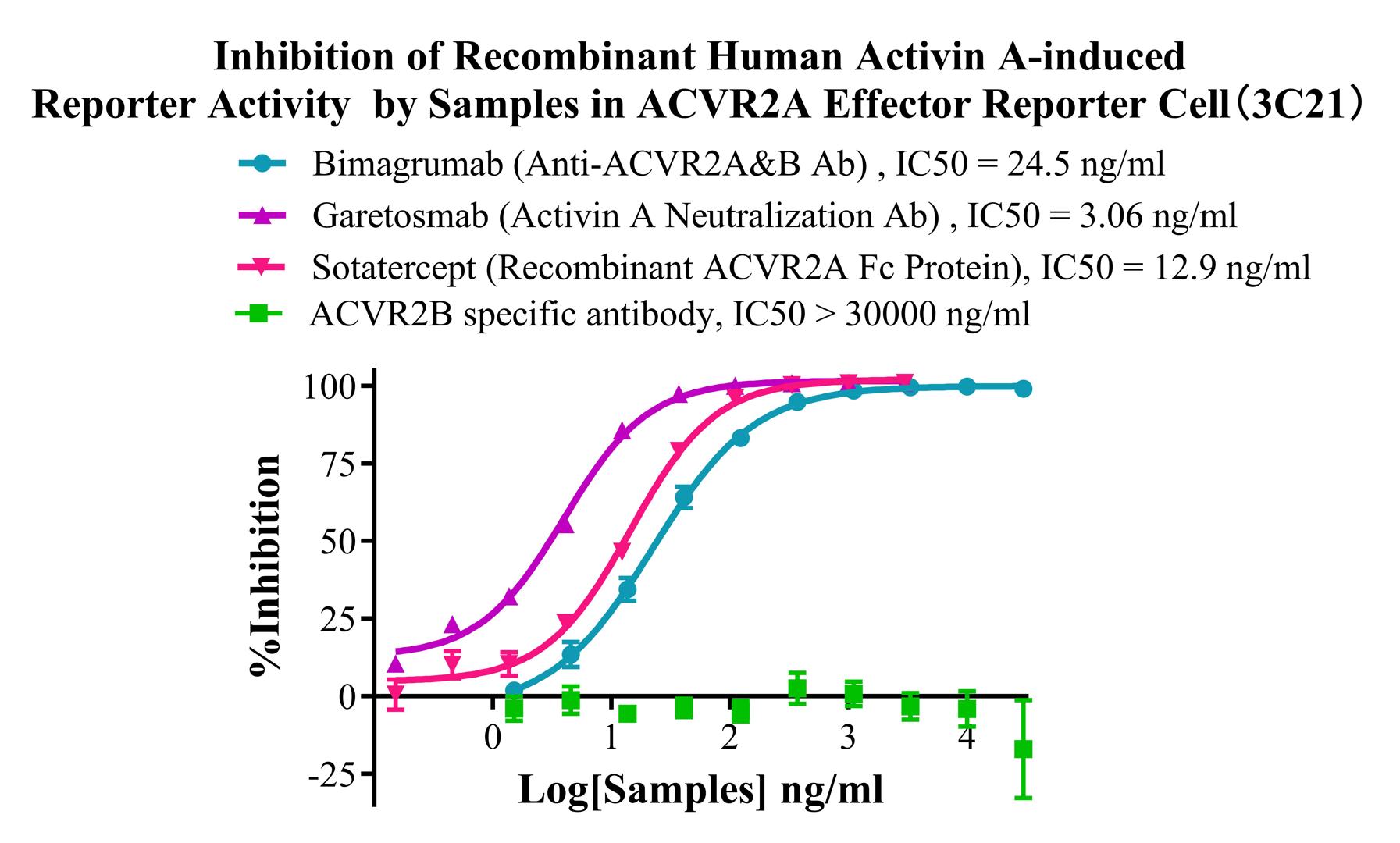
Figure 5. Inhibition of Recombinant Human Activin A-induced Reporter Activity by Samples in ACVR2A Effector Reporter Cell (3C21).
Cell Resuscitation
1)Rapidly thaw the frozen cells in a 37 °C water bath for approximately 60 seconds. Once thawed (which may take slightly less or more than 60 seconds), immediately transfer the cell suspension from the cryovial into a 15 mL centrifuge tube containing 10 mL of pre-warmed HEK293 Human ACVR2A Effector Reporter Cell complete culture medium.
2)Centrifuge cells at 1000 rpm for 5 min to remove medium, then resuspend cells in 5 mL of pre-warmed complete medium.
3)Transfer the cell suspension into a T25 culture flask and incubate at 37 °C with 5% CO₂.
4)After approximately 24–36 hours, replace the medium or passage the cells to remove non-adherent dead cells.
Subculturing procedure
1)When the cell density reaches the appropriate confluency for passaging, wash the cells with PBS, then add 1 mL trypsin to detach the cells. When more than 80% of the cells detach upon gently tapping the culture flask, add complete culture medium to terminate digestion. Gently pipette to obtain a single-cell suspension, transfer to a 15 mL centrifuge tube, and centrifuge at 1000 rpm for 5 minutes.
2)Discard supernatant after centrifugation. Resuspend cells in fresh medium to a single-cell suspension and transfer to a new culture flask for continued growth.
Cell Freezing
After trypsinization and centrifugation of cells from each T75 flask or 10 cm culture dish, discard the supernatant. Add 2 mL of cryopreservation medium (90% FBS + 10% DMSO), gently resuspend thoroughly, and aliquot into two cryovials. Immediately place the cryovials into a controlled-rate freezing container (e.g., Nalgene 5100-0001), fill with isopropanol to the indicated level, and store at −80 °C. After 24 hours, transfer the cryovials to liquid nitrogen for long-term storage.
Related products
CHO-K1 Human NTSR2 Cell Line
CHO-K1 Human CCR4 Cell Line
HEK293 Human NK1R CRE-Luc Cell Line
Raji-Luc-GFP
Jurkat E6.1-Luc
We Are Pleased to Announce: Global Commercial Licensing Rights for Jurkat E6.1, CHO-K1, HEK293, THP-1 and RAJI Cell Lines Officially Secured.
ExploreWe Are Pleased to Announce: Global Commercial Licensing Rights for Jurkat E6.1, CHO-K1, HEK293, THP-1 and RAJI Cell Lines Officially Secured.
Explore