

CHO-K1 Human MC1R CRE-Luc Cell Line
Cat. No: RQP71618
Size: 1 vial of frozen cells (>1E6 per vial in 1 mL)
Unit Price: Contact For Pricing
Description
Product Info
Biological Info
Assay Data
Cell Culture
This product is a CHO-K1 stable cell line that stably expresses tag-free full-length human MC1R receptor with CRE-Luc reporter. Upon agonist binding to human MC1R, the receptor activates Gαs, triggering adenylate cyclase activation and cAMP accumulation, leading to PKA-CREB pathway activation and luciferase expression. Therefore, receptor activity can be easily quantified in live cells in a dose-response manner by measuring luminescence or cAMP level. The cell line has been functionally validated with positive agonists. It is suitable for HTS and receptor functional evaluation assays.
| Cat. No | RQP71618 |
| Product Name | CHO-K1 Human MC1R CRE-Luc Cell Line |
| Engineering method | plasmid transfection |
| Culture Properties | Adherent |
| Stability | 32passages (in-house test, that not means the cell line will be instable beyond the passages we tested.) |
| Mycoplasma Status | Negative |
| Culture Medium | F12K+10%FBS+5 μg/ml Puromycin + 600μg/ml Hygromycin B |
| Freeze Medium | 90% FBS+10% DMSO |
| Storage Conditions | Liquid nitrogen immediately upon delivery |
| Transducer | Gs |
| Application | Functional assay for MC1R |
| Safety Precautions |
Wear lab coat, disposable gloves to avoid direct sample contact. For spills: apply disinfectant, allow full inactivation, then clean up. |
For research use only. Not intended for human or animal clinical trials, therapeutic or diagnostic use.
| Target Class | GPCR |
| Family | Melanocortin receptor |
| Sub Family | Class A(Rhodopsin) |
| Gene Name | MC1R |
| Gene Aliases | MSHR, MC1-R, melanotropin receptor |
| Gene ID | 4157 |
| Accession Number | NM_002386 |
| UniProt Number | Q01726 |
| Protein Name | Melanocyte-stimulating hormone receptor |
| Protein Aliases | Melanocortin receptor 1 (MC1-R), MSH-R |
| Target Species | Human |
| Host cell | CHO-K1 |
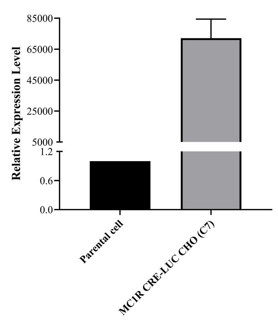
Figure 1. qPCR of MC1R CRE-Luc CHO-K1
The MC1R CRE-Luc CHO-K1 (C7) stable cell line shows significantly higher MC1R mRNA expression compared with parental CHO-K1 cells.
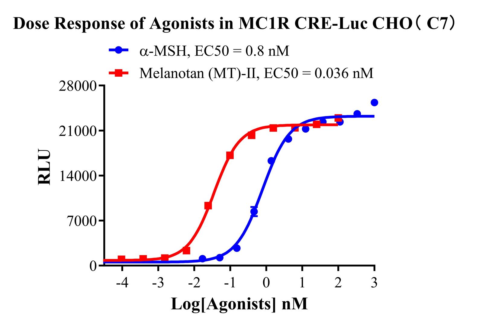
Figure 2. Dose Response of Agonists in MC1R CRE-Luc CHO-K1(C7)
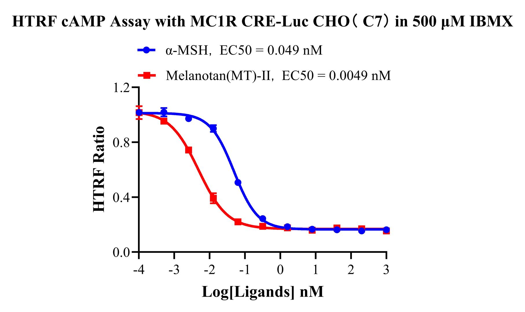
Figure 3. HTRF cAMP Assay with MC1R CRE-Luc CHO-K1(C7) in 500 μM IBMX
HTRF cAMP assay shows dose-dependent MC1R activation by α-MSH and Melanotan (MT)-II. EC₅₀ values are 0.049 nM and 0.0049 nM.
Cell Resuscitation
1)Rapidly thaw the frozen cells in a 37 °C water bath for approximately 60 seconds. Once thawed (which may take slightly less or more than 60 seconds), immediately transfer the cell suspension from the cryovial into a 15 mL centrifuge tube containing 10 mL of pre-warmed CHO-K1 Human MC1R CRE-Luc Cell Line complete culture medium.
2)Centrifuge cells at 1000 rpm for 5 min to remove medium, then resuspend cells in 5 mL of pre-warmed complete medium.
3)Transfer the cell suspension into a T25 culture flask and incubate at 37 °C with 5% CO₂.
4)After approximately 24–36 hours, replace the medium or passage the cells to remove non-adherent dead cells.
Subculturing procedure
1)When the cell density reaches the appropriate confluency for passaging, wash the cells with PBS, then add 1 mL trypsin to detach the cells. When more than 80% of the cells detach upon gently tapping the culture flask, add complete culture medium to terminate digestion. Gently pipette to obtain a single-cell suspension, transfer to a 15 mL centrifuge tube, and centrifuge at 1000 rpm for 5 minutes.
2)Discard supernatant after centrifugation. Resuspend cells in fresh medium to a single-cell suspension and transfer to a new culture flask for continued growth.
Cell Freezing
After trypsinization and centrifugation of cells from each T75 flask or 10 cm culture dish, discard the supernatant. Add 2 mL of cryopreservation medium (90% FBS + 10% DMSO), gently resuspend thoroughly, and aliquot into two cryovials. Immediately place the cryovials into a controlled-rate freezing container (e.g., Nalgene 5100-0001), fill with isopropanol to the indicated level, and store at −80 °C. After 24 hours, transfer the cryovials to liquid nitrogen for long-term storage.