

Home>
Resources>
Learning>
Articles>
PAC1R Target: A Core GPCR and Cell Models for Neuromodulation, Pain, and Neuroprotection
PAC1R Target: A Core GPCR and Cell Models for Neuromodulation, Pain, and Neuroprotection
Within the class B G protein-coupled receptor (GPCR) family, PAC1R (pituitary adenylate cyclase-activating polypeptide receptor 1) is a highly specific core functional target. It has a binding preference for the endogenous ligand PACAP (pituitary adenylate cyclase-activating polypeptide) that is more than 1,000-fold greater than that for the peptide VIP from the same family. This extremely high ligand selectivity enables PAC1R to independently mediate PACAP-specific neurobiological effects. PAC1R is mainly enriched in the nervous system and acts as an irreplaceable molecular switch in neuronal development, central stress responses, nociceptive signaling, and neural injury repair. In recent years, with a deeper understanding of the mechanisms of diseases such as migraine, neuropathic pain, and stroke, PAC1R has become a popular target in neuroscience drug development, and both agonist and antagonist development tracks have shown substantial potential for clinical translation. Relying on its established GPCR drug screening platform, Reqbio has successfully developed a series of PAC1R cell models covering the G protein pathway (CRE-Luc) and the β-arrestin pathway, providing researchers worldwide with efficient and comprehensive tools for targeted drug screening.
I. PAC1R: A Specific ‘Receiver’ of PACAP Signaling
PAC1R is encoded by the human ADCYAP1R1 gene and is a specific, high-affinity receptor for PACAP-27 and PACAP-38. Its ligand binding is highly selective: it has nanomolar affinity for PACAP-27/PACAP-38, whereas its affinity for the peptide VIP from the same family is more than 1,000-fold lower. This property enables PAC1R to independently mediate the specific biological effects of PACAP without cross-interference from VPAC receptor signaling pathways—this is the core feature that distinguishes PAC1R from other receptors in the same family.
PAC1R expression is highly tissue-specific. It is mainly enriched in the central nervous system (hypothalamus, hippocampus, amygdala, and brainstem) and peripheral nervous system (trigeminal ganglia and dorsal root ganglia), with limited distribution in endocrine and immune tissues. This precise expression pattern determines that the core functions of PAC1R are focused on neuronal development, stress responses, pain modulation, and neural repair, making it a ‘precision target’ for neuroscience drug development.
II. PAC1R Structure and Signal Transduction
PAC1R has a typical class B1 GPCR structure and consists of three parts, each with highly specific functions:
|
Domain |
Composition |
Function |
|
Extracellular N-terminal ECD |
Contains a unique amino acid insertion sequence |
Determines ligand selectivity: specifically binds PACAP and excludes VIP, forming the structural basis for the uniqueness of the target |
|
Seven transmembrane TM helices |
Hydrophobic core composed of seven α-helices |
Senses ligand-binding signals and undergoes conformational remodeling, mediates G protein coupling, and serves as a key binding region for small-molecule drugs |
|
Intracellular ICD |
Contains alternatively spliced regions (Hip/Hop isoforms) |
Regulates downstream signaling bias, determines the coupling efficiency of G proteins and β-arrestin, and mediates internalization and desensitization |
PAC1R follows the classical two-site activation model of class B GPCRs. After activation, it can couple to multiple downstream pathways, forming a multidimensional neuromodulatory network:
|
Signaling Pathway |
Coupled G Protein |
Core Molecular Events |
Biological Function |
|
Gs-cAMP-PKA-CREB |
Gs |
AC activation → cAMP ↑ → PKA activation → CREB phosphorylation |
Dominant pathway: neuronal differentiation, synaptic plasticity, antioxidation, and anti-apoptosis—the core of neuroprotection and repair |
|
Gq-PLC-IP3/DAG-Ca²⁺/PKC |
Gq |
PLC activation → IP3/DAG production → Ca²⁺ release and PKC activation |
Pain sensitization, regulation of neuronal excitability, and activation of neuroinflammation—key mechanisms of chronic pain and migraine |
|
β-arrestin-ERK/Akt |
β-arrestin |
β-arrestin recruitment → ERK1/2/Akt activation |
Cell proliferation, inhibition of apoptosis, and promotion of neurite regeneration—key to post-ischemic repair and neural regeneration |
III. PAC1R-Targeted Drug Development: Dual Tracks of Agonism and Antagonism
With the advantages of high ligand specificity, precise tissue functions, and clear disease associations, PAC1R has become a core R&D target in the fields of neuropathic pain, stress disorders, and neural injury repair. Current global development directions are mainly divided into two tracks: agonists (for neuroprotection, stroke, and cognitive disorders) and antagonists (for migraine, neuropathic pain, and anxiety).
Representative Investigational Pipelines:
|
Name |
Developer |
Stage |
Type |
Target |
Indication |
|
MT-002 |
Paragon (Mentari) |
Preclinical |
Bispecific antibody |
PAC1R × CGRP |
Migraine |
|
MT-001 |
Paragon (Mentari) |
Preclinical |
Monoclonal antibody |
PAC1R |
Migraine |
|
Anti-CGRPR/PAC1R bispecific antibody |
Amgen |
Preclinical |
Bispecific antibody |
PAC1R × CGRPR |
Headache |
|
BAY 2686013 |
Bayer |
Preclinical |
Small molecule |
PAC1R |
Pain |
Although most PAC1R-targeted drugs are currently at the preclinical stage, the strong scientific evidence for the PACAP/PAC1R pathway in migraine, neuropathic pain, and stroke, together with the exploration of new drug formats such as bispecific antibodies, has made this target highly anticipated.
IV. Accelerating PAC1R-Targeted Drug Development: Reqbio Cell Models and Data Advantages
In GPCR-targeted drug screening and mechanistic studies, the ability to evaluate the activities of both the G protein pathway and the β-arrestin pathway is key to determining the functional profile and bias of candidate molecules. For the PAC1R target, Reqbio has developed four complementary cell models that cover the full range of screening needs, including cAMP signaling, CREB transcriptional activity, β-arrestin recruitment, and receptor-binding specificity.
Reqbio PAC1R Cell Models:
|
Cell Line |
Product No. |
Host Cell |
Assay Format |
Signaling Pathway |
Core Application |
|
HEK293 Human PAC1R CRE-Luc Cell Line |
RQP71454 |
HEK293 |
CRE luciferase reporter gene |
Gs-cAMP-CREB |
PAC1R agonist screening and Gs pathway activity evaluation (Figures 3 and 4) |
CHO-K1 Human PAC1R CRE-Luc Cell Line |
RQP71565 |
CHO |
CRE luciferase reporter gene |
Gs-cAMP-CREB |
PAC1R agonist/antagonist screening and neutralizing antibody evaluation (Figures 5, 6, and 7) |
|
CHO-K1 Human PAC1R β-Arrestin Receptor Cell Line |
RQP71577 |
CHO |
β-Arrestin recruitment assay |
β-arrestin |
Biased ligand screening and receptor internalization/desensitization studies (Figures 8, 9, and 10) |
|
CHO-K1 Human PAC1R Cell Line |
RQP71455 |
CHO |
HTRF cAMP assay |
Gs-cAMP |
Agonist/antagonist screening and high-throughput cAMP quantification (Figures 11 and 12) |
Validation Data and Analysis of Advantages:
1. HEK293 Human PAC1R CRE-Luc Cell Line RQP71454
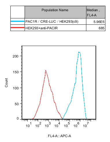
Figure 1. Flow cytometry or Western blot confirmed stable PAC1R expression in HEK293 cells.
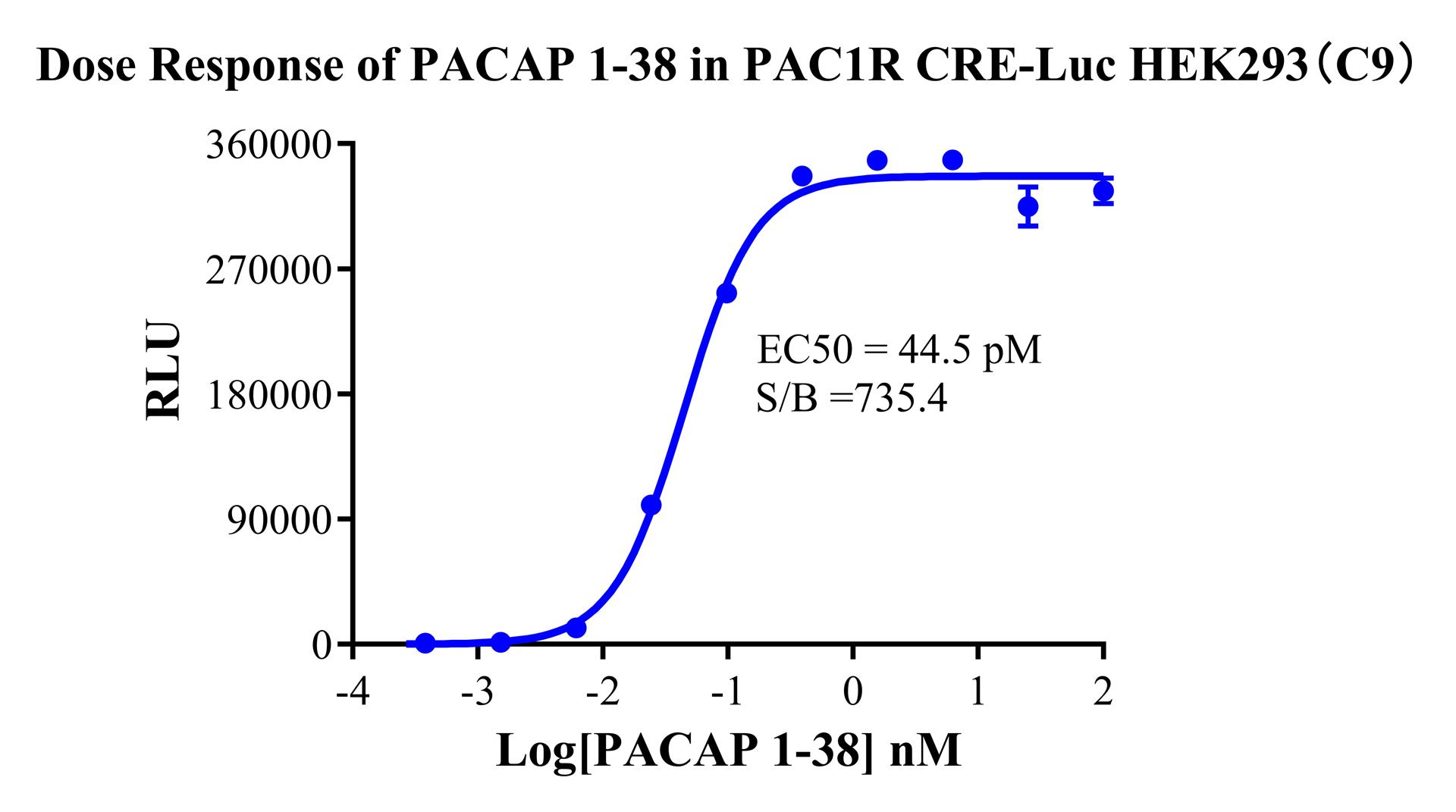
Figure 2 (functional validation): PACAP 1-38 produced a typical dose-dependent sigmoidal activation curve, with a stable EC₅₀ (generally at the pM level) and a wide signal window (>10-fold), demonstrating that this cell line responds to PAC1R agonists with high sensitivity.
Analysis of advantages: HEK293 cells have high transfection efficiency and stable protein expression levels, making them an ideal host for studies of the Gs-cAMP-CREB pathway. The CRE-Luc reporter system directly reflects the phosphorylation level of the transcription factor CREB and most closely represents the physiological endpoint of the PAC1R neuroprotection pathway.
2. CHO-K1 Human PAC1R CRE-Luc Cell Line RQP71565
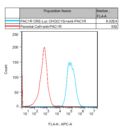
Figure 3: Flow cytometry confirmed stable, high PAC1R expression in CHO cells.
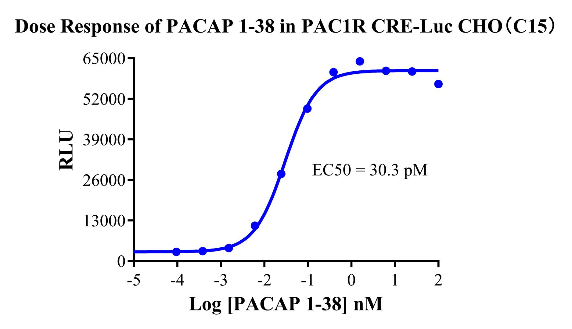
Figure 4: PACAP 1-38 activated the reporter gene in a dose-dependent manner, with a stable EC₅₀, validating the robustness and reproducibility of this model in a different host-cell background.
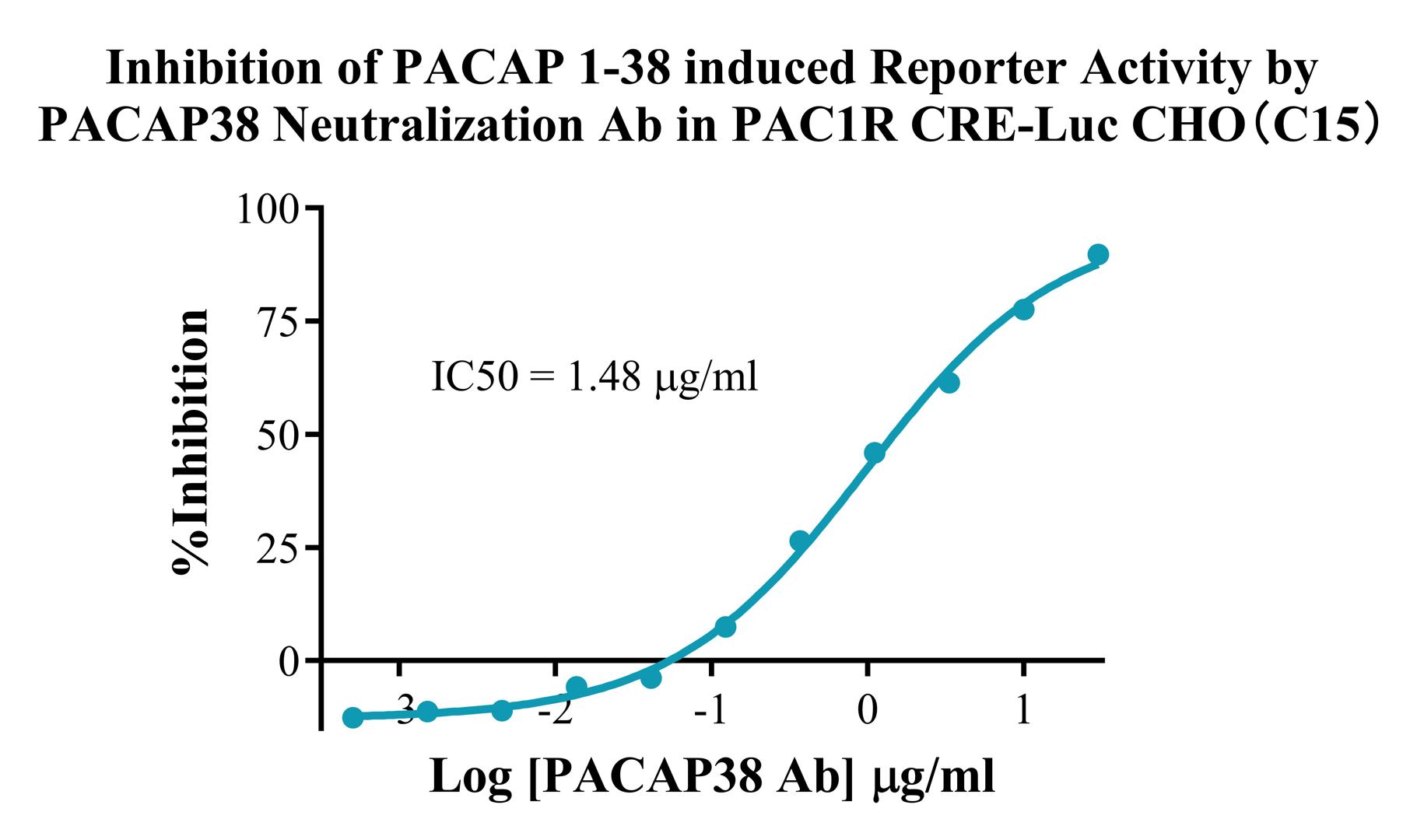
Figure 5 : The PACAP38 neutralizing antibody inhibited PACAP 1-38-induced reporter activity in a dose-dependent manner, demonstrating that this model can be used for potency determination of anti-PACAP antibodies and screening of PAC1R antagonists.
Analysis of advantages: CHO-K1 cells have the advantages of no endogenous PAC1R expression, simple culture, and high lot-to-lot consistency, making them the preferred host for medium- and high-throughput screening. The parallel availability of HEK293 and CHO-K1 host cells allows the activity of candidate molecules to be cross-validated and host-dependent effects to be excluded.
3. CHO-K1 Human PAC1R β-Arrestin Receptor Cell Line RQP71577
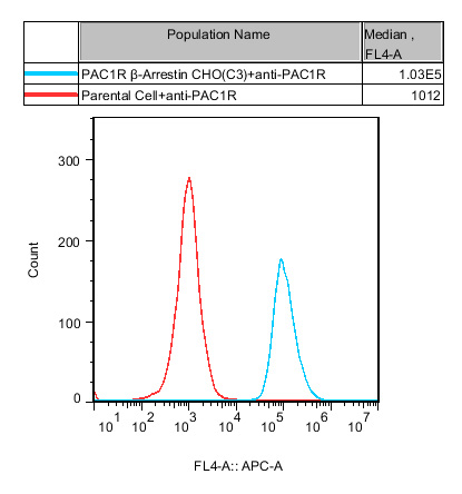
Figure 6: Flow cytometry confirmed stable co-expression of PAC1R and the β-arrestin reporter system in CHO cells.
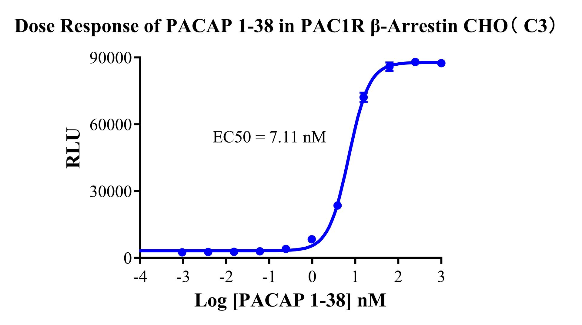
Figure 7: PACAP 1-38 induced β-arrestin recruitment in a dose-dependent manner, validating the sensitive response of this model to biased signaling.
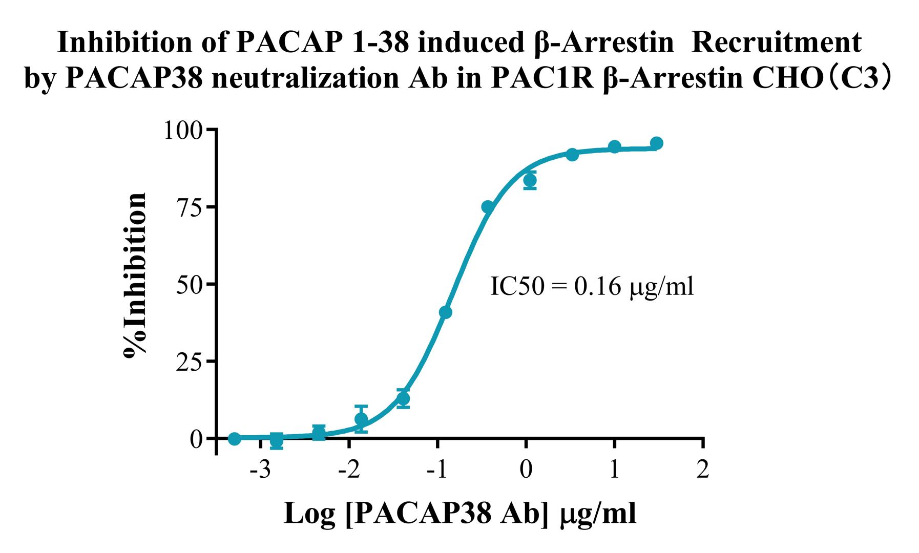
Figure 8: The PACAP38 neutralizing antibody inhibited β-arrestin recruitment in a dose-dependent manner, demonstrating that this model is also suitable for evaluating antibody neutralizing activity through the β-arrestin pathway.
Analysis of advantages: This is a core tool for biased ligand screening—it can distinguish G protein-biased agonists (strong Gs pathway activation and weak β-arrestin activation), β-arrestin-biased agonists (strong β-arrestin activation and weak Gs activation), and balanced agonists, providing a direct basis for screening candidate molecules with specific functional profiles. It can also be used to study the mechanisms of receptor internalization and desensitization.
4. CHO-K1 Human PAC1R Cell Line RQP71455
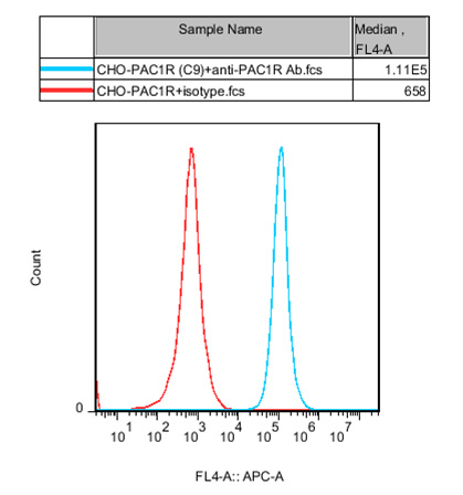
Figure 9: Flow cytometry confirmed stable, high PAC1R expression in CHO cells.
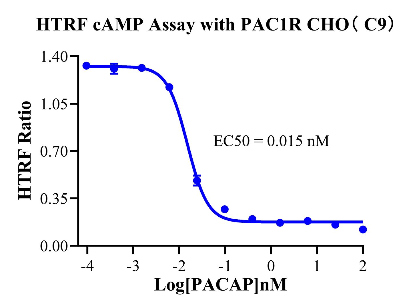
Figure 10: An HTRF (homogeneous time-resolved fluorescence) cAMP assay platform was used to measure PACAP 1-38-induced cAMP accumulation, producing a typical dose-response curve.
Analysis of advantages: The HTRF cAMP assay is the gold standard for studying the GPCR-Gs pathway and has the advantages of being wash-free and homogeneous, with a high signal-to-noise ratio, making it suitable for medium- and high-throughput screening. Compared with the CRE-Luc reporter system, the cAMP assay directly quantifies second-messenger levels and reflects more upstream and immediate signaling events.
Coordinated Application Strategy for the Four Models:
|
Development Stage |
Recommended Model |
Purpose |
|
Primary screening |
PAC1R CRE-Luc CHO |
High-throughput evaluation of the Gs pathway agonist/antagonist activity of compounds |
|
Mechanism confirmation |
PAC1R/CHO (cAMP assay) |
Verify the effects of candidate molecules on cAMP levels and exclude indirect effects of the reporter gene system |
|
Biased screening |
PAC1R β-Arrestin CHO |
Distinguish G protein pathway activity from β-arrestin pathway activity and screen biased ligands |
|
Cross-validation |
PAC1R CRE-Luc HEK293 |
Validate activity in different host backgrounds and exclude host dependence |
|
Antibody/fusion protein evaluation |
PAC1R CRE-Luc CHO + PAC1R β-Arrestin CHO |
Evaluate the effects of neutralizing antibodies on the Gs pathway and β-arrestin pathway in parallel |
Summary of Core Product Advantages:
|
Advantage |
Detailed Description |
|
High target specificity |
PAC1R has a 1,000-fold binding preference for PACAP over VIP and is a highly distinctive GPCR target in neuroscience. |
|
Complete signaling pathway coverage |
Three assay formats are provided simultaneously—Gs-cAMP-CREB (CRE-Luc), Gs-cAMP quantification (cAMP HTRF), and β-arrestin—to comprehensively analyze ligand functional profiles. |
|
Cross-validation in two host cells |
HEK293 and CHO host cells are provided in parallel to eliminate interference from the host background and increase data reliability. |
|
Biased screening capability |
The β-arrestin model can distinguish differences in activity between the G protein pathway and the β-arrestin pathway, helping to screen next-generation drugs with specific functional profiles. |
|
Broad application scenarios |
Covers multiple scenarios, including agonist screening, antagonist screening, neutralizing antibody potency determination, biased ligand development, and receptor internalization studies. |
|
High sensitivity and stability |
Each model has undergone monoclonal screening and functional validation, with stable EC₅₀ values and wide assay windows, making it suitable for high-throughput screening and experiments with high requirements for data reproducibility. |
Ⅴ. Typical Application Scenarios
PAC1R agonist screening (neuroprotection): Use PAC1R CRE-Luc CHO or PAC1R/CHO (cAMP assay) to screen small molecules or antibodies that can activate the Gs-cAMP-CREB pathway for neuroprotective treatment of stroke and neurodegenerative diseases.
PAC1R antagonist screening (pain/migraine): Using PACAP 1-38 as the agonist background, use CRE-Luc or cAMP assay models to screen small molecules or antibodies that can block PAC1R signaling for the treatment of migraine and neuropathic pain.
Biased ligand development: Use the CRE-Luc model and β-Arrestin model in parallel to screen G protein-biased agonists that activate only the Gs pathway (neuroprotection) without activating the β-arrestin pathway (which may cause receptor internalization and desensitization).
Potency determination of anti-PACAP neutralizing antibodies: Use PAC1R CRE-Luc CHO with PACAP 1-38 as the stimulus to evaluate the neutralizing activity of anti-PACAP antibodies for quality control of antibody drugs for migraine.
Bispecific antibody evaluation (e.g., PAC1R × CGRP): Use PAC1R-related models to evaluate the binding and functional activity of the PAC1R-targeting arm of a bispecific antibody.
Conclusion
With its extreme selectivity for PACAP, precise expression in the nervous system, and diverse downstream signaling network, PAC1R has become a highly attractive GPCR target in neuroscience drug development. From migraine to neuropathic pain and from stroke to neurodegenerative diseases, both PAC1R agonist and antagonist pathways have shown clear prospects for translation. The four PAC1R cell models launched by Reqbio—covering three major assay formats, namely the CRE-Luc reporter gene, HTRF cAMP quantification, and β-arrestin recruitment, together with HEK293 and CHO host backgrounds—provide drug developers worldwide with a complete, flexible, and reliable combination of evaluation tools.
Relying on cell functional engineering technology and a high-precision gene-editing platform, Reqbio continuously provides drug-testing cell models covering multiple target classes, including GPCRs, immunotherapy targets, and kinases, while also providing high-quality cell-based bioassay services.


news recommendation
CaSR: The Calcium Homeostasis Sentinel and a Core GPCR Target for Metabolic and Kidney Disease Research
CD3E: The T-Cell Activation Switch and Cell Models for Targeted Therapeutics
NFAT Signaling Pathway: A Critical Target for Drug R&D and Cell Models
TSHR Cell Model for Thyroid Cancer Drug Screening | GPCR Assay Platform-Reqbio
We Are Pleased to Announce: Global Commercial Licensing Rights for Jurkat E6.1, CHO-K1, HEK293, THP-1 and RAJI Cell Lines Officially Secured.
Explore