

Home>
Resources>
Learning>
Articles>
IL-36/IL-36R: A Rising Star in the Treatment of Pustular Psoriasis and Broad-Spectrum Inflammatory Diseases
IL-36/IL-36R: A Rising Star in the Treatment of Pustular Psoriasis and Broad-Spectrum Inflammatory Diseases
Within the IL-1 cytokine superfamily, Interleukin-36 (IL-36) is rapidly emerging from a relatively understudied member to a key target in inflammatory diseases. This cytokine family comprises three agonists (IL-36α, IL-36β, and IL-36γ) and one antagonist (IL-36 receptor antagonist, IL-36Ra), and plays an important immunoregulatory role in barrier tissues such as the skin, lungs, and intestine. Aberrant activation of IL-36 signaling is closely associated with severe inflammatory diseases, including generalized pustular psoriasis (GPP), palmoplantar pustulosis (PPP), pyoderma gangrenosum (PG), and inflammatory bowel disease (IBD). In 2022, the IL-36R-targeting monoclonal antibody Spesolimab received FDA approval, becoming the first approved therapy for GPP flares and validating the feasibility and clinical value of the IL-36/IL-36R axis as a drug target. Leveraging its established cell engineering platform, Reqbio has developed adherent and suspension IL-36 effector reporter cell models, providing researchers with reliable and flexible cell-based tools for target-based drug screening.
I. The IL-36 Cytokine Family: A Quartet within the IL-1 Superfamily
The IL-36 cytokine family consists of four members whose gene cluster is located in the q14 region of human chromosome 2. These four members were first identified in 1999 and, because of their high sequence homology with IL-1, were named IL-1F6, IL-1F8, IL-1F9, and IL-1F5. In 2011, they were formally assigned the unified names IL-36α, IL-36β, IL-36γ, and IL-36Ra, respectively.
|
Member |
Former name |
Functional type |
Key features |
|
IL-36α |
IL-1F6 |
Agonist |
Requires proteolytic cleavage for activation; mainly expressed in the epithelium of the skin, lungs, and intestine |
|
IL-36β |
IL-1F8 |
Agonist |
Requires proteolytic cleavage for activation; activated by cathepsin G |
|
IL-36γ |
IL-1F9 |
Agonist |
Requires proteolytic cleavage for activation; activated by elastase |
|
IL-36Ra |
IL-1F5 |
Antagonist |
The only endogenous antagonist; competitively blocks receptor binding |
A distinctive feature of the IL-36 cytokine family is that all of its agonists require proteolytic cleavage for activation. Neutrophil-derived cathepsin G can activate IL-36α and IL-36β, whereas elastase can activate IL-36β and IL-36γ. This cleavage-dependent activation mechanism closely couples IL-36 signaling to neutrophil infiltration: at sites of inflammation, neutrophils release proteases that convert inactive precursors into highly active ligands, creating an inflammatory amplification loop. Proteolytic cleavage can increase the affinity of IL-36 ligands for their receptor by several hundred-fold, providing an efficient trigger-like regulatory mechanism.
IL-36 is expressed in a variety of human tissues and cell types, including skin keratinocytes, bronchial epithelial cells, intestinal epithelial cells, and immune cells such as neutrophils, monocytes/macrophages, and T cells. This broad expression profile underlies its extensive involvement in inflammation across multiple organs.
II. The IL-36/IL-36R Axis: Signaling and Disease Associations
All IL-36 agonists share the same receptor complex: a heterodimer formed by the IL-36 receptor (IL-36R; IL1Rrp2) and the IL-1 receptor accessory protein (IL-1RAcP).
The central role of IL-36 in inflammatory diseases:
Psoriasis and GPP: IL-36 is a key driver of the inflammatory loop in psoriasis. In patients with GPP, loss-of-function mutations in IL36RN, the gene encoding IL-36Ra, lead to uncontrolled IL-36 signaling and represent a central genetic cause of the disease.
Palmoplantar pustulosis (PPP): Aberrant activation of IL-36 signaling in the palms and soles drives the formation of sterile pustules.
Pyoderma gangrenosum (PG): IL-36 contributes to the maintenance of ulcerative inflammation in the skin.
Inflammatory bowel disease (IBD): Aberrant IL-36 expression in the intestinal epithelial barrier contributes to chronic intestinal inflammation.
Pulmonary inflammation: IL-36 is involved in the pathological processes of inflammatory airway diseases such as asthma and chronic obstructive pulmonary disease (COPD).
III. Drug Development Targeting IL-36/IL-36R: Expanding beyond GPP
The clinical druggability of the IL-36/IL-36R axis has been well validated. Current drug development efforts targeting this axis focus primarily on anti-IL-36R monoclonal antibodies, while strategies that modulate the balance between agonists and antagonists are also receiving attention.
|
Name |
Developer |
Stage |
Target |
Drug type |
Indications |
|
Spesolimab |
Boehringer Ingelheim |
Approved (FDA, 2022) |
IL-36R |
Monoclonal antibody |
GPP, hidradenitis suppurativa, pyoderma gangrenosum, palmoplantar pustulosis |
|
Imsidolimab |
AnaptysBio |
Phase II/III |
IL-36R |
Monoclonal antibody |
GPP, palmoplantar pustulosis, hidradenitis suppurativa, atopic dermatitis |
|
HB0034 |
HuotaBio |
Phase I |
IL-36R |
Monoclonal antibody |
GPP and other inflammatory diseases |
|
IMG008 |
Inmagene Bio |
Preclinical |
IL-36R |
Monoclonal antibody |
Inflammatory diseases |
Drug development trends:
Expansion from a single target to multiple indications: GPP was the first validated indication, and clinical development is gradually expanding to hidradenitis suppurativa, pyoderma gangrenosum, palmoplantar pustulosis, and atopic dermatitis.
Biologics predominate: All IL-36-targeted therapies currently in clinical development are monoclonal antibodies, while small-molecule candidates remain at earlier stages.
Agonist-antagonist balance: The identification of IL-36Ra as an endogenous antagonist provides a natural template and mechanistic reference for the development of IL-36 signaling modulators.
IV. Advancing IL-36-Targeted Drug Development: Reqbio Cell Models and Data Advantages
Functional cell models that sensitively and specifically detect ligand-receptor binding and downstream signal activation are essential tools for screening and bioactivity assays of IL-36/IL-36R-targeted therapeutics. Reqbio has developed both adherent and suspension IL-36 effector reporter cell models for the IL-36 signaling pathway, supporting different experimental settings and throughput requirements.
Reqbio IL-36 cell models:
|
Cell name |
Catalog no. |
Cell background |
Cell format |
Assay format |
Core applications |
|
IL36 Effector Reporter Cell (Adherent) |
RQP74511 |
Adherent cells |
Adherent |
Luciferase reporter gene |
IL-36 agonist activity and blocking antibody screening (Figure 1) |
|
Jurkat E6.1 Human IL36 Effector Reporter Cell |
RQP74119 |
Jurkat E6.1 (human T-cell lymphoma) |
Suspension |
Luciferase reporter gene |
IL-36 activity assays and blocking antibody potency determination (Figure 3) |
Validation data and analysis of advantages:
1. IL36 Effector Reporter Cell (Adherent) (RQP74511)
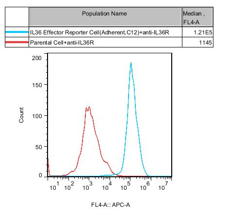
Figure 1. Recombinant IL36 Effector Reporter Cell(Adherent) stably expressing IL36R.
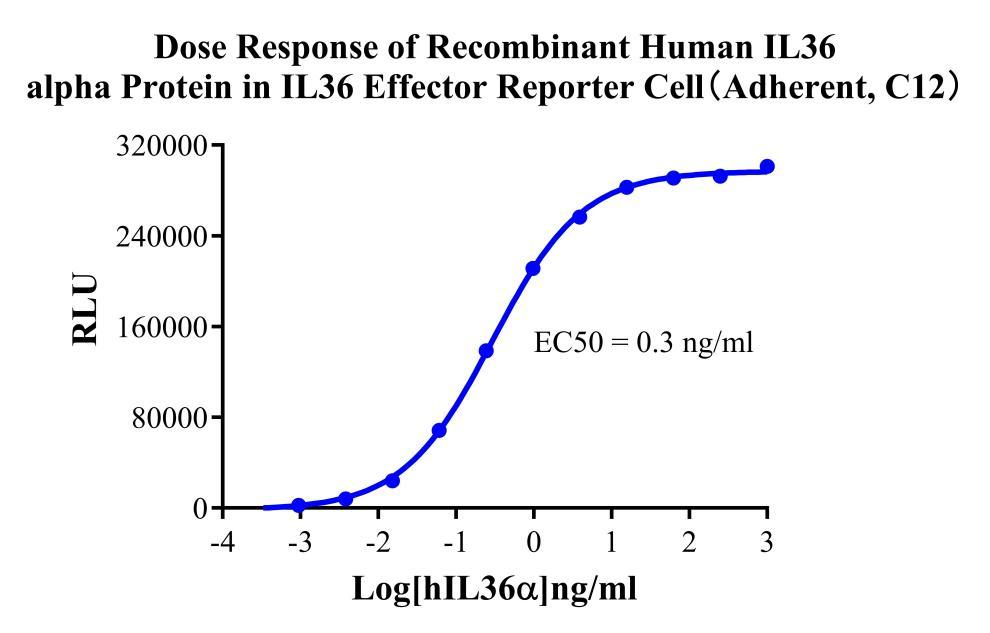
Figure 2. Dose Response of Recombinant Human IL36 alpha Protein in IL36 Effector Reporter Cell(Adherent, C12).
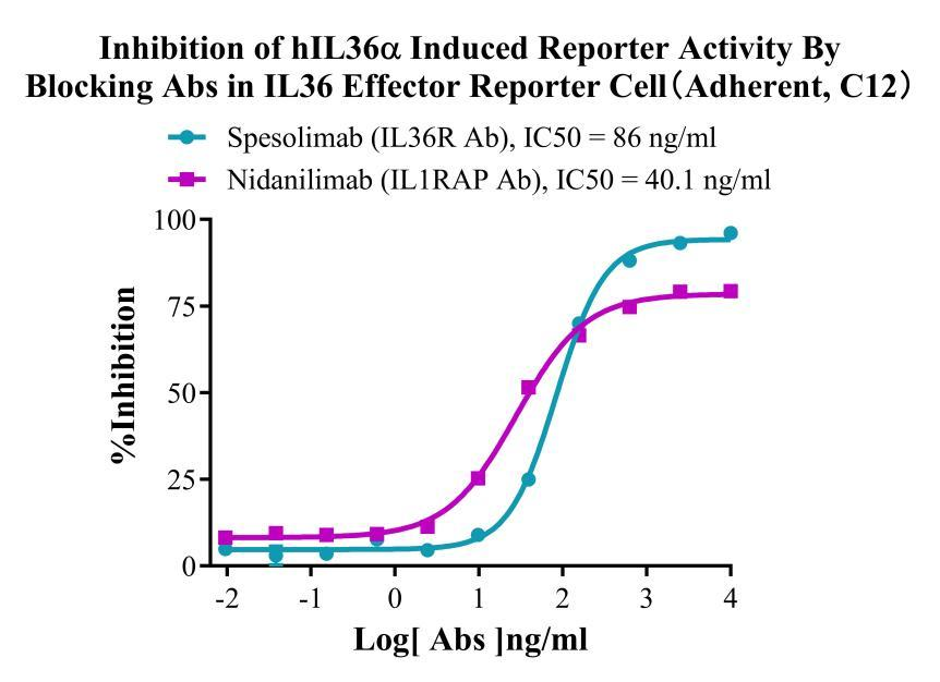
Figure 3. Inhibition of hIL36α Induced Reporter Activity By Blocking Abs in IL36 Effector Reporter Cell(Adherent, C12).
Assay principle: The adherent reporter cells stably express IL-36R and a luciferase reporter gene driven by an NF-κB response element. Following the addition of IL-36α (or IL-36β/γ), reporter gene expression is activated through the IL-36R/IL-1RAcP/NF-κB signaling pathway.
Data interpretation: The figure shows that IL-36α-induced reporter activity is inhibited by multiple blocking antibodies in a dose-dependent manner, producing clear S-shaped inhibition curves.
Advantages:
• Broad target coverage: The model can evaluate blocking antibodies directed against different components of the pathway, including anti-IL-36 ligand, anti-IL-36R, and anti-IL-1RAcP antibodies, covering the principal therapeutic approaches targeting this pathway.
• Physiological relevance: The model uses the natural IL-36 agonists IL-36α, IL-36β, and IL-36γ as stimuli, more closely reflecting the in vivo mechanism.
• Adherent format: The model is suitable for conventional manual laboratory workflows. Strong cell adherence facilitates medium changes and sample addition.
2. Jurkat E6.1 Human IL36 Effector Reporter Cell (RQP74119)
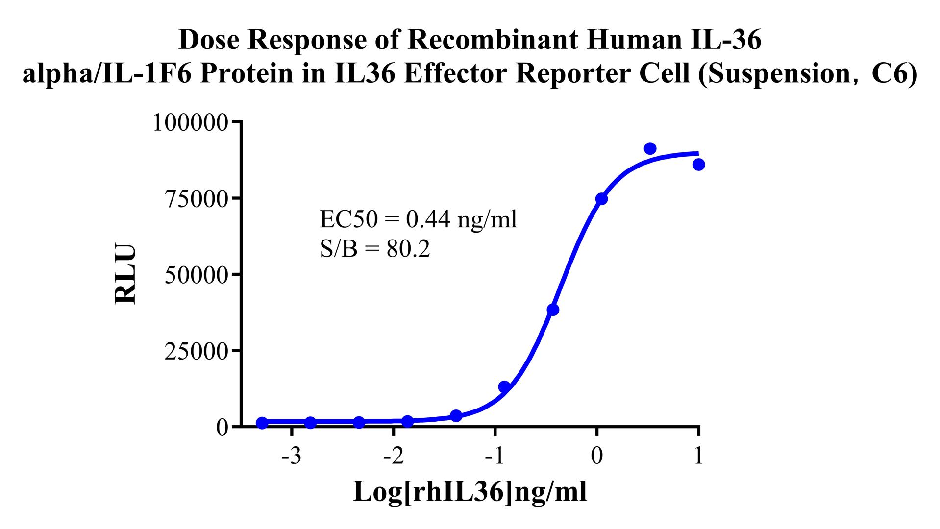
Figure 4. Dose Response of Recombinant Human IL-36 alpha/IL-1F6 Protein in IL36 Effector Reporter Cell(Suspension, C6).
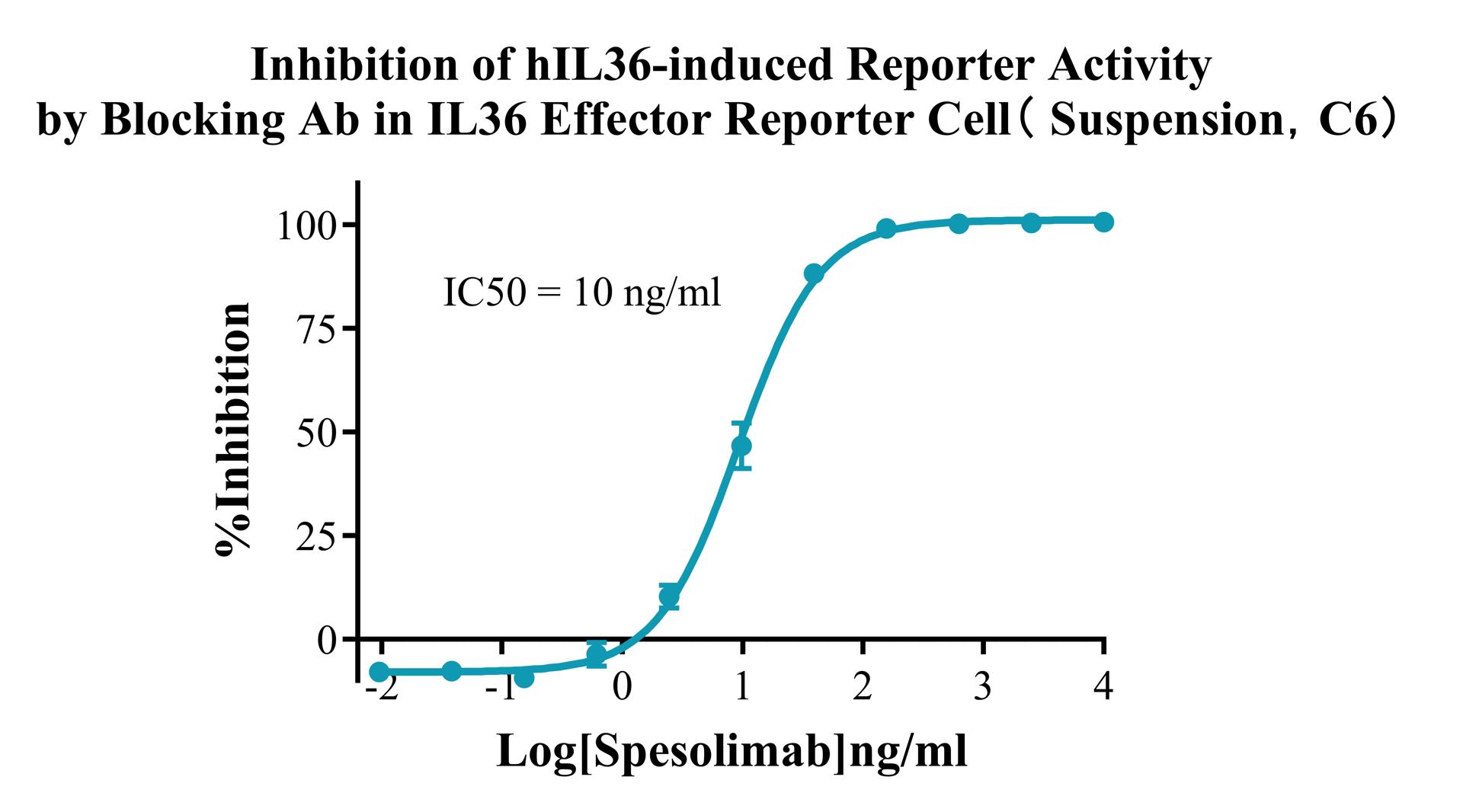
Figure 5. Inhibition of hIL36-induced Reporter Activity by Blocking Ab in IL36 Effector Reporter Cell(Suspension, C6).
Assay principle: Based on the human Jurkat E6.1 T-cell lymphoma background, the model stably integrates IL-36R and an NF-κB reporter system and is maintained in suspension culture. The addition of an IL-36 agonist activates downstream signaling.
Data interpretation: The figure shows dose-dependent inhibition of IL-36-induced reporter activity by a blocking antibody.
Advantages:
• Suspension format: No trypsinization is required for passaging, simplifying routine handling. The model is fully compatible with automated liquid-handling workstations and high-throughput screening platforms, making it a preferred tool for HTS.
• Human T-cell background: Jurkat E6.1 is a widely used T-cell line in immunology research and more closely reflects the signaling characteristics of IL-36 in lymphocytes.
• Ready for direct assay setup: Suspension cells can be sampled directly from the culture flask and seeded into assay plates, substantially reducing experiment preparation time.
• Robust blocking validation: The clear dose-dependent blocking curve shown in Figure 4 demonstrates that the model can be used for precise potency determination of neutralizing antibodies, with accurate quantification of IC₅₀ values.
Selection guide: adherent vs. suspension models
|
Application |
Recommended model |
Reason |
|
Small-scale screening in conventional laboratories |
Adherent model (RQP74511) |
Broad operating window; cell status is easy to monitor; suitable for method development |
|
Automated high-throughput screening |
Suspension model (RQP74119) |
No dissociation required; compatible with automated equipment; high throughput and good reproducibility |
|
Drug lot-release testing |
Either model |
The suspension model is easier to standardize and scale |
|
Blocking antibody potency determination |
Suspension model preferred |
Simple operation and high precision in IC₅₀ determination |
|
Cross-evaluation of multiple ligands |
Adherent model |
Facilitates the design of ligand concentration gradients in multiwell plates |
Summary of core product advantages:
|
Advantage |
Description |
|
Clinically validated target |
The FDA approval of Spesolimab makes IL-36/IL-36R a successful example of translation from genomics to the clinic in inflammatory diseases, reducing target-related drug development risk. |
|
Flexible dual-format options |
Adherent and suspension cell models are available to support manual workflows and automated high-throughput screening, respectively. |
|
Pathway-specific design |
Designed around the native IL-36R/MyD88/NF-κB signaling axis to directly reflect the pharmacological effects of IL-36 agonists and blocking agents. |
|
Compatibility with cleavage-activated ligands |
The models use mature (post-cleavage) IL-36 ligands, bypassing the proteolytic cleavage step and focusing on the evaluation of receptor binding and signal inhibition. |
|
Comprehensive blocking validation |
Figures 1 and 3 show clear dose-dependent inhibition curves, enabling direct IC₅₀ determination and comparison of candidate blocking antibodies. |
|
Ready-to-use products |
Following monoclonal screening and functional validation, the cells can be used after recovery, substantially shortening research and development timelines. |
V. Representative Applications
Screening and potency determination of anti-IL-36R monoclonal antibodies: Use suspension or adherent reporter cells with IL-36α, IL-36β, or IL-36γ as the agonist to evaluate the receptor-blocking activity of anti-IL-36R antibodies, such as Spesolimab biosimilars or novel molecules.
Development of neutralizing antibodies against IL-36 ligands: Use the same assay system with a specific IL-36 ligand as the target to screen monoclonal antibodies that selectively neutralize IL-36α, IL-36β, or IL-36γ.
Comparison of IL-36 agonist activity: Use reporter cells to compare the relative activities of IL-36α, IL-36β, and IL-36γ in parallel for ligand quality control and structure-activity relationship studies.
Screening of IL-36Ra analogs or mimetics: Screen small molecules or peptide therapeutics that mimic the function of endogenous IL-36Ra by competitively blocking the association of IL-36R with IL-1RAcP and inhibiting inflammatory signaling.
Evaluation of multispecific antibodies: Assess the activity of the IL-36R-targeting arm in bispecific or multispecific antibodies directed against IL-36R and other targets, such as IL-17 or IL-23.
Drug development for IL-36-related inflammatory diseases: Support candidate screening and activity testing for indications including GPP, palmoplantar pustulosis, pyoderma gangrenosum, and hidradenitis suppurativa.
VI. Conclusion
The IL-36/IL-36R axis is a central IL-1 superfamily pathway regulating inflammation in barrier tissues, and the successful approval of Spesolimab has validated its clinical druggability. The range of indications under investigation continues to expand, from GPP and hidradenitis suppurativa to palmoplantar pustulosis and inflammatory bowel disease. With pathway-specific design, flexible experimental formats, and blocking validation, Reqbio's adherent and suspension IL-36 effector reporter cell models provide drug developers worldwide with reliable and efficient evaluation tools.


news recommendation
CaSR: The Calcium Homeostasis Sentinel and a Core GPCR Target for Metabolic and Kidney Disease Research
CD3E: The T-Cell Activation Switch and Cell Models for Targeted Therapeutics
NFAT Signaling Pathway: A Critical Target for Drug R&D and Cell Models
TSHR Cell Model for Thyroid Cancer Drug Screening | GPCR Assay Platform-Reqbio
We Are Pleased to Announce: Global Commercial Licensing Rights for Jurkat E6.1, CHO-K1, HEK293, THP-1 and RAJI Cell Lines Officially Secured.
Explore