

Raji Human TCR Activator Cell
Cat. No: RQP74015
Size: 1 vial of frozen cells (>1E6 per vial in 1 mL)
Unit Price: Contact For Pricing
Product Info
Description
Assay Data
Cell Culture
| Cat. No | RQP74015 |
| Product Name | Raji Human TCR Activator Cell |
| Product Type | Reporter Cell |
| Culture Properties | suspension |
| Stability | 32passages (in-house test, that not means the cell line will be instable beyond the passages we tested.) |
| Mycoplasma Status | Negative |
| Culture Medium | RPMI-1640+10%FBS+1μg/ml puromycin |
| Freeze Medium | 90% FBS+10% DMSO |
| Storage Conditions | Liquid nitrogen immediately upon delivery |
| Application | Functional(Report Gene) Assay |
For research use only. Not intended for human or animal clinical trials, therapeutic or diagnostic use.
T cells play a central role in cell-mediated immunity, mediating long-lasting, antigen-specific effector and memory responses. T cells serve as targets for numerous immunotherapies, including checkpoint inhibitors, bispecific T-cell engagers, and immune agonists. Furthermore, T cells themselves have been investigated as therapeutic agents—specifically through chimeric antigen receptor (CAR) engineering or autologous adoptive transfer—for the treatment of hematological malignancies. In recent years, various immunotherapeutic strategies aimed at inducing, enhancing, and/or engineering T-cell responses have emerged as promising approaches for treating diseases such as cancer and autoimmune disorders.
T-cell activation is initiated by the engagement of the T-cell receptor (TCR) and CD3 complex, followed by the engagement of co-stimulatory molecules, such as the CD28 receptor. The concerted action of these receptors on the cell surface triggers intracellular signaling events and the activation of nuclear transcription factors, including NFAT, NFκB, and AP-1. Specifically, engagement of the TCR/CD3 complex induces the phosphorylation of PLC-γ, leading to an increase in intracellular Ca²⁺ levels and the transcriptional activation of NFAT; conversely, the combined action of the TCR/CD3 complex and the co-stimulatory receptor CD28 leads to the activation of ERK/JNK and IκB kinase (IKK), which in turn regulate the transcriptional activation of the AP-1 and NFκB pathways, respectively. The IL-2 promoter contains DNA-binding sites for NFAT, NF-κB, and AP-1. Consequently, the combined action of the TCR/CD3 complex and CD28 results in the production of IL-2, which can therefore serve as a measurable indicator of T-cell activation.
The Raji Human TCR Activator Cell model effectively mimics the in vivo CD28&CD80/CD86 or CTLA4 signaling transduction process. The principle is illustrated in the figure below.
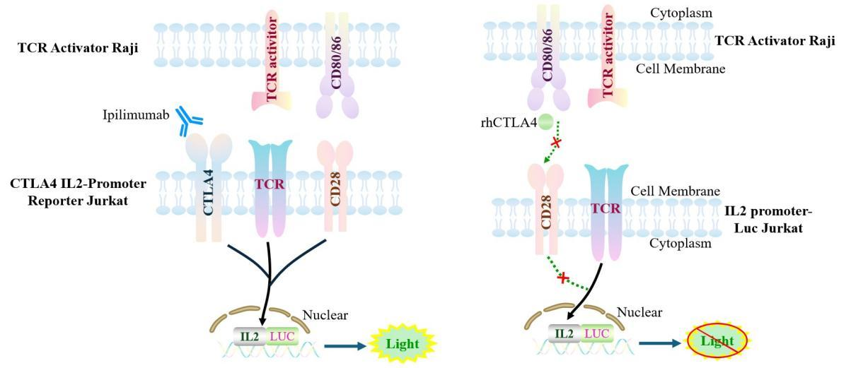
Figure 1. Schematic diagram of the Raji Human TCR Activator Cell cell model.
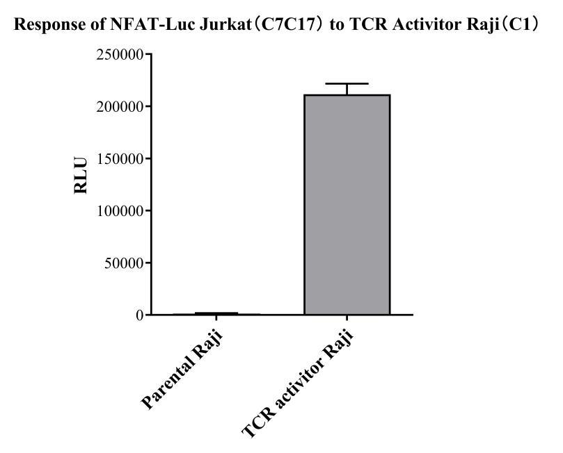
Figure 2. Response of NFAT-Luc Jurkat.(C7C17) to TCR Activitor Raji(C1).
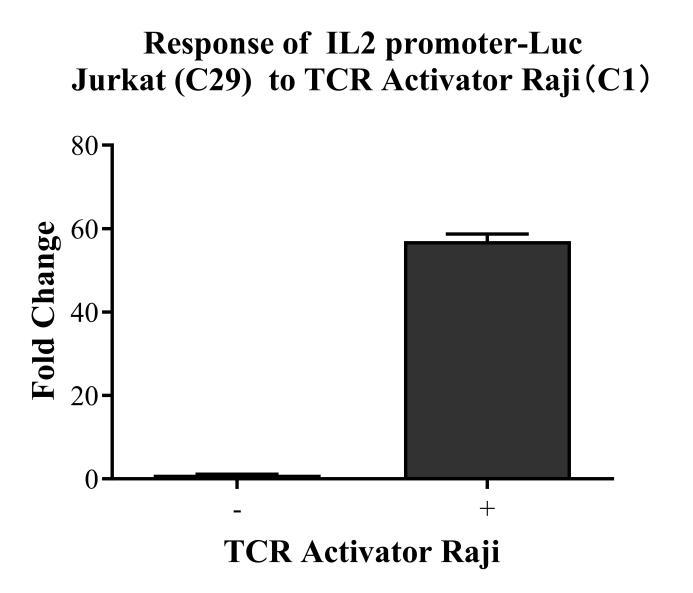
Figure 3. Response of IL2 promoter-Luc Jurkat (C29) to TCR Activator Raji (C1).
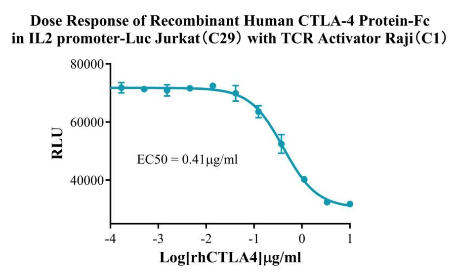
Figure 4. Dose Response of Recombinant Human CTLA-4 Protein-Fc in IL2 Promoter-Luc Jurkat (C29)with TCR Activitor Raji (C1).
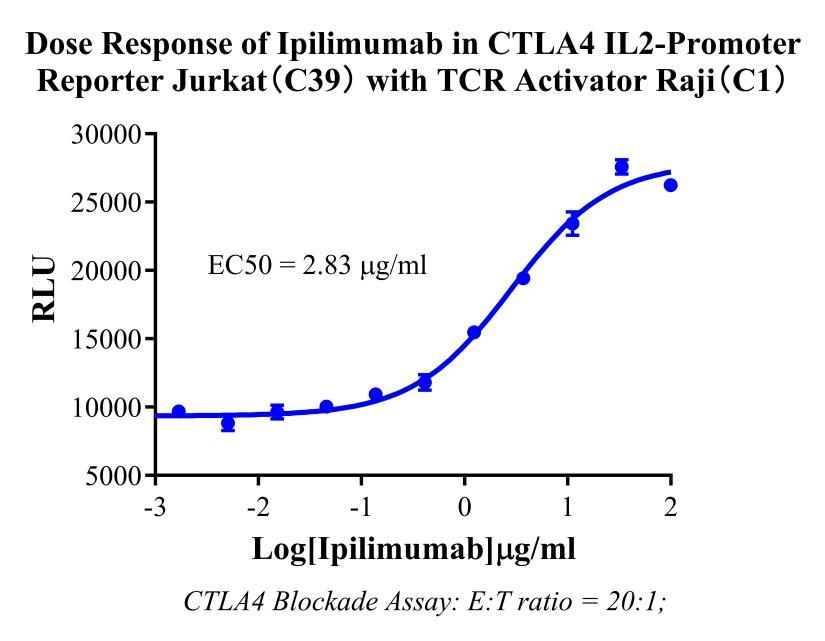
Figure 5. Dose Response of Ipilimumab in CTLA4 IL-2 Promoter Reporter-Jurkat (C39)with TCR Activitor Raji (C1).
Cell Passage Procedures
1.This cell line grows in suspension.
2.Upon receipt, cells should be thawed immediately or stored in liquid nitrogen until use.
3.Before thawing, pre-warm the water bath and culture medium to 37 °C, and prepare a small amount of dry ice.
4.Remove the cryovial from storage and transport it to the cell culture laboratory on dry ice.
5.Rapidly thaw the cells in a 37 °C water bath. Once the cells are completely thawed, spray the cryovial with 70% ethanol for disinfection and transfer it to a biosafety cabinet.
6.Add 10 mL of pre-warmed culture medium into a 15 mL centrifuge tube. Transfer the contents of the cryovial into the tube and centrifuge at 1000 rpm for 5 minutes.
7.Carefully discard the supernatant. Resuspend the cell pellet in 5 mL of pre-warmed culture medium by gentle pipetting. Immediately perform cell counting and adjust the cell density to 3–6 × 10⁵ cells/mL based on the counting results, then transfer the cells into a culture flask.
8.Count the cells every 1–2 days. When the cell density exceeds 1 × 10⁶ cells/mL, passage the cells promptly or add fresh culture medium. Maintain the cell density between 2 × 10⁵ and 1 × 10⁶ cells/mL.
Suspension Cell Cryopreservation Procedure:
1.Collect 8 × 10⁶ cells, centrifuge, and discard the supernatant.
2.Add 1 mL of cell freezing medium (90% FBS + 10% DMSO) and gently pipette to mix thoroughly. Transfer the suspension into a cryovial.
3.Immediately place the cryovial into a controlled-rate freezing container (Nalgene 5100-0001), fill with isopropanol up to the indicated level, and store at −80 °C.
4.After 24 hours, transfer the cryovial to liquid nitrogen for long-term storage.
Related products
CHO-K1 Human CCR4 Cell Line
HEK293 Human NK1R CRE-Luc Cell Line
Raji-Luc-GFP
Jurkat E6.1-Luc
THP-1-GFP
THP-1-Luc
Raji-GFP
Raji-Luc
Jurkat E6.1-GFP
HEK293 Human GAL4-Luc Cell
We Are Pleased to Announce: Global Commercial Licensing Rights for Jurkat E6.1, CHO-K1, and HEK293 Cell Lines Officially Secured.
Explore