

Jurkat E6.1 Human NFAT-Luc Cell
Cat. No: RQP74025
Size: 1 vial of frozen cells (>1E6 per vial in 1 mL)
Unit Price: Contact For Pricing
Product Info
Description
Assay Data
Cell Culture
| Cat. No | RQP74025 |
| Product Name | Jurkat E6.1 Human NFAT-Luc Cell |
| Product Type | Reporter Cell |
| Culture Properties | suspension |
| Stability | 32passages (in-house test, that not means the cell line will be instable beyond the passages we tested.) |
| Mycoplasma Status | Negative |
| Culture Medium | RPMI-1640+10%FBS+800μg/ml Hygromycin B |
| Freeze Medium | 90% FBS+10% DMSO |
| Storage Conditions | Liquid nitrogen immediately upon delivery |
| Application | Functional(Report Gene) Assay |
For research use only. Not intended for human or animal clinical trials, therapeutic or diagnostic use.
Intracellular calcium (Ca²⁺) regulates a wide range of cellular functions by activating various downstream cellular mediators. Elevated levels of intracellular Ca²⁺ lead to the activation of calcineurin—a Ca²⁺- and calmodulin-dependent phosphatase. Calcineurin dephosphorylates and activates the Nuclear Factor of Activated T-cells (NFAT) family of transcription factors, a family that plays pivotal roles in immune function, inflammatory responses, angiogenesis, and cell proliferation and differentiation.
The NFAT family comprises five members: NFAT1, NFAT2, NFAT3, NFAT4, and NFAT5. With the exception of NFAT5, all other members of this family are regulated by changes in intracellular Ca²⁺ levels. NFAT proteins possess a conserved N-terminal regulatory domain containing multiple serine residues, which remain phosphorylated under resting conditions. The nuclear translocation of NFAT proteins is inhibited when this N-terminal regulatory domain is phosphorylated. Upon cellular stimulation, the dephosphorylation of these serine residues is mediated by the Ca²⁺-dependent phosphatase, calcineurin. This dephosphorylation exposes nuclear localization signals, thereby facilitating nuclear translocation and the transcriptional induction of NFAT-regulated genes. In response to calcineurin-mediated Ca²⁺ activation, NFAT proteins can either independently induce gene expression or act synergistically with other transcription factors to do so. NFAT proteins bind to and function synergistically with other transcription factors—such as Activator Protein 1 (AP-1)—to induce gene transcription. Through these interactions with other transcription factors, NFAT proteins are able to integrate signals originating from diverse pathways and execute a multitude of cellular functions. One of the critical functions of NFAT proteins within immune cells is the induction of inflammatory cytokine expression.
The Jurkat E6.1 Human NFAT-Luc Cell model effectively mimics the in vivo NFAT signaling transduction process. The principle is illustrated in the figure below.
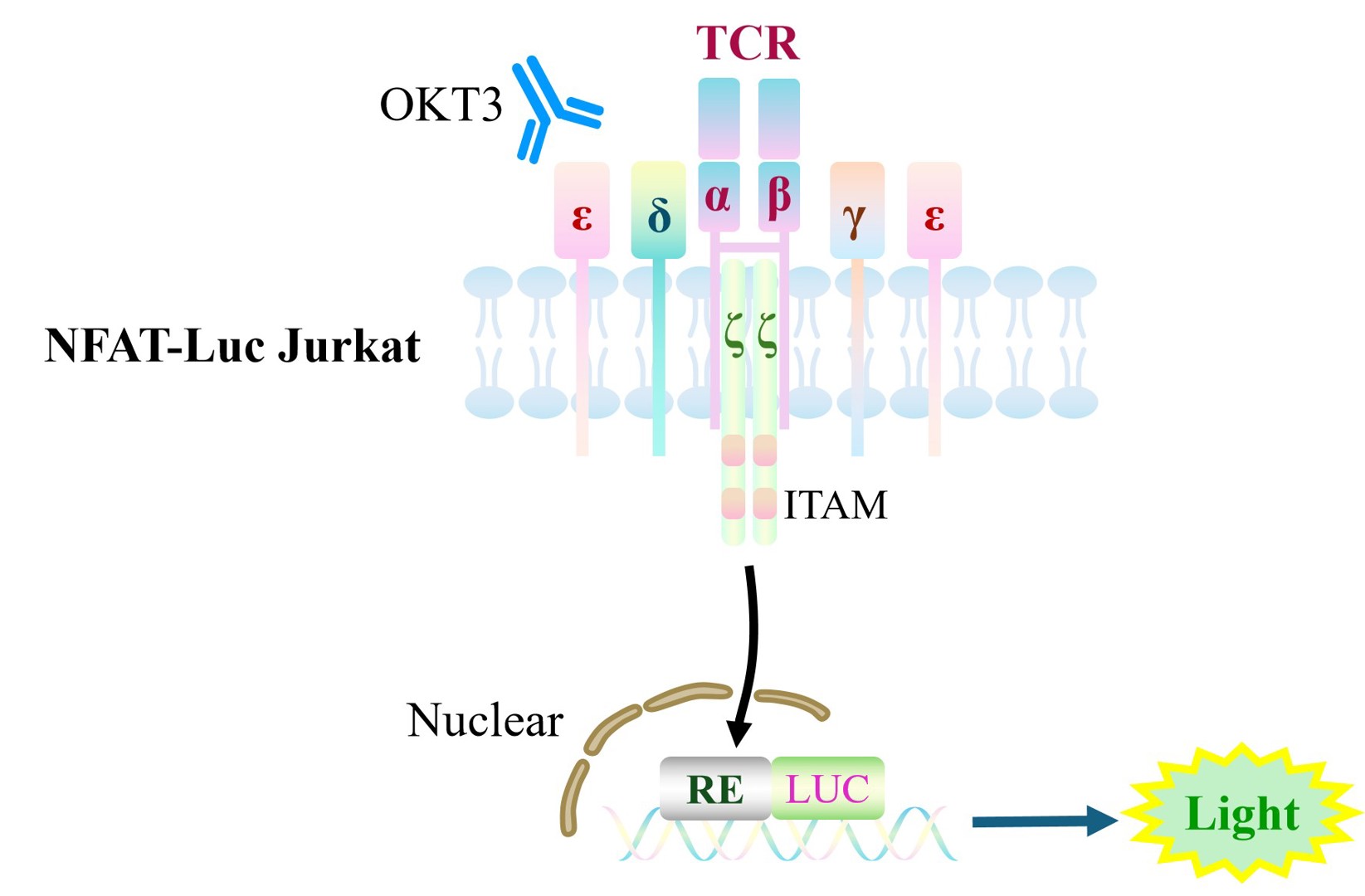
Figure 1. Schematic diagram of the Jurkat E6.1 Human NFAT-Luc Cell model.
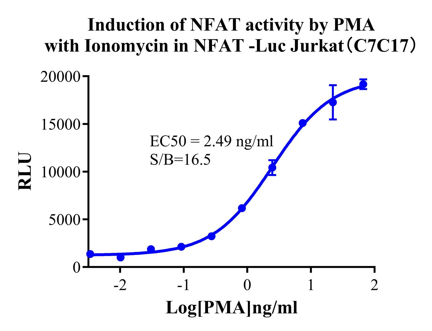
Figure 2. Induction of NFAT activity by PMA with Ionomycin in NFAT-Luc Jurkat(C7C17).
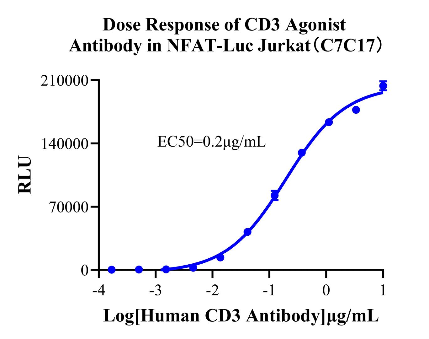
Figure 3. Dose Response of CD3 Agonist Antibody in NFAT-Luc Jurkat(C7C17).
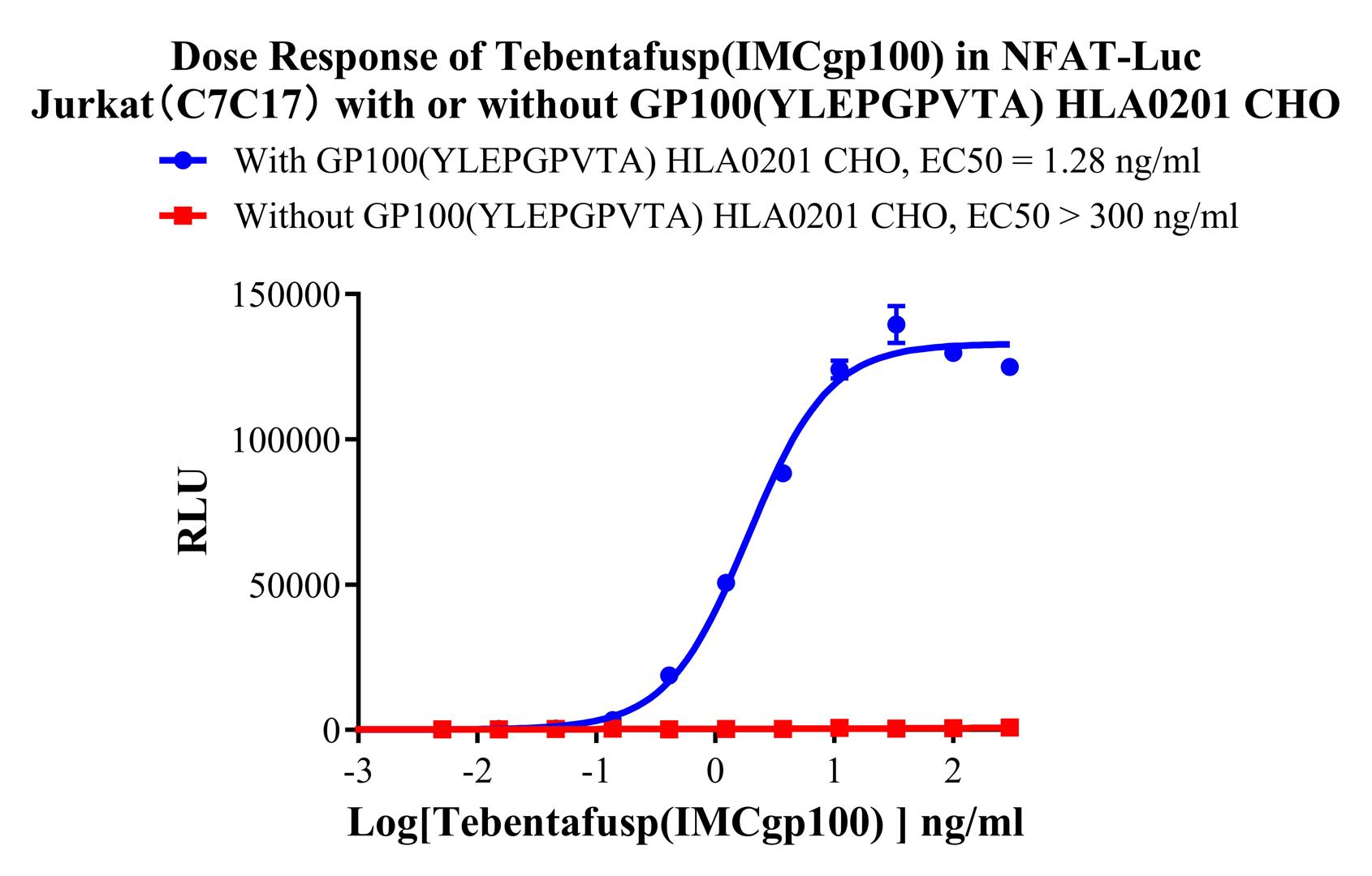
Figure 4. Dose Response of Tebentafusp (IMCgp100) in NFAT-Luc Jurkat(C7C17) with or without GP100(YLEPGPVTA) HLA0201 CHO.
Cell Passage Procedures
1.This cell line grows in suspension.
2.Upon receipt, cells should be thawed immediately or stored in liquid nitrogen until use.
3.Before thawing, pre-warm the water bath and culture medium to 37 °C, and prepare a small amount of dry ice.
4.Remove the cryovial from storage and transport it to the cell culture laboratory on dry ice.
5.Rapidly thaw the cells in a 37 °C water bath. Once the cells are completely thawed, spray the cryovial with 70% ethanol for disinfection and transfer it to a biosafety cabinet.
6.Add 10 mL of pre-warmed culture medium into a 15 mL centrifuge tube. Transfer the contents of the cryovial into the tube and centrifuge at 1000 rpm for 5 minutes.
7.Carefully discard the supernatant. Resuspend the cell pellet in 5 mL of pre-warmed culture medium by gentle pipetting. Immediately perform cell counting and adjust the cell density to 3–6 × 10⁵ cells/mL based on the counting results, then transfer the cells into a culture flask.
8.Count the cells every 1–2 days. When the cell density exceeds 1 × 10⁶ cells/mL, passage the cells promptly or add fresh culture medium. Maintain the cell density between 2 × 10⁵ and 1 × 10⁶ cells/mL.
Suspension Cell Cryopreservation Procedure:
1.Collect 8 × 10⁶ cells, centrifuge, and discard the supernatant.
2.Add 1 mL of cell freezing medium (90% FBS + 10% DMSO) and gently pipette to mix thoroughly. Transfer the suspension into a cryovial.
3.Immediately place the cryovial into a controlled-rate freezing container (Nalgene 5100-0001), fill with isopropanol up to the indicated level, and store at −80 °C.
4.After 24 hours, transfer the cryovial to liquid nitrogen for long-term storage.
Related products
CHO-K1 Human CCR4 Cell Line
HEK293 Human NK1R CRE-Luc Cell Line
Raji-Luc-GFP
Jurkat E6.1-Luc
THP-1-GFP
THP-1-Luc
Raji-GFP
Raji-Luc
Jurkat E6.1-GFP
HEK293 Human GAL4-Luc Cell
We Are Pleased to Announce: Global Commercial Licensing Rights for Jurkat E6.1, CHO-K1, and HEK293 Cell Lines Officially Secured.
Explore