

Jurkat E6.1 Human TNFR2(TNFR1 KO) Effector Reporter Cell
Cat. No: RQP74539
Size: 1 vial of frozen cells (>1E6 per vial in 1 mL)
Unit Price: Contact For Pricing
Product Info
Description
Biological Information
Assay Data
Cell Culture
| Cat. No | RQP74539 |
| Product Name | Jurkat E6.1 Human TNFR2(TNFR1 KO) Effector Reporter Cell |
| Product Type | Reporter Cell |
| Culture Properties | Suspension |
| Stability | 32passages (in-house test, that not means the cell line will be instable beyond the passages we tested.) |
| Mycoplasma Status | Negative |
| Culture Medium | RPMI-1640+10%FBS+800 μg/ml Hygromycin B+1 μg/ml Puromycin |
| Freeze Medium | 90% FBS+10% DMSO |
| Storage Conditions | Liquid nitrogen immediately upon delivery |
| Application | Functional(Report Gene) Assay |
For research use only. Not intended for human or animal clinical trials, therapeutic or diagnostic use.
TNFR2 (Tumor Necrosis Factor Receptor 2; also known as TNFRSF1B or CD120b) is a key member of the tumor necrosis factor receptor superfamily, functioning primarily through its binding to the ligand TNF-α. Unlike TNFR1—which is expressed on virtually all cells—TNFR2 exhibits highly cell-specific expression. It is predominantly and highly expressed on the surface of immunosuppressive cells, such as regulatory T cells (Tregs) and myeloid-derived suppressor cells (MDSCs); additionally, its expression can be induced in certain tumor cells, activated effector T cells, and endothelial cells. Lacking an intracellular "death domain," TNFR2 does not directly mediate apoptosis; instead, it serves as a regulatory hub within the immune system. In the context of disease, TNFR2 plays a dual and seemingly contradictory role: on one hand, signaling defects associated with TNFR2 are implicated in autoimmune diseases such as rheumatoid arthritis and inflammatory bowel disease; on the other hand, in various cancers (including lung, breast, and esophageal cancers), high expression of TNFR2 often correlates with a poor prognosis, as it facilitates the creation of an immunosuppressive tumor microenvironment and promotes tumor growth and immune evasion.
TNFR2 exerts its functions primarily by recruiting adaptor proteins—such as TRAF2—to activate multiple downstream signaling pathways. Its core mechanisms include: activating the NF-κB pathway (encompassing both canonical and non-canonical routes) to promote cell survival, proliferation, and the expression of inflammation-related genes; activating the PI3K-Akt pathway to protect cells from apoptosis and sustain cell growth; and activating the MAPK pathway to participate in the regulation of cell proliferation and differentiation. TNFR2 signaling significantly promotes the activation, expansion, and immunosuppressive functions of Tregs, while also activating MDSCs, thereby maintaining immune tolerance and tissue homeostasis. Within the tumor microenvironment, TNFR2 acts in two ways: first, it directly promotes the proliferation and apoptosis resistance of tumor cells themselves via the aforementioned survival pathways; and second, it establishes an immunosuppressive barrier by expanding populations of Tregs and MDSCs, thereby suppressing the anti-tumor responses of effector T cells.
The Jurkat E6.1 Human TNFR2(TNFR1 KO) Effector Reporter Cell model accurately simulates the in vivo TNFR2 signal transduction process, the underlying principle is illustrated in the figure below.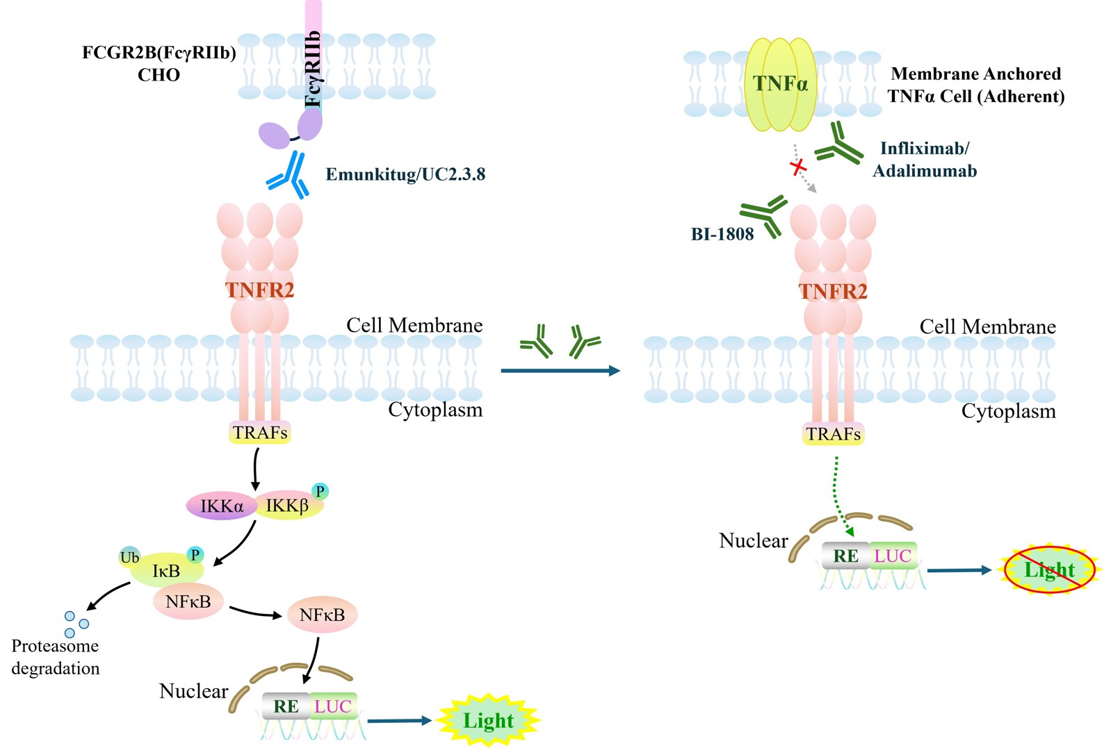
Figure 1. Schematic diagram of the Jurkat E6.1 Human TNFR2(TNFR1 KO) Effector Reporter Cell model
| Classification | Cytokine&Growth Factor |
| Family | Tumor necrosis factor receptor superfamily |
| Gene Name | TNFRSF1B |
| Gene Aliases |
p75; TBPII; TNFBR; TNFR2; CD120b; TNFR1B; TNFR80; TNF-R75; p75TNFR; TNF-R-II
|
| Gene ID | 7133 |
| Accession Number | NM_001066.3 |
| UniProt Number | P20333 |
| Protein Name | Tumor necrosis factor receptor superfamily member 1B |
| Protein Aliases | TNFR2; TNF-R2; TNF-RII; p75; CD120b |
| Target Species | Human |
| Host cell | Jurkat E6.1 |
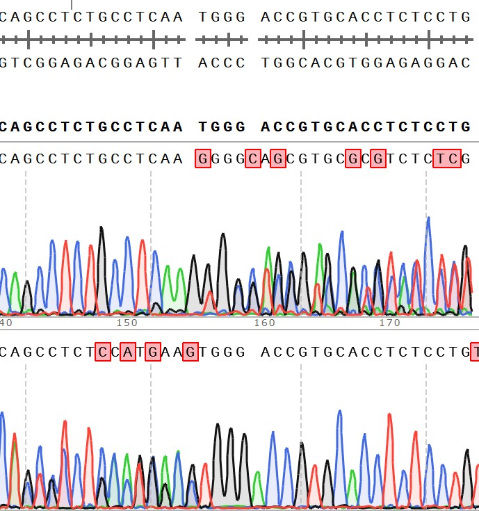
Figure 2. Sanger of TNFR2( TNFR1 KO) Effector Reporter Cell.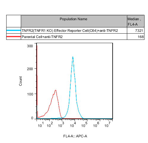
Figure 3. Recombinant TNFR2( TNFR1 KO) Effector Reporter Cell stably expressing TNFR2.
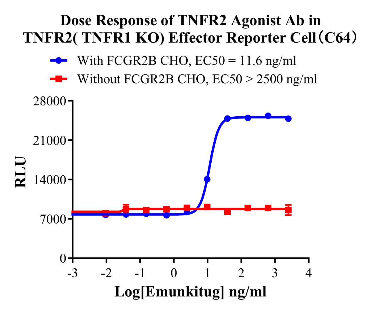
Figure 4. Dose Response of TNFR2 Agonist Ab in TNFR2(TNFR1 KO) Effector Reporter Cell(C64).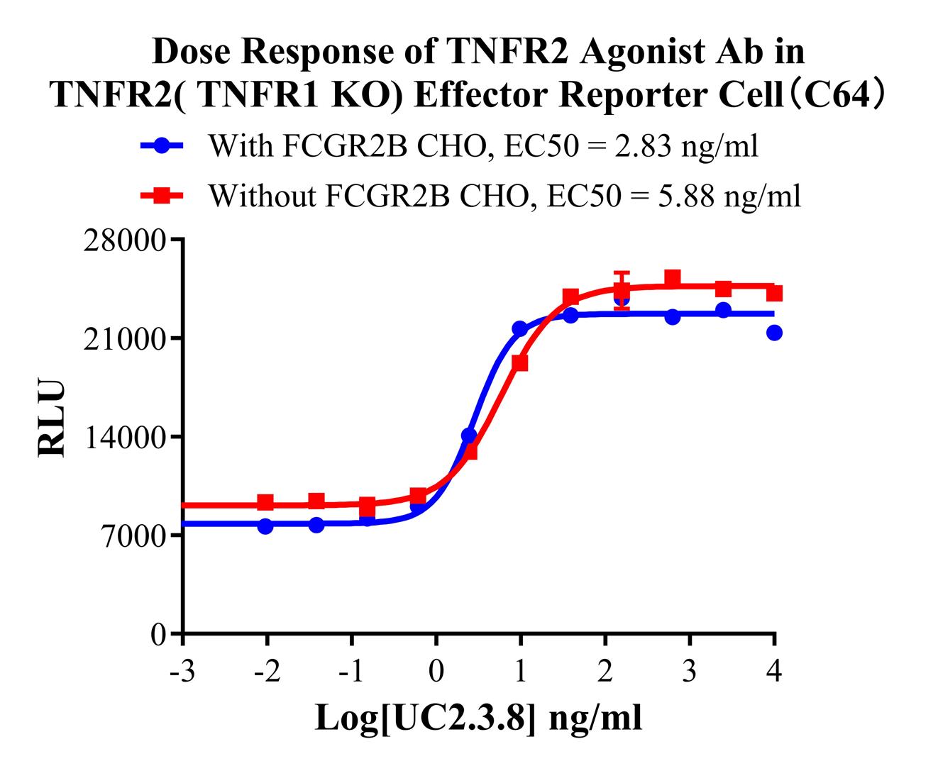
Figure 5. Dose Response of TNFR2 Agonist Ab in TNFR2(TNFR1 KO) Effector Reporter Cell(C64).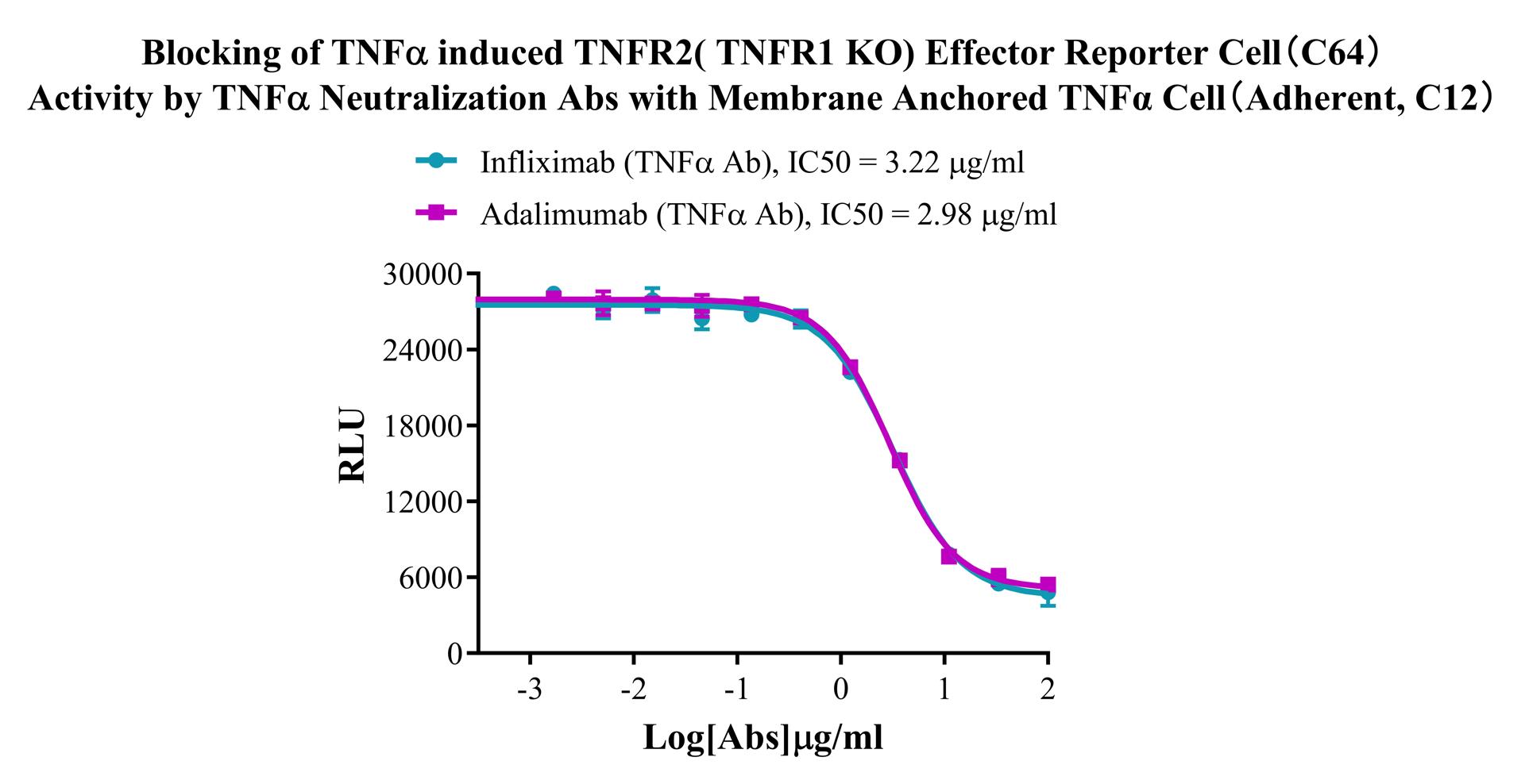
Figure 6. Blocking of TNFα induced TNFR2( TNFR1 KO) Effector Reporter Cell(C64) Activity by TNFα Neutralization Abs with Membrane Anchored TNFα Cell (Adherent, C12).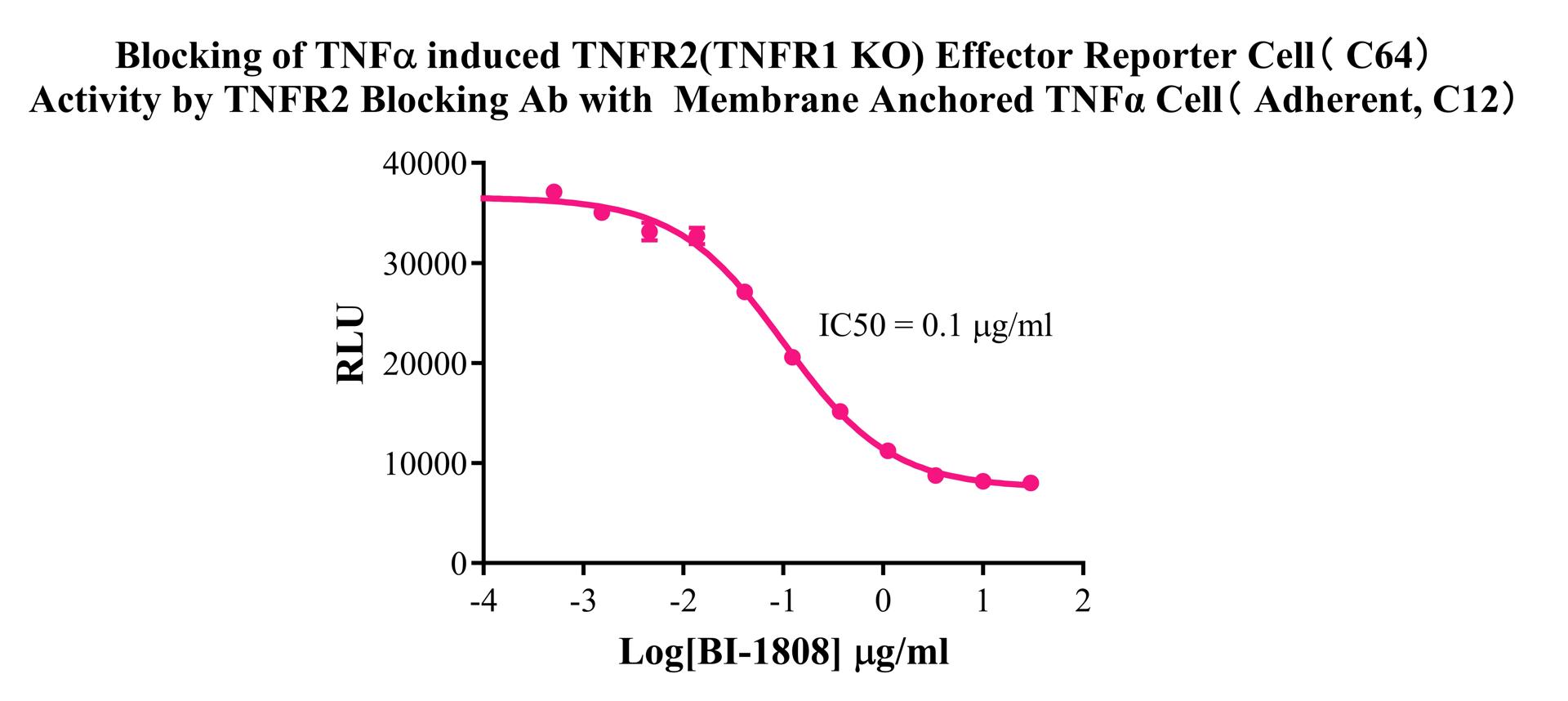
Figure 7. Blocking of TNFα induced TNFR2(TNFR1 KO) Effector Reporter Cell( C64) Activity by TNFR2 Blocking Ab with Membrane Anchored TNFα Cell (Adherent, C12).
Cell Passage Procedures
1.This cell line grows in suspension.
2.Upon receipt, cells should be thawed immediately or stored in liquid nitrogen until use.
3.Before thawing, pre-warm the water bath and culture medium to 37 °C, and prepare a small amount of dry ice.
4.Remove the cryovial from storage and transport it to the cell culture laboratory on dry ice.
5.Rapidly thaw the cells in a 37 °C water bath. Once the cells are completely thawed, spray the cryovial with 70% ethanol for disinfection and transfer it to a biosafety cabinet.
6.Add 10 mL of pre-warmed culture medium into a 15 mL centrifuge tube. Transfer the contents of the cryovial into the tube and centrifuge at 1000 rpm for 5 minutes.
7.Carefully discard the supernatant. Resuspend the cell pellet in 5 mL of pre-warmed culture medium by gentle pipetting. Immediately perform cell counting and adjust the cell density to 3–6 × 10⁵ cells/mL based on the counting results, then transfer the cells into a culture flask.
8.Count the cells every 1–2 days. When the cell density exceeds 1 × 10⁶ cells/mL, passage the cells promptly or add fresh culture medium. Maintain the cell density between 2 × 10⁵ and 1 × 10⁶ cells/mL.
Suspension Cell Cryopreservation Procedure:
1.Collect 8 × 10⁶ cells, centrifuge, and discard the supernatant.
2.Add 1 mL of cell freezing medium (90% FBS + 10% DMSO) and gently pipette to mix thoroughly. Transfer the suspension into a cryovial.
3.Immediately place the cryovial into a controlled-rate freezing container (Nalgene 5100-0001), fill with isopropanol up to the indicated level, and store at −80 °C.
4.After 24 hours, transfer the cryovial to liquid nitrogen for long-term storage.
Related products
CHO-K1 Human CCR4 Cell Line
HEK293 Human NK1R CRE-Luc Cell Line
Raji-Luc-GFP
Jurkat E6.1-Luc
THP-1-GFP
THP-1-Luc
Raji-GFP
Raji-Luc
Jurkat E6.1-GFP
HEK293 Human GAL4-Luc Cell
We Are Pleased to Announce: Global Commercial Licensing Rights for Jurkat E6.1, CHO-K1, and HEK293 Cell Lines Officially Secured.
Explore