

CHO-K1 Human Membrane Anchored TNFα Cell (Adherent)
Cat. No: RQP74152
Size: 1 vial of frozen cells (>1E6 per vial in 1 mL)
Unit Price: Contact For Pricing
Product Info
Description
Biological Information
Assay Data
Cell Culture
| Cat. No | RQP74152 |
| Product Name | CHO-K1 Human Membrane Anchored TNFα Cell (Adherent) Cell |
| Product Type | Reporter Cell |
| Culture Properties | Adherent |
| Stability | 32passages (in-house test, that not means the cell line will be instable beyond the passages we tested.) |
| Mycoplasma Status | Negative |
| Culture Medium | F12K+10%FBS+5μg/ml puromycin |
| Freeze Medium | 90% FBS+10% DMSO |
| Storage Conditions | Liquid nitrogen immediately upon delivery |
| Application | Functional(Report Gene) Assay |
For research use only. Not intended for human or animal clinical trials, therapeutic or diagnostic use.
Tumor Necrosis Factor-alpha (TNF-α) is a transmembrane protein with a molecular weight of 26 kDa; its precursor peptide is secreted as a soluble 17 kDa molecule following cleavage by TNF-α-converting enzyme (TACE). Although activated macrophages are the primary source of TNF-α, it can also be produced by a variety of other cell types—such as fibroblasts, astrocytes, smooth muscle cells, keratinocytes, and various tumor cells—including those associated with B-cell lymphoma, breast cancer, and colon cancer.
Acting as a tumor promoter, TNF-α participates in every stage of tumorigenesis, including cellular transformation, proliferation, angiogenesis, invasion, and metastasis. TNF-α signaling also constitutes a critical component of the immune system, capable of suppressing tumorigenesis and inhibiting viral replication; furthermore, it functions as an endogenous pyrogen, capable of inducing fever and apoptosis.
The Membrane-Anchored TNFα Cell (Adherent) serves as the target cell for the ADCC Bioassay Effector Cell V variant (High Affinity)—Fcγ-NFAT Jurkat or the TNFR2 (TNFR1 KO) Effector Reporter Cell, effectively mimicking *in vivo* ADCC effector functions or TNFR2 signal transduction processes.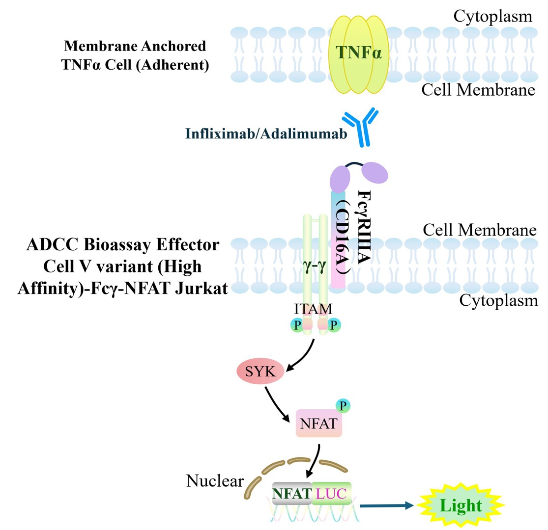
Figure 1. Schematic Diagram of the Cellular Mechanism of the ADCC Effect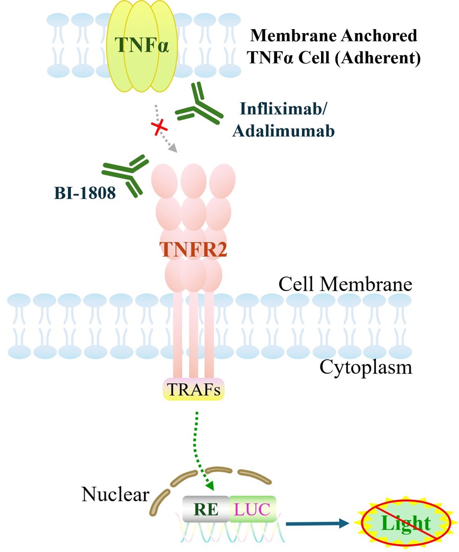
Figure 2. Schematic of the Cellular Model for TNFR2 Signal Transduction
| Classification | Cytokine&Growth Factor |
| Family | Tumor necrosis factor superfamily |
| Gene Name | TNF |
| Gene Aliases | TNFA;TNFSF2;DIF;TNF-alpha |
| Gene ID | 7124 |
| Accession Number | NM_000594.4 |
| UniProt Number | P01375 |
| Protein Name | Tumor necrosis factor |
| Protein Aliases | Cachectin;TNF-alpha;Tumor necrosis factor ligand superfamily member 2 (TNF-a) |
| Target Species | Human |
| Host cell | CHO-K1 |
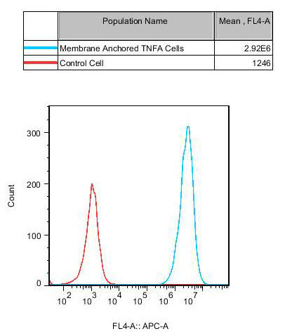
Figure 3. Recombinant Membrane Anchored TNFα Cell (Adherent) constitutively expressing TNFα.
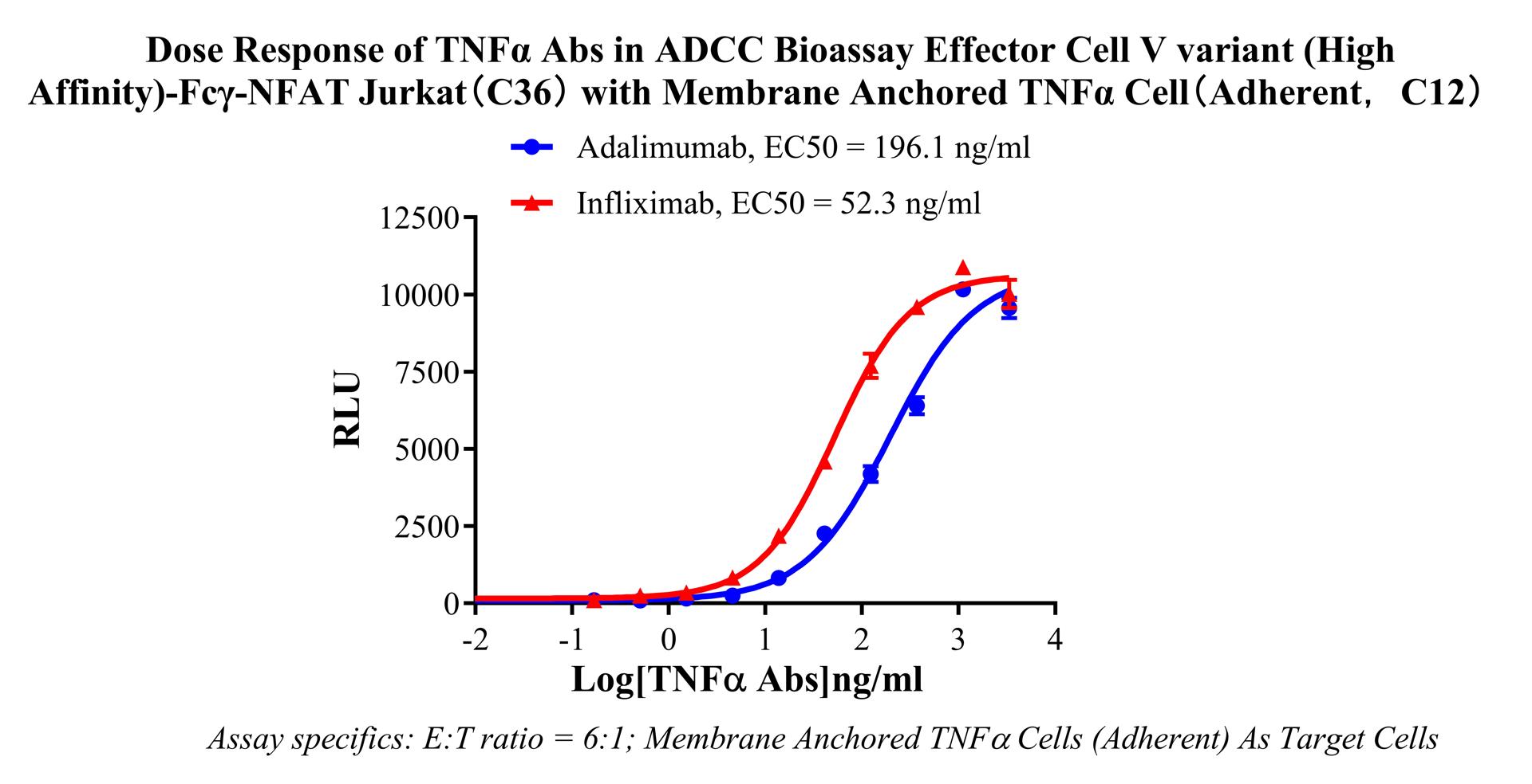
Figure 4. Dose Response of TNFα Abs in ADCC Bioassay Effector Cell V variant (High Affinity)-Fcγ-NFAT-Jurkat (C36) with Membrane Anchored TNFα Cell (Adherent, C12).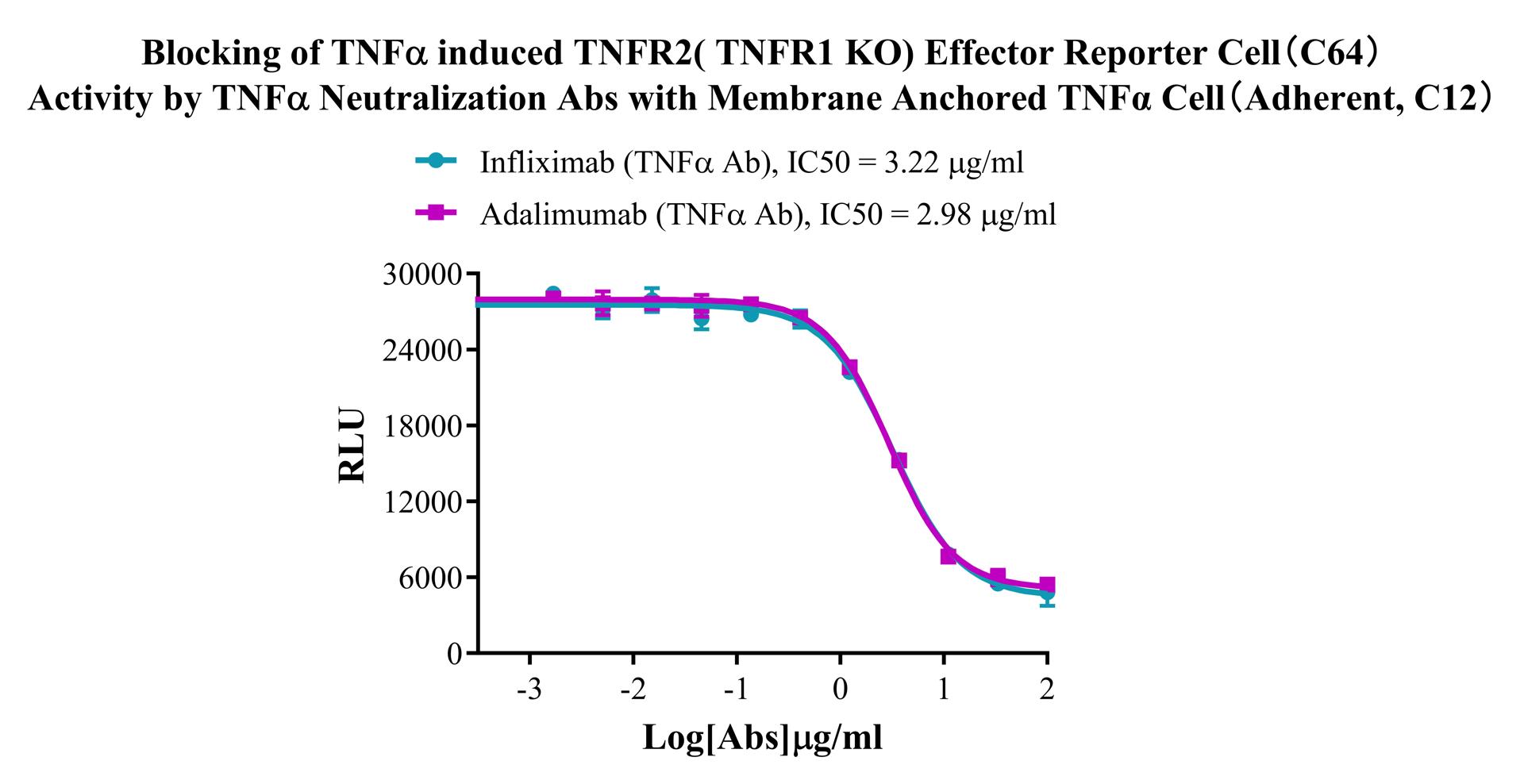
Figure 5. Blocking of TNFα induced TNFR2( TNFR1 KO) Effector Reporter Cell(C64) Activity by TNFα Neutralization Abs with Membrane Anchored TNFα Cell(Adherent, C12).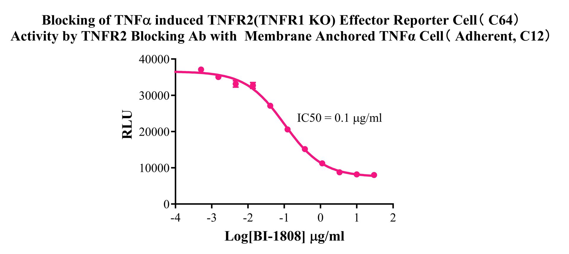
Figure 6. Blocking of TNFα induced TNFR2(TNFR1 KO) Effector Reporter Cell( C64) Activity by TNFR2 Blocking Ab with Membrane Anchored TNFα Cell ( Adherent, C12).
Cell Resuscitation
1)Rapidly thaw the frozen cells in a 37 °C water bath for approximately 60 seconds. Once thawed (which may take slightly less or more than 60 seconds), immediately transfer the cell suspension from the cryovial into a 15 mL centrifuge tube containing 10 mL of pre-warmed CHO-K1 Human Membrane Anchored TNFα Cell (Adherent) Cell complete culture medium.
2)Centrifuge cells at 1000 rpm for 5 min to remove medium, then resuspend cells in 5 mL of pre-warmed complete medium.
3)Transfer the cell suspension into a T25 culture flask and incubate at 37 °C with 5% CO₂.
4)After approximately 24–36 hours, replace the medium or passage the cells to remove non-adherent dead cells.
Subculturing procedure
1)When the cell density reaches the appropriate confluency for passaging, wash the cells with PBS, then add 1 mL trypsin to detach the cells. When more than 80% of the cells detach upon gently tapping the culture flask, add complete culture medium to terminate digestion. Gently pipette to obtain a single-cell suspension, transfer to a 15 mL centrifuge tube, and centrifuge at 1000 rpm for 5 minutes.
2)Discard supernatant after centrifugation. Resuspend cells in fresh medium to a single-cell suspension and transfer to a new culture flask for continued growth.
Cell Freezing
After trypsinization and centrifugation of cells from each T75 flask or 10 cm culture dish, discard the supernatant. Add 2 mL of cryopreservation medium (90% FBS + 10% DMSO), gently resuspend thoroughly, and aliquot into two cryovials. Immediately place the cryovials into a controlled-rate freezing container (e.g., Nalgene 5100-0001), fill with isopropanol to the indicated level, and store at −80 °C. After 24 hours, transfer the cryovials to liquid nitrogen for long-term storage.
Related products
CHO-K1 Human CCR4 Cell Line
HEK293 Human NK1R CRE-Luc Cell Line
Raji-Luc-GFP
Jurkat E6.1-Luc
THP-1-GFP
THP-1-Luc
Raji-GFP
Raji-Luc
Jurkat E6.1-GFP
HEK293 Human GAL4-Luc Cell
We Are Pleased to Announce: Global Commercial Licensing Rights for Jurkat E6.1, CHO-K1, and HEK293 Cell Lines Officially Secured.
Explore