

Jurkat E6.1 Human TIGIT/NFAT-Luc Cell
Cat. No: RQP74020
Size: 1 vial of frozen cells (>1E6 per vial in 1 mL)
Unit Price: Contact For Pricing
Product Info
Description
Biological Information
Assay Data
Cell Culture
| Cat. No | RQP74020 |
| Product Name | Jurkat E6.1 Human TIGIT/NFAT-Luc Cell |
| Product Type | Reporter Cell |
| Culture Properties | suspension |
| Stability | 32passages (in-house test, that not means the cell line will be instable beyond the passages we tested.) |
| Mycoplasma Status | Negative |
| Culture Medium | RPMI-1640+10%FBS+800μg/ml Hygromycin B+1μg/ml puromycin |
| Freeze Medium | 90% FBS+10% DMSO |
| Storage Conditions | Liquid nitrogen immediately upon delivery |
| Application | Functional(Report Gene) Assay |
For research use only. Not intended for human or animal clinical trials, therapeutic or diagnostic use.
TIGIT—also known as WUCAM, Vstm3, or VSIG9—is a co-inhibitory receptor belonging to the immunoglobulin superfamily. TIGIT consists of an extracellular immunoglobulin variable (IgV) domain, a type I transmembrane domain, and an intracellular domain containing both an ITIM motif and an Ig-tail tyrosine-like motif (ITT). TIGIT is expressed on activated conventional αβ T cells, as well as on memory T cells, regulatory T cells (Tregs), follicular helper T cells, and NKT cells. By activating inhibitory receptors within T cells, natural killer (NK) cells, and Tregs, TIGIT has emerged as a promising target for immunotherapy.
The ligands for TIGIT include CD112 and the poliovirus receptor (PVR—also known as CD155, Necl-5, and Tage4), with PVR serving as TIGIT's high-affinity cognate receptor. Furthermore, TIGIT competes with CD226 (DNAM-1) and CD96 (TACTILE) for ligand binding. TIGIT effectively blocks the binding of CD155 to either CD96 or CD226, thereby providing further evidence that TIGIT possesses the highest affinity for CD155. TIGIT not only competes with CD226 for ligands but can also directly bind to CD226 *in cis*, thereby preventing its homodimerization and rendering CD226 unable to bind to CD155 to exert its co-stimulatory function.
CD155 is primarily expressed on the surface of dendritic cells (DCs), T cells, B cells, and macrophages; it is also expressed to varying degrees in non-hematopoietic tissues, such as the kidneys, the nervous system, and the intestines. Additionally, CD155 has been reported to be highly expressed in various human malignancies, including melanoma, pancreatic cancer, colon cancer, lung adenocarcinoma, and glioblastoma. As a cell adhesion molecule, CD155 influences cellular proliferation, migration, invasion, and adhesion through tumor-associated signaling pathways.
The Jurkat E6.1 Human TIGIT/NFAT-Luc Cell effectively simulates the TIGIT/CD155 or TIGIT/Nectin-4 signal transduction processes occurring in vivo. The principle is illustrated in the figure below.
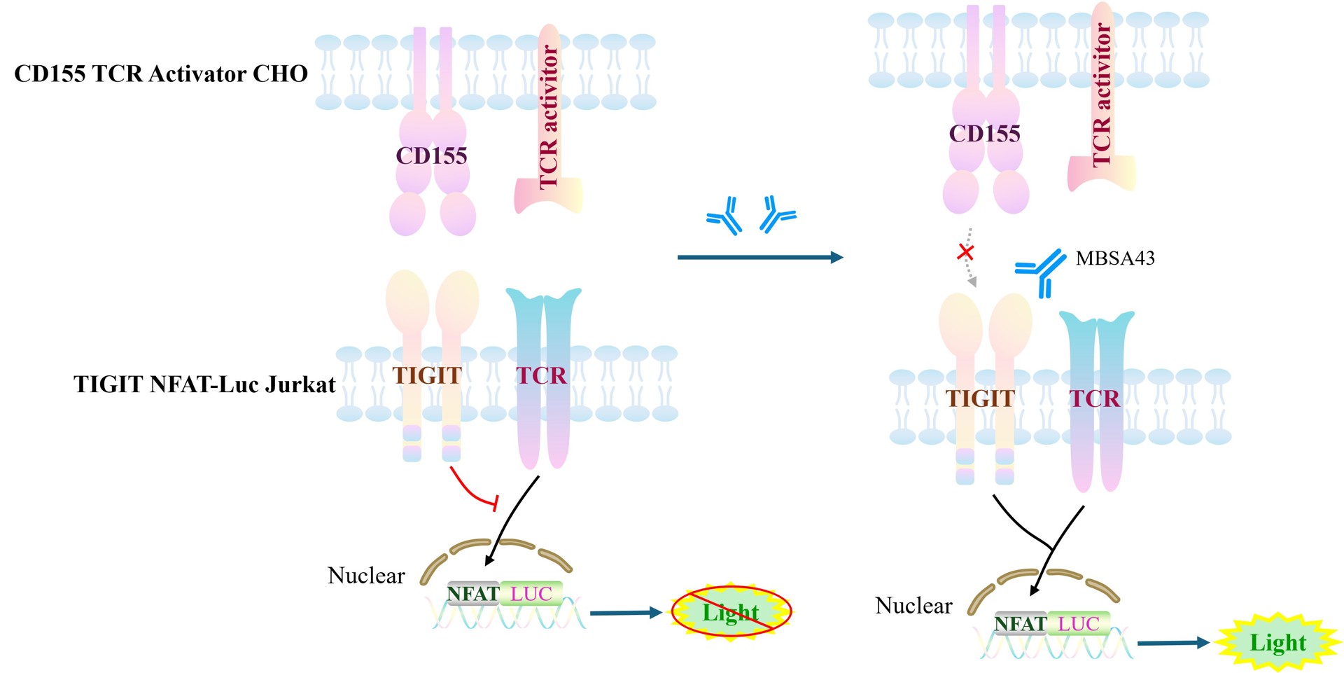
Figure 1. Schematic diagram of the cellular model for TIGIT/CD155 signal transduction
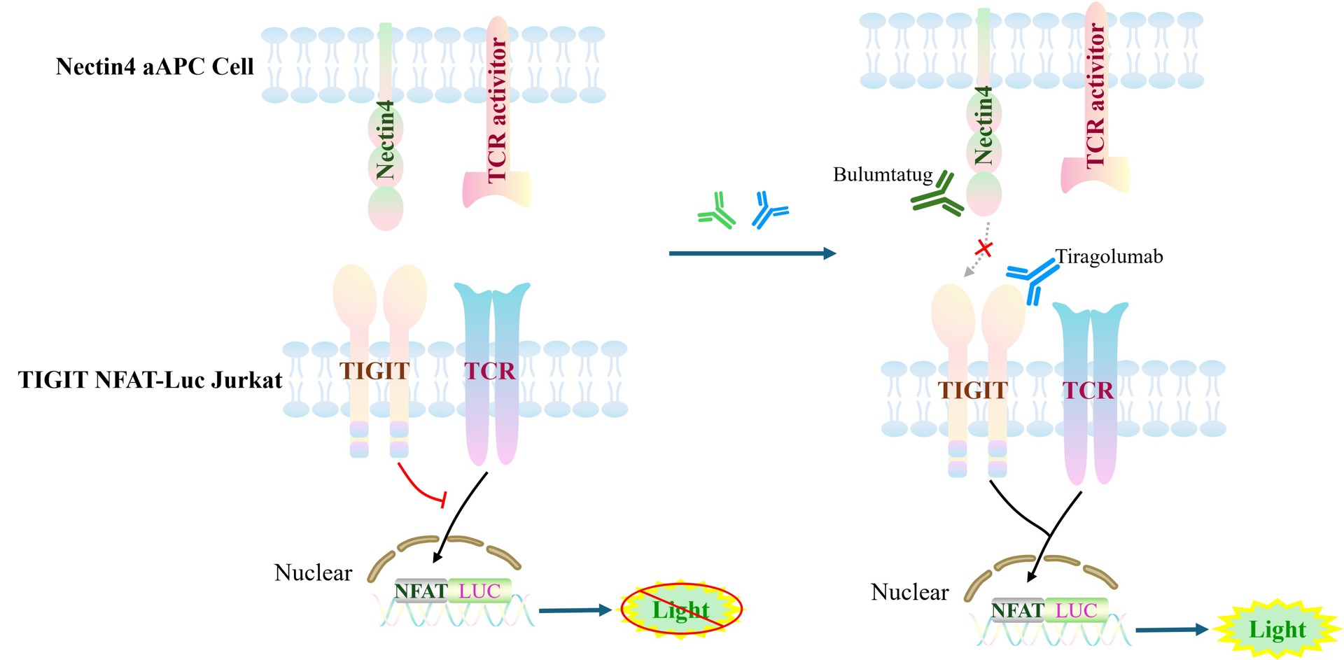
Figure 2. Schematic diagram of the cellular model for TIGIT and Nectin-4 signaling transduction.
| Classification | Co-Inhibitory |
| Family | Nectin and nectin-like family |
| Gene Name | TIGIT |
| Gene Aliases | VSIG9;VSTM3 |
| Gene ID | 201633 |
| Accession Number | NM_173799.4 |
| UniProt Number | Q495A1 |
| Protein Name | T-cell immunoreceptor with Ig and ITIM domains |
| Protein Aliases | V-set and immunoglobulin domain-containing protein 9;V-set and transmembrane domain-containing protein 3 |
| Target Species | Human |
| Host cell | Jurkat E6.1 |
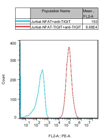
Figure 3. Recombinant TIGIT/NFAT-Luc/Jurkat stably expressing TIGIT.
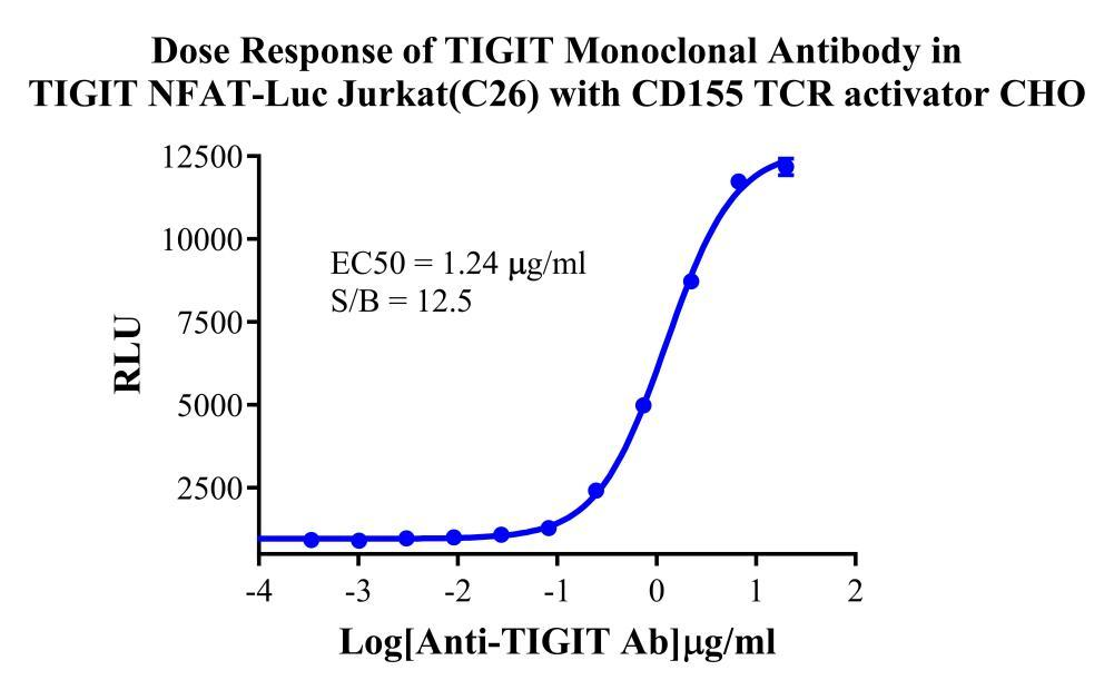
Figure 4. Dose Response of TIGIT Monoclonal Antibody in TIGIT NFAT-Luc Jurkat(C26) with CD155/TCR activator-CHO.
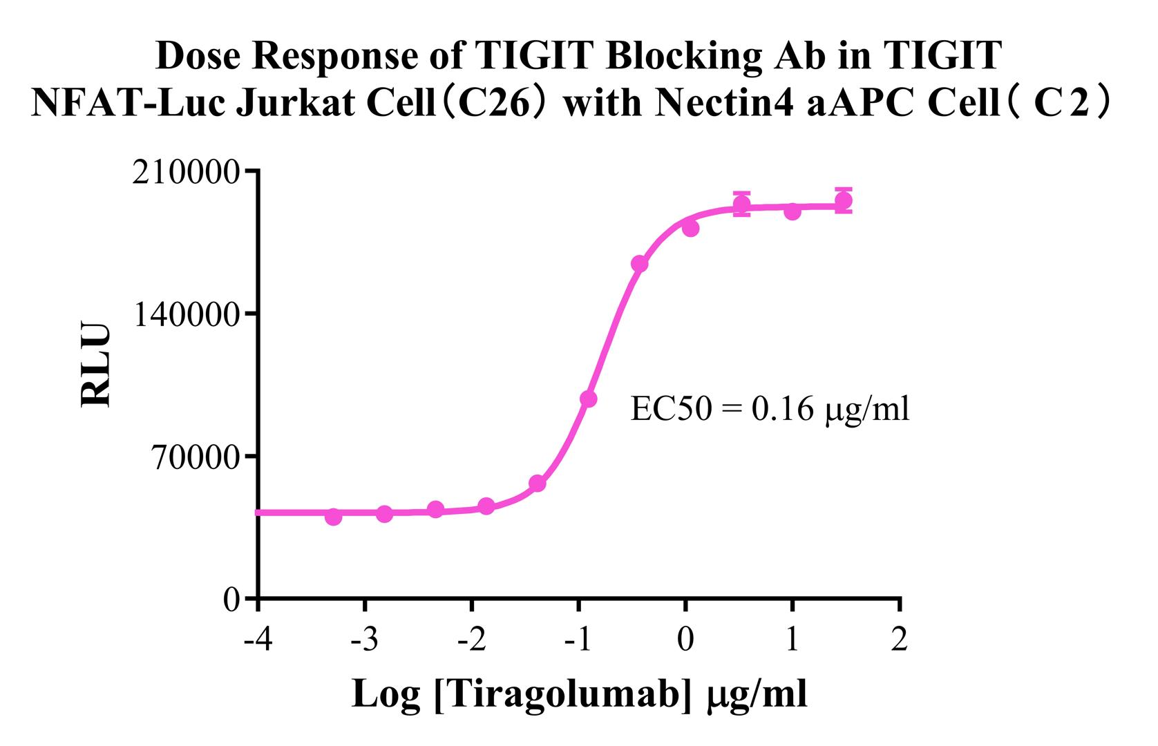
Figure 5. Dose Response of TIGIT Blocking Ab in TIGIT NFAT-Luc Jurkat Cell(C26) with Nectin4 aAPC Cell(C2).

Figure 6. Dose Response of Nectin4 Blocking Ab in TIGIT NFAT-Luc Jurkat Cell(C26) with Nectin4 aAPC Cell(C2).
Cell Passage Procedures
1.This cell line grows in suspension.
2.Upon receipt, cells should be thawed immediately or stored in liquid nitrogen until use.
3.Before thawing, pre-warm the water bath and culture medium to 37 °C, and prepare a small amount of dry ice.
4.Remove the cryovial from storage and transport it to the cell culture laboratory on dry ice.
5.Rapidly thaw the cells in a 37 °C water bath. Once the cells are completely thawed, spray the cryovial with 70% ethanol for disinfection and transfer it to a biosafety cabinet.
6.Add 10 mL of pre-warmed culture medium into a 15 mL centrifuge tube. Transfer the contents of the cryovial into the tube and centrifuge at 1000 rpm for 5 minutes.
7.Carefully discard the supernatant. Resuspend the cell pellet in 5 mL of pre-warmed culture medium by gentle pipetting. Immediately perform cell counting and adjust the cell density to 3–6 × 10⁵ cells/mL based on the counting results, then transfer the cells into a culture flask.
8.Count the cells every 1–2 days. When the cell density exceeds 1 × 10⁶ cells/mL, passage the cells promptly or add fresh culture medium. Maintain the cell density between 2 × 10⁵ and 1 × 10⁶ cells/mL.
Suspension Cell Cryopreservation Procedure:
1.Collect 8 × 10⁶ cells, centrifuge, and discard the supernatant.
2.Add 1 mL of cell freezing medium (90% FBS + 10% DMSO) and gently pipette to mix thoroughly. Transfer the suspension into a cryovial.
3.Immediately place the cryovial into a controlled-rate freezing container (Nalgene 5100-0001), fill with isopropanol up to the indicated level, and store at −80 °C.
4.After 24 hours, transfer the cryovial to liquid nitrogen for long-term storage.
Related products
CHO-K1 Human CCR4 Cell Line
HEK293 Human NK1R CRE-Luc Cell Line
Raji-Luc-GFP
Jurkat E6.1-Luc
THP-1-GFP
THP-1-Luc
Raji-GFP
Raji-Luc
Jurkat E6.1-GFP
HEK293 Human GAL4-Luc Cell
We Are Pleased to Announce: Global Commercial Licensing Rights for Jurkat E6.1, CHO-K1, and HEK293 Cell Lines Officially Secured.
Explore