
CHO-K1 Human Nectin4 aAPC Cell
Cat. No: RQP74553
Size: 1 vial of frozen cells (>1E6 per vial in 1 mL)
Unit Price: Contact For Pricing
Product Info
Description
Biological Information
Assay Data
Cell Culture
| Cat. No | RQP74553 |
| Product Name | CHO-K1 Human Nectin4 aAPC Cell |
| Product Type | Reporter Cell line |
| Culture Properties | Adherent |
| Stability | 32passages (in-house test, that not means the cell line will be instable beyond the passages we tested.) |
| Mycoplasma Status | Negative |
| Culture Medium | F12K+10%FBS+3 μg/ml Puromycin+600 μg/ml Hygromycin B |
| Freeze Medium | 90% FBS+10% DMSO |
| Storage Conditions | Liquid nitrogen immediately upon delivery |
| Application | Functional(Report Gene) Assay |
For research use only. Not intended for human or animal clinical trials, therapeutic or diagnostic use.
TIGIT—also known as WUCAM, Vstm3, or VSIG9—is a co-inhibitory receptor belonging to the immunoglobulin superfamily. It consists of an extracellular immunoglobulin variable (IgV) domain, a type I transmembrane domain, and an intracellular domain containing an ITIM and an Ig-tail tyrosine-like motif (ITT). TIGIT is expressed on activated conventional αβ T cells, as well as on memory T cells, Tregs, follicular helper T cells, and NKT cells. By activating inhibitory signaling pathways in T cells, natural killer (NK) cells, and regulatory T cells (Tregs), TIGIT has emerged as a promising target for immunotherapy.
Nectin cell adhesion molecule 4 (Nectin-4) is a type I transmembrane glycoprotein belonging to the Nectin family within the immunoglobulin superfamily. In addition to Nectin-4, the family includes three other members: Nectin-1, Nectin-2, and Nectin-3. All Nectins share a common structural organization, comprising three extracellular immunoglobulin-like domains (two Ig-like C2-type domains and one Ig-like V-type domain), a transmembrane region, and an intracellular domain. A soluble form of the Nectin-4 protein is generated through the proteolytic cleavage of the cell-surface protein by the metalloproteinase ADAM17/TACE.
TIGIT functions as an immunosuppressive receptor on the surface of T cells and NK cells, with Nectin-4 serving as one of its ligands. Upon binding, the intracellular inhibitory motifs of TIGIT undergo phosphorylation and recruit inhibitory molecules such as SHIP1; this blocks downstream signaling pathways—including PI3K-AKT and MAPK—thereby directly impairing the cytotoxic function of immune cells and facilitating tumor immune escape. Concurrently, Nectin-4 possesses intrinsic oncogenic signaling capabilities within tumor cells: it can activate pathways such as PI3K-AKT and Rac1 via the adaptor protein Afadin, thereby promoting tumor cell proliferation, migration, and survival. Therefore, the TIGIT-Nectin4 signaling axis tightly links immunosuppression with malignant tumor progression, representing a combination of anti-tumor targets with potential for synergistic intervention.
CHO-K1 Human Nectin4 aAPC Cell for TIGIT NFAT-Luc Jurkat cells, effectively simulating the TIGIT/Nectin4 signal transduction process that occurs in vivo, the principle is illustrated in the figure below.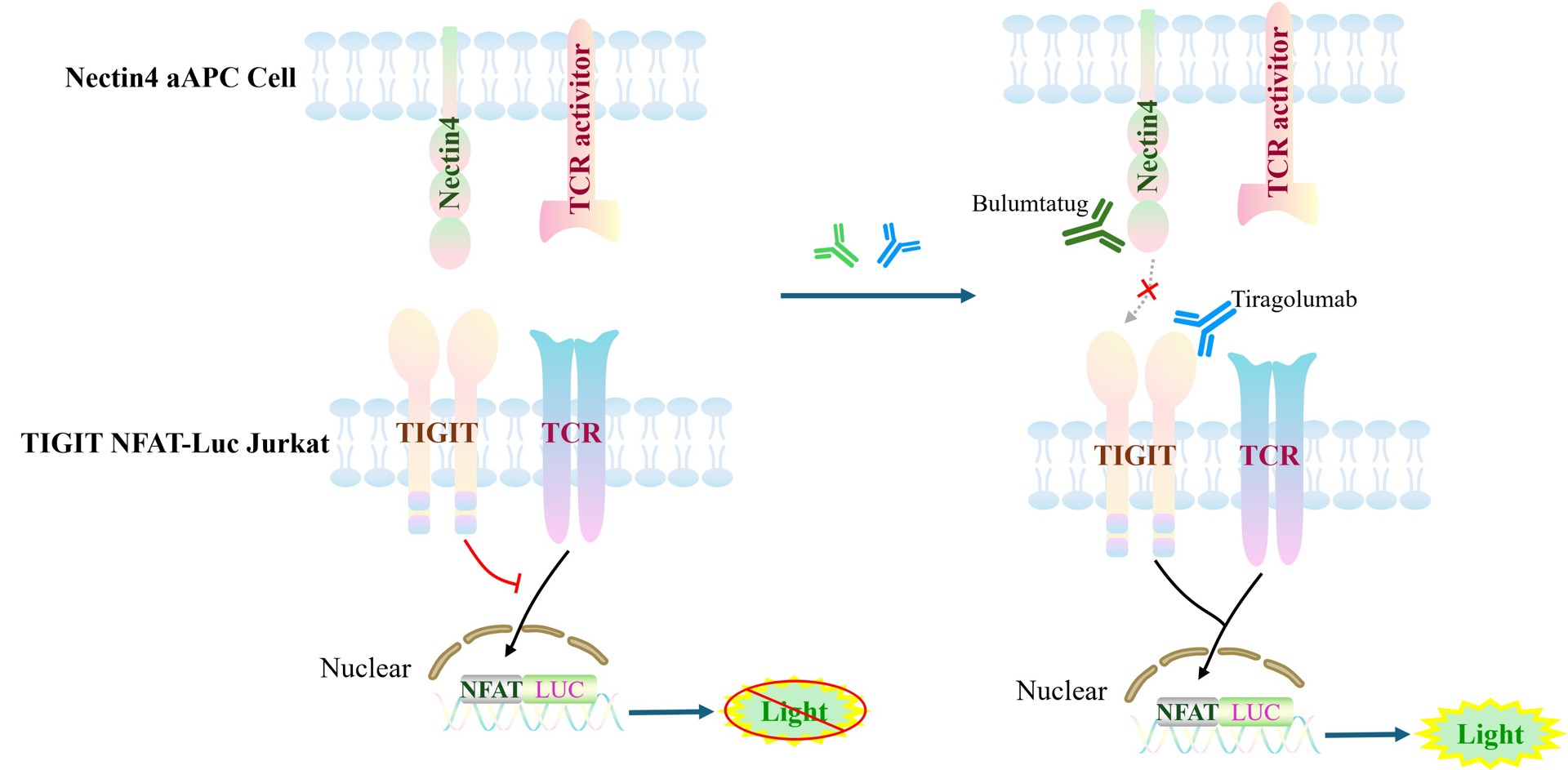
Figure 1. Schematic diagram of the CHO-K1 Human Nectin4 aAPC Cell model
| Classification | TAA |
| Family | nectin family |
| Gene Name | NECTIN4 |
| Gene Aliases | nectin-4;PVRL4;PRR4;LNIR |
| Gene ID | 81607 |
| Accession Number | NM_030916.3 |
| UniProt Number | Q96NY8 |
| Protein Name | Nectin-4 |
| Protein Aliases | Ig superfamily receptor LNIR;Nectin cell adhesion molecule 4;Poliovirus receptor-related protein 4 |
| Target Species | Human |
| Host cell | CHO-K1 |
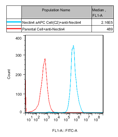
Figure 2. Recombinant Nectin4 aAPC Cell stably expressing Nectin4.
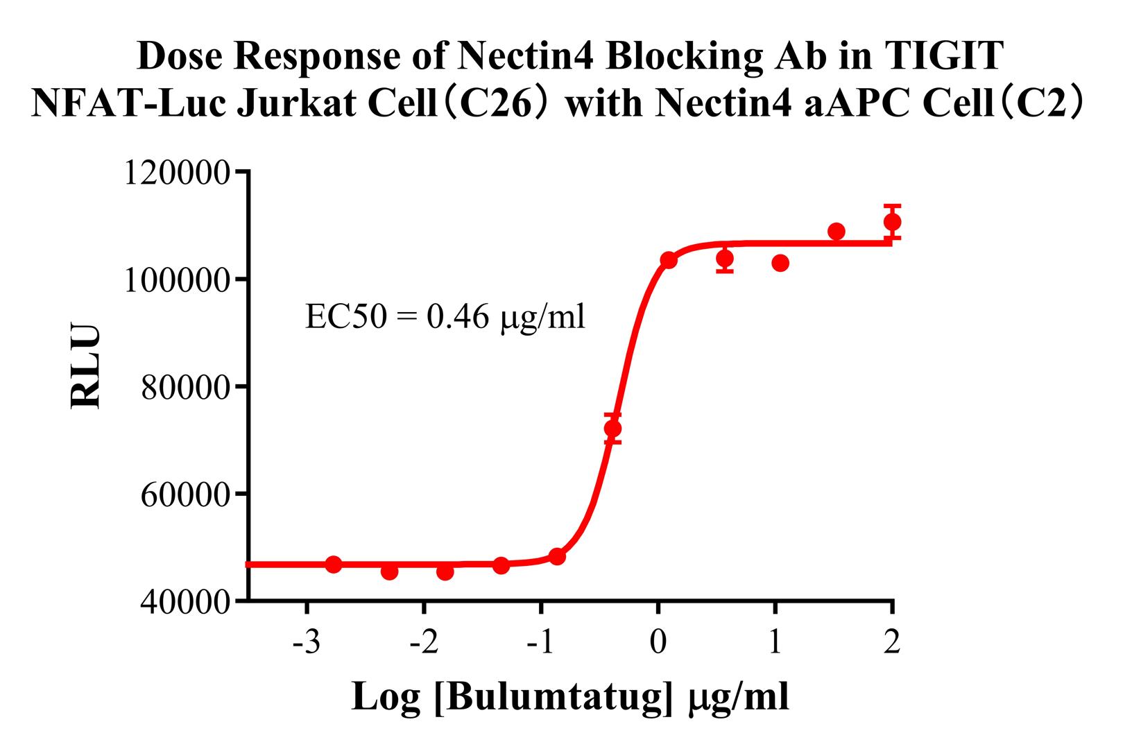
Figure 3. Dose Response of Nectin4 Blocking Ab in TIGIT NFAT-Luc Jurkat Cell(C26) with Nectin4 aAPC Cell(C2).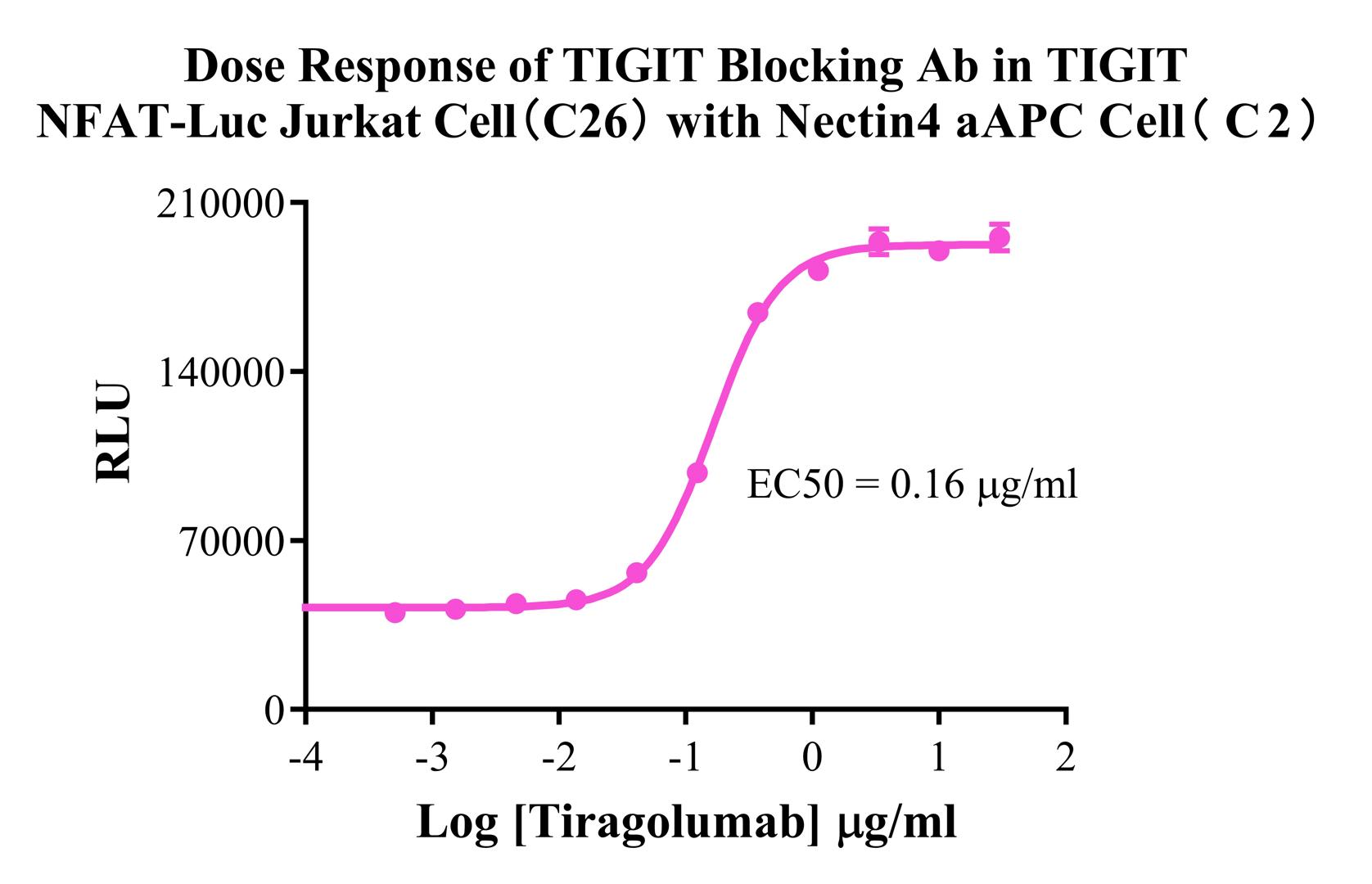
Figure 4. Dose Response of TIGIT Blocking Ab in TIGIT NFAT-Luc Jurkat Cell(C26) with Nectin4 aAPC Cell(C2).
Cell Resuscitation
1)Rapidly thaw the frozen cells in a 37 °C water bath for approximately 60 seconds. Once thawed (which may take slightly less or more than 60 seconds), immediately transfer the cell suspension from the cryovial into a 15 mL centrifuge tube containing 10 mL of pre-warmed CHO-K1 Human Nectin4 aAPC Cell complete culture medium.
2)Centrifuge cells at 1000 rpm for 5 min to remove medium, then resuspend cells in 5 mL of pre-warmed complete medium.
3)Transfer the cell suspension into a T25 culture flask and incubate at 37 °C with 5% CO₂.
4)After approximately 24–36 hours, replace the medium or passage the cells to remove non-adherent dead cells.
Subculturing procedure
1)When the cell density reaches the appropriate confluency for passaging, wash the cells with PBS, then add 1 mL trypsin to detach the cells. When more than 80% of the cells detach upon gently tapping the culture flask, add complete culture medium to terminate digestion. Gently pipette to obtain a single-cell suspension, transfer to a 15 mL centrifuge tube, and centrifuge at 1000 rpm for 5 minutes.
2)Discard supernatant after centrifugation. Resuspend cells in fresh medium to a single-cell suspension and transfer to a new culture flask for continued growth.
Cell Freezing
After trypsinization and centrifugation of cells from each T75 flask or 10 cm culture dish, discard the supernatant. Add 2 mL of cryopreservation medium (90% FBS + 10% DMSO), gently resuspend thoroughly, and aliquot into two cryovials. Immediately place the cryovials into a controlled-rate freezing container (e.g., Nalgene 5100-0001), fill with isopropanol to the indicated level, and store at −80 °C. After 24 hours, transfer the cryovials to liquid nitrogen for long-term storage.