

Jurkat E6.1 Human PD1/NFAT-Luc Cell
Cat. No: RQP74018
Size: 1 vial of frozen cells (>1E6 per vial in 1 mL)
Unit Price: Contact For Pricing
Product Info
Description
Biological Information
Assay Data
Cell Culture
| Cat. No | RQP74018 |
| Product Name | Jurkat E6.1 Human PD1/NFAT-Luc Cell |
| Product Type | Reporter Cell |
| Culture Properties | suspension |
| Stability | 32passages (in-house test, that not means the cell line will be instable beyond the passages we tested.) |
| Mycoplasma Status | Negative |
| Culture Medium | RPMI-1640+10%FBS+800μg/ml Hygromycin B+1μg/ml puromycin |
| Freeze Medium | 90% FBS+10% DMSO |
| Storage Conditions | Liquid nitrogen immediately upon delivery |
| Application | Functional(Report Gene) Assay |
For research use only. Not intended for human or animal clinical trials, therapeutic or diagnostic use.
Tumor cells can evade recognition and elimination by the host immune system by utilizing immune checkpoint receptors; therefore, blocking these receptors holds promise as a broadly effective approach to cancer immunotherapy. Currently, while anti-PD-1/PD-L1 antibodies—much like anti-CTLA-4 antibodies—represent a relatively mature therapeutic modality, their overall response rates in patients remain low due to the prevalence of drug resistance. Consequently, the identification of novel targets for cancer immunotherapy has become a matter of urgent necessity.
Programmed Cell Death Protein 1 (PD-1) is a receptor expressed on activated T cells that negatively regulates immune responses through its binding to its ligands, PD-L1 and PD-L2. PD-1 ligands are present in the majority of cancers; the interaction between PD-1 and PD-L1/2 suppresses T-cell activity and enables cancer cells to evade immune surveillance. The PD-1/PD-L1 signaling pathway constitutes a critical component of tumor-induced immunosuppression, serving to dampen T-lymphocyte activation and enhance immune tolerance toward tumor cells, thereby facilitating tumor immune evasion. The binding of PD-1 to PD-L1 attenuates T-cell-mediated immune surveillance, leading to a compromised immune response or even T-cell apoptosis. PD-1/PD-L1 inhibitors function by releasing anti-tumor T cells from this state of immunosuppression, thereby promoting T-cell proliferation, infiltration into the tumor microenvironment, and the subsequent induction of an anti-tumor immune response. The PD-1/PD-L1/2 pathway also plays a role in regulating autoimmune responses, rendering these proteins promising therapeutic targets for a wide range of conditions, including various cancers as well as autoimmune diseases such as multiple sclerosis, arthritis, lupus, and Type 1 diabetes. The tyrosine phosphatase SHP2 is a key regulator of T-cell function, mediating both the downstream activation signals initiated by the T-cell receptor (TCR) and the downstream inhibitory signals triggered by PD-1.
The Jurkat E6.1 Human PD1/NFAT-Luc Cell Model—effectively simulates the signal transduction process of PD1 *in vivo*. The underlying principle is illustrated in the figure below.
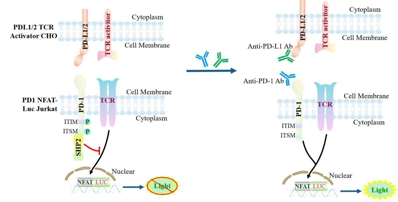
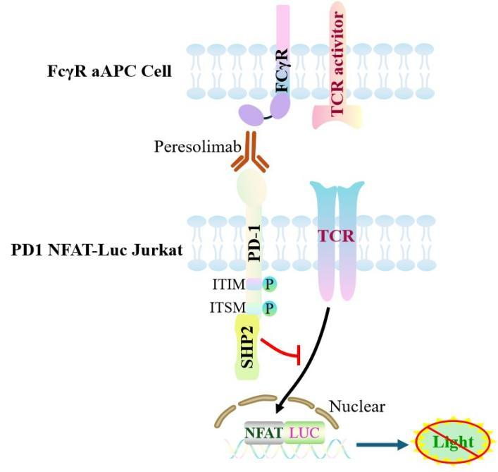
Figure 1. Schematic Diagram of the Jurkat E6.1 Human PD1/NFAT-Luc Cell Model
| Classification | Co-Inhibitory |
| Family | CD28 family |
| Gene Name | PDCD1 |
| Gene Aliases | CD279;PD1;hSLE1;PD-1 |
| Gene ID | 5133 |
| Accession Number | NM_005018.3 |
| UniProt Number | Q15116 |
| Protein Name | Protein PD-1;hPD-1 |
| Protein Aliases | N/A |
| Target Species | Human |
| Host cell | Jurkat E6.1 |
Jurkat E6.1 cell line expressing full length human PD1. Expression is confirmed by flow cytometry.
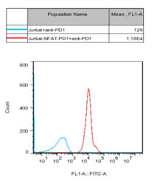
Figure 2. Recombinant PD1/NFAT-Luc/Jurkat stably expressing PD1.
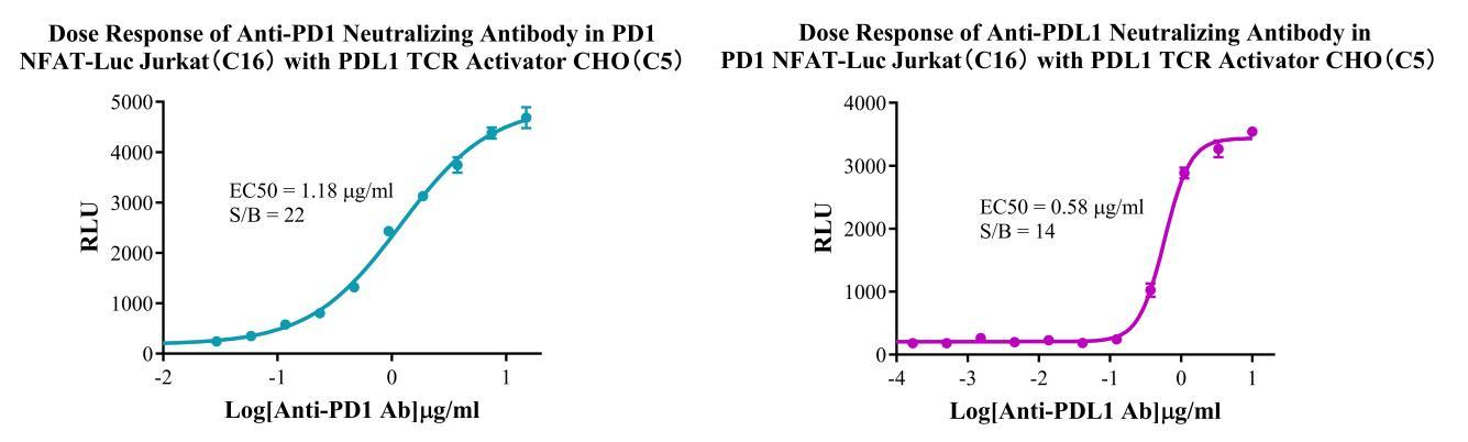
Figure 3. Dose response of Anti-PD-1 Neutralizing Antibody in PD1 NFAT-Luc Jurkat(C16) with PDL1 TCR Activator CHO (C5).Dose response of Anti-PDL1 Neutralizing Antibody in PD1 NFAT-Luc Jurkat(C16) with PDL1 TCR Activator CHO (C5).
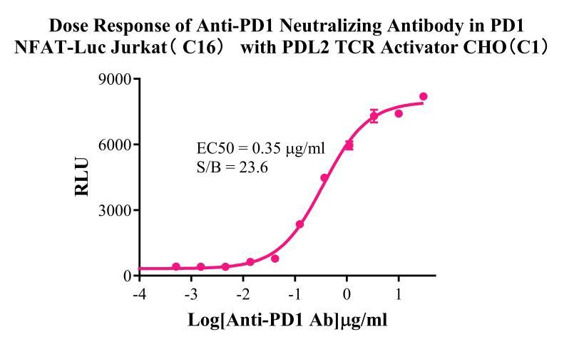
Figure 4. Dose response of Anti-PD1 Neutralizing Antibody in PD1 NFAT-Luc Jurkat(C16) with PDL2 TCR Activator CHO(C1).
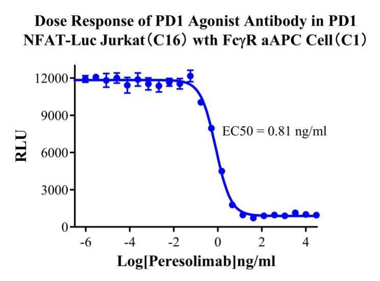
Figure 5. Dose response of PD1 Agonist Antibody in PD1 NFAT-Luc Jurkat(C16) with FcγR aAPC Cell(C1).
Cell Passage Procedures
1.This cell line grows in suspension.
2.Upon receipt, cells should be thawed immediately or stored in liquid nitrogen until use.
3.Before thawing, pre-warm the water bath and culture medium to 37 °C, and prepare a small amount of dry ice.
4.Remove the cryovial from storage and transport it to the cell culture laboratory on dry ice.
5.Rapidly thaw the cells in a 37 °C water bath. Once the cells are completely thawed, spray the cryovial with 70% ethanol for disinfection and transfer it to a biosafety cabinet.
6.Add 10 mL of pre-warmed culture medium into a 15 mL centrifuge tube. Transfer the contents of the cryovial into the tube and centrifuge at 1000 rpm for 5 minutes.
7.Carefully discard the supernatant. Resuspend the cell pellet in 5 mL of pre-warmed culture medium by gentle pipetting. Immediately perform cell counting and adjust the cell density to 3–6 × 10⁵ cells/mL based on the counting results, then transfer the cells into a culture flask.
8.Count the cells every 1–2 days. When the cell density exceeds 1 × 10⁶ cells/mL, passage the cells promptly or add fresh culture medium. Maintain the cell density between 2 × 10⁵ and 1 × 10⁶ cells/mL.
Suspension Cell Cryopreservation Procedure:
1.Collect 8 × 10⁶ cells, centrifuge, and discard the supernatant.
2.Add 1 mL of cell freezing medium (90% FBS + 10% DMSO) and gently pipette to mix thoroughly. Transfer the suspension into a cryovial.
3.Immediately place the cryovial into a controlled-rate freezing container (Nalgene 5100-0001), fill with isopropanol up to the indicated level, and store at −80 °C.
4.After 24 hours, transfer the cryovial to liquid nitrogen for long-term storage.
Related products
CHO-K1 Human CCR4 Cell Line
HEK293 Human NK1R CRE-Luc Cell Line
Raji-Luc-GFP
Jurkat E6.1-Luc
THP-1-GFP
THP-1-Luc
Raji-GFP
Raji-Luc
Jurkat E6.1-GFP
HEK293 Human GAL4-Luc Cell
We Are Pleased to Announce: Global Commercial Licensing Rights for Jurkat E6.1, CHO-K1, and HEK293 Cell Lines Officially Secured.
Explore