

CHO-K1 Human FCγR aAPC Cell
Cat. No: RQP74185
Size: 1 vial of frozen cells (>1E6 per vial in 1 mL)
Unit Price: Contact For Pricing
Product Info
Description
Biological Information
Assay Data
Cell Culture
| Cat. No | RQP74185 |
| Product Name | CHO-K1 Human FCγR aAPC Cell |
| Product Type | Reporter Cell |
| Culture Properties | Adherent |
| Stability | 32passages (in-house test, that not means the cell line will be instable beyond the passages we tested.) |
| Mycoplasma Status | Negative |
| Culture Medium | F12K+10%FBS+3μg/ml puromycin+600μg/ml Hygromycin B |
| Freeze Medium | 90% FBS+10% DMSO |
| Storage Conditions | Liquid nitrogen immediately upon delivery |
| Application | Functional(Report Gene) Assay |
For research use only. Not intended for human or animal clinical trials, therapeutic or diagnostic use.
In humans, FcγR receptors (FcγRI, FcγRIIa, FcγRIIb, FcγRIIc, FcγRIIIa, and FcγRIIIb) are widely expressed on immune cells, including macrophages, neutrophils, natural killer (NK) cells, eosinophils, basophils, and dendritic cells. These cell types exhibit distinct receptor expression profiles and participate in various effector functions, such as ADCC, to counteract viral entry into cells, as well as phagocytosis of antibody-virus immune complexes or infected cells.
The functional relationship between receptors and cell types is complex, as the different actions of activating and inhibitory FcγRs modulate cellular effector functions. Receptors may also play additional roles in antigen presentation, B-cell activation and suppression, and potentially in HIV neutralization or suppression of infection in susceptible cells. Classic FcγRs comprise a family of cell surface receptors belonging to the immunoglobulin superfamily that are specific for IgG, with varying affinities, IgG subclass specificities, expressions, signaling mechanisms, and binding outcomes. FcγRI is an activating receptor consisting of an α-chain with an intracellular domain that forms a complex with the common FcRγ-chain dimer for signal transduction. FcγRIIa is an activating receptor containing an intracellular tyrosine-based immune receptor activation motif (ITAM). FcγRIIb is an inhibitory receptor containing an intracellular immune receptor tyrosine-based inhibitory motif (ITIM). FcγRIIc is an activating receptor whose extracellular domain is identical to that of FcγRIIb, and whose intracellular ITAM domain is identical to that of FcγRIIa. FcγRIIIa is an activating receptor that relies on the common FcRγ chain for signal transduction. FcγRIIIb is expressed exclusively on neutrophils and is anchored to the cell membrane via phosphatidylinositol; despite lacking an intracellular domain, FcγRIIIb is capable of triggering the release of oxidants, driving phagocytosis, and impairing FcγRIIa-mediated ADCC.
FcγR aAPC Cells stably express FcγR and aAPC on basal cells. The schematic diagram illustrating the principle of using FcγR aAPC Cells as target cells for functional assays with PD1 NFAT-Luc Jurkat Cells/BTLA Effector Reporter Cells is shown below.
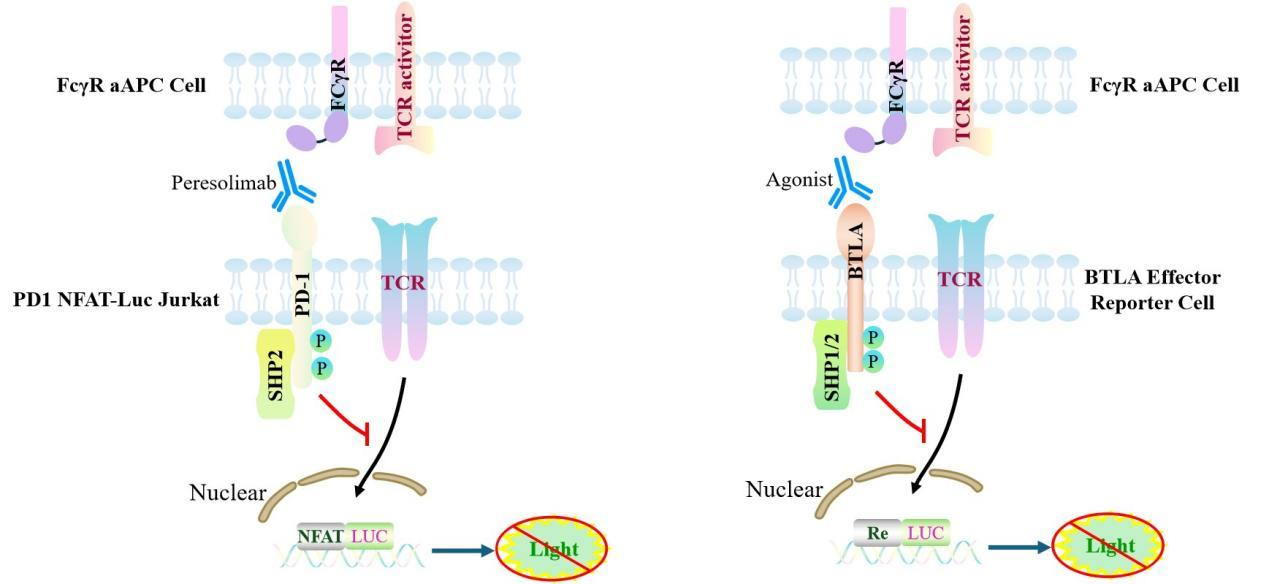
Figure 1. Schematic diagram of an FcγR-expressing antigen-presenting cell (aAPC)
| Classification | Fc Effector |
| Family | Fc receptor family (FcγR family) |
| Gene Name | FCGR1A |
| Gene Aliases | CD64;CD64A;FcgammaRI;FcgammaRIa;FCG1;FCGR1 |
| Gene ID | 2209 |
| Accession Number | NM_000566.4 |
| UniProt Number | P12314 |
| Protein Name | IgG Fc receptor I |
| Protein Aliases | Fc-gamma RI (FcRI);Fc-gamma RIA (FcgammaRIa) |
| Target Species | Human |
| Host cell | CHO-K1 |

Figure 2. Dose Response of PD1 Agonist Antibody in PD1/NFAT-Luc/Jurkat Cells With FCγR aAPC Cell(C1).
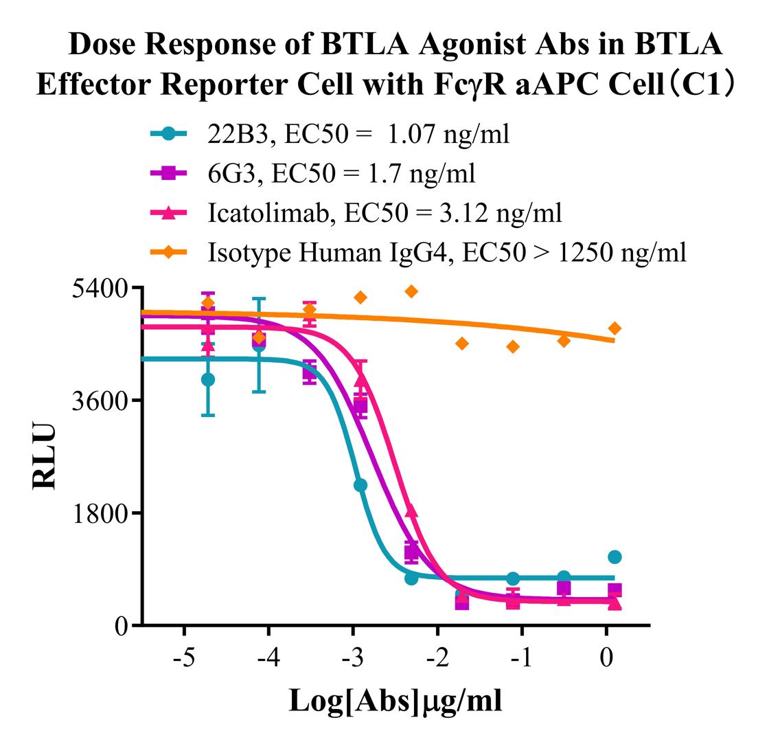
Figure 3. Dose Response of BTLA Agonist Abs in BTLA Effector Reporter Cell with FCγR aAPC Cell(C1).
Cell Resuscitation
1)Rapidly thaw the frozen cells in a 37 °C water bath for approximately 60 seconds. Once thawed (which may take slightly less or more than 60 seconds), immediately transfer the cell suspension from the cryovial into a 15 mL centrifuge tube containing 10 mL of pre-warmed CHO-K1 Human FCγR aAPC Cell complete culture medium.
2)Centrifuge cells at 1000 rpm for 5 min to remove medium, then resuspend cells in 5 mL of pre-warmed complete medium.
3)Transfer the cell suspension into a T25 culture flask and incubate at 37 °C with 5% CO₂.
4)After approximately 24–36 hours, replace the medium or passage the cells to remove non-adherent dead cells.
Subculturing procedure
1)When the cell density reaches the appropriate confluency for passaging, wash the cells with PBS, then add 1 mL trypsin to detach the cells. When more than 80% of the cells detach upon gently tapping the culture flask, add complete culture medium to terminate digestion. Gently pipette to obtain a single-cell suspension, transfer to a 15 mL centrifuge tube, and centrifuge at 1000 rpm for 5 minutes.
2)Discard supernatant after centrifugation. Resuspend cells in fresh medium to a single-cell suspension and transfer to a new culture flask for continued growth.
Cell Freezing
After trypsinization and centrifugation of cells from each T75 flask or 10 cm culture dish, discard the supernatant. Add 2 mL of cryopreservation medium (90% FBS + 10% DMSO), gently resuspend thoroughly, and aliquot into two cryovials. Immediately place the cryovials into a controlled-rate freezing container (e.g., Nalgene 5100-0001), fill with isopropanol to the indicated level, and store at −80 °C. After 24 hours, transfer the cryovials to liquid nitrogen for long-term storage.
Related products
CHO-K1 Human CCR4 Cell Line
HEK293 Human NK1R CRE-Luc Cell Line
Raji-Luc-GFP
Jurkat E6.1-Luc
THP-1-GFP
THP-1-Luc
Raji-GFP
Raji-Luc
Jurkat E6.1-GFP
HEK293 Human GAL4-Luc Cell
We Are Pleased to Announce: Global Commercial Licensing Rights for Jurkat E6.1, CHO-K1, and HEK293 Cell Lines Officially Secured.
Explore