

CHO-K1 Human PDL2/TCR Activator Cell
Cat. No: RQP74065
Size: 1 vial of frozen cells (>1E6 per vial in 1 mL)
Unit Price: Contact For Pricing
Product Info
Description
Biological Information
Assay Data
Cell Culture
| Cat. No | RQP74065 |
| Product Name | CHO-K1 Human PDL2/TCR Activator Cell |
| Product Type | Reporter Cell |
| Culture Properties | Adherent |
| Stability | 32passages (in-house test, that not means the cell line will be instable beyond the passages we tested.) |
| Mycoplasma Status | Negative |
| Culture Medium | F12K+10%FBS+2μg/ml puromycin+500μg/ml Hygromycin B |
| Freeze Medium | 90% FBS+10% DMSO |
| Storage Conditions | Liquid nitrogen immediately upon delivery |
| Application | Functional(Report Gene) Assay |
For research use only. Not intended for human or animal clinical trials, therapeutic or diagnostic use.
Programmed Cell Death Protein 1 (PD-1), a receptor expressed on activated T cells, binds to its ligands—PD-L1 and PD-L2—to negatively regulate immune responses. PD-1 ligands are present in most cancers; the interaction between PD-1 and PD-L1/2 suppresses T-cell activity and enables cancer cells to evade immune surveillance. The PD-1/PD-L1 signaling pathway constitutes a critical component of tumor-induced immunosuppression, inhibiting T-lymphocyte activation and enhancing immune tolerance within the tumor, thereby facilitating tumor immune evasion. The binding of PD-1 to PD-L1 attenuates T-cell-mediated immune surveillance, leading to a compromised immune response or even T-cell apoptosis. PD-1/PD-L1 inhibitors can reverse the immunosuppression of anti-tumor T cells, thereby promoting T-cell proliferation, infiltration into the tumor microenvironment, and the induction of anti-tumor responses. The PD-1/PD-L1/2 pathway is also involved in regulating autoimmune responses, making these proteins promising therapeutic targets for various cancers, as well as for conditions such as multiple sclerosis, arthritis, lupus, and type 1 diabetes. The tyrosine phosphatase SHP2 is a key regulator of T-cell function, mediating both downstream activation signals from the T-cell receptor (TCR) and downstream inhibitory signals from PD-1.
The CHO-K1 Human PDL2/TCR Activator Cell Model—effectively simulates the signal transduction process of PD1&PDL2 *in vivo*. The underlying principle is illustrated in the figure below.
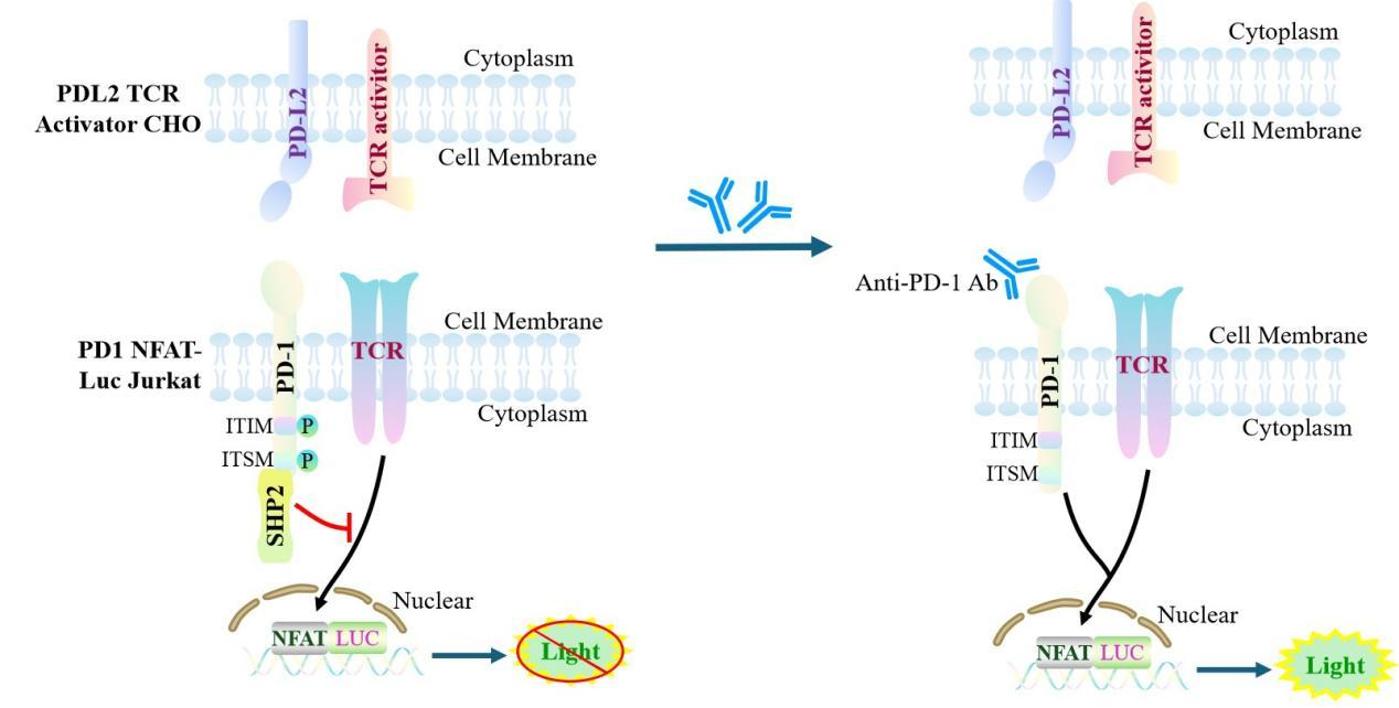
Figure 1. Schematic Diagram of the CHO-K1 Human PDL2/TCR Activator Cell Model
| Classification | Co-Inhibitory |
| Family | immunoglobulin superfamily. BTN/MOG family |
| Gene Name | PDCD1LG2 |
| Gene Aliases | PD-L2;Btdc;PDL2;bA574F11.2;CD273;B7-DC;B7DC |
| Gene ID | 80380 |
| Accession Number | NM_025239.4 |
| UniProt Number | Q9BQ51 |
| Protein Name | PD-1 ligand 2; PD-L2; PDCD1 ligand 2; Programmed death ligand 2 |
| Protein Aliases | Butyrophilin B7-DC (B7-DC) |
| Target Species | Human |
| Host cell | CHO-K1 |
CHO cell line expressing full length human PDL2. Expression is confirmed by flow cytometry.
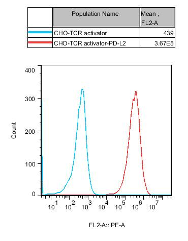
Figure 2.Recombinant PDL2/TCR Activator/CHO constitutively expressing PDL2.
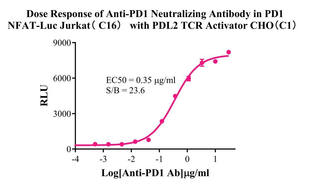
Figure 3. Dose Responese of anti-PD1 neutralizing antibody in PD1 NFAT-Luc Jurkat (C16) with PDL2 TCR Activator CHO(C1).
Cell Resuscitation
1)Rapidly thaw the frozen cells in a 37 °C water bath for approximately 60 seconds. Once thawed (which may take slightly less or more than 60 seconds), immediately transfer the cell suspension from the cryovial into a 15 mL centrifuge tube containing 10 mL of pre-warmed CHO-K1 Human PDL2/TCR Activator Cell complete culture medium.
2)Centrifuge cells at 1000 rpm for 5 min to remove medium, then resuspend cells in 5 mL of pre-warmed complete medium.
3)Transfer the cell suspension into a T25 culture flask and incubate at 37 °C with 5% CO₂.
4)After approximately 24–36 hours, replace the medium or passage the cells to remove non-adherent dead cells.
Subculturing procedure
1)When the cell density reaches the appropriate confluency for passaging, wash the cells with PBS, then add 1 mL trypsin to detach the cells. When more than 80% of the cells detach upon gently tapping the culture flask, add complete culture medium to terminate digestion. Gently pipette to obtain a single-cell suspension, transfer to a 15 mL centrifuge tube, and centrifuge at 1000 rpm for 5 minutes.
2)Discard supernatant after centrifugation. Resuspend cells in fresh medium to a single-cell suspension and transfer to a new culture flask for continued growth.
Cell Freezing
After trypsinization and centrifugation of cells from each T75 flask or 10 cm culture dish, discard the supernatant. Add 2 mL of cryopreservation medium (90% FBS + 10% DMSO), gently resuspend thoroughly, and aliquot into two cryovials. Immediately place the cryovials into a controlled-rate freezing container (e.g., Nalgene 5100-0001), fill with isopropanol to the indicated level, and store at −80 °C. After 24 hours, transfer the cryovials to liquid nitrogen for long-term storage.
Related products
CHO-K1 Human CCR4 Cell Line
HEK293 Human NK1R CRE-Luc Cell Line
Raji-Luc-GFP
Jurkat E6.1-Luc
THP-1-GFP
THP-1-Luc
Raji-GFP
Raji-Luc
Jurkat E6.1-GFP
HEK293 Human GAL4-Luc Cell
We Are Pleased to Announce: Global Commercial Licensing Rights for Jurkat E6.1, CHO-K1, and HEK293 Cell Lines Officially Secured.
Explore