

Jurkat E6.1 Human TMIGD2 Effector Reporter Cell
Cat. No: RQP74202
Size: 1 vial of frozen cells (>1E6 per vial in 1 mL)
Unit Price: Contact For Pricing
Product Info
Description
Biological Information
Assay Data
Cell Culture
| Cat. No | RQP74202 |
| Product Name | Jurkat E6.1 Human TMIGD2 Effector Reporter Cell |
| Product Type | Reporter Cell |
| Culture Properties | suspension |
| Stability | 32passages (in-house test, that not means the cell line will be instable beyond the passages we tested.) |
| Mycoplasma Status | Negative |
| Culture Medium | RPMI-1640+10%FBS+1μg/ml puromycin+800μg/ml Hygromycin B |
| Freeze Medium | 90% FBS+10% DMSO |
| Storage Conditions | Liquid nitrogen immediately upon delivery |
| Application | Functional(Report Gene) Assay |
For research use only. Not intended for human or animal clinical trials, therapeutic or diagnostic use.
TMIGD2 (also known as CD28H or IGPR1) is a member of the Immunoglobulin Superfamily (IgSF) and a novel immune molecule belonging to the CD28 family. Acting as a co-stimulatory receptor for HHLA2—a member of the B7 family—it is primarily expressed on activated CD8⁺ T cells, NK cells, and intestinal intraepithelial lymphocytes. TMIGD2 is a transmembrane protein predominantly expressed on T cells and Natural Killer (NK) cells. Upon binding to HHLA2, TMIGD2 co-stimulates human T and NK cells, leading to cellular proliferation and cytokine production. Its principle of action relies on the activation of the PI3K-AKT-mTOR signaling pathway via an intracellular YXXM motif following HHLA2 binding; this activation promotes T-cell metabolic reprogramming (enhancing both glycolysis and oxidative phosphorylation), thereby driving T-cell proliferation, survival, and memory differentiation, while simultaneously antagonizing PD-1-mediated exhaustion signals. Within the tumor microenvironment, TMIGD2 signaling can enhance anti-tumor immune responses; however, its interactions with specific ligand isoforms may exert inhibitory effects. This highlights its dual potential in immune regulation and identifies it as a novel therapeutic target for the development of combination immunotherapies (e.g., in synergy with PD-1 inhibitors).
The TMIGD2 Effector Reporter Cell model accurately simulates the in vivo signal transduction processes of TMIGD2; the underlying principle is illustrated in the figure below.
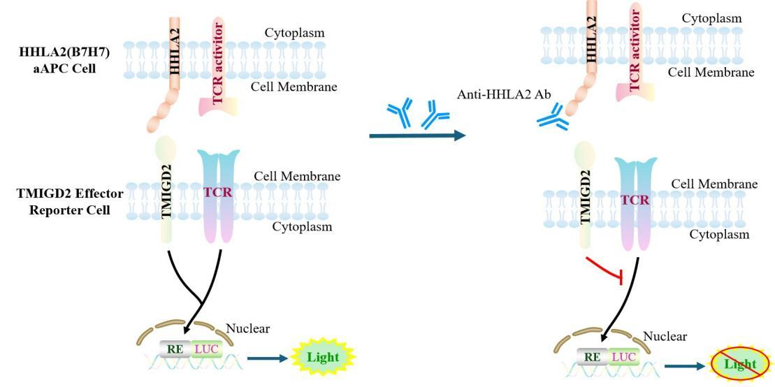
Figure 1. Schematic Diagram of the Jurkat E6.1 Human TMIGD2 Effector Reporter Cell Model
| Classification | Co-Stimulatory |
| Family | Immunoglobulin superfamily (IgSF) |
| Gene Name | TMIGD2 |
| Gene Aliases | CD28H;IGPR-1 |
| Gene ID | 126259 |
| Accession Number | NM_001169126.2 |
| UniProt Number | Q96BF3 |
| Protein Name | Transmembrane and immunoglobulin domain-containing protein 2 |
| Protein Aliases | CD28 homolog;Immunoglobulin and proline-rich receptor 1 (IGPR-1) |
| Target Species | Human |
| Host cell | Jurkat E6.1 |
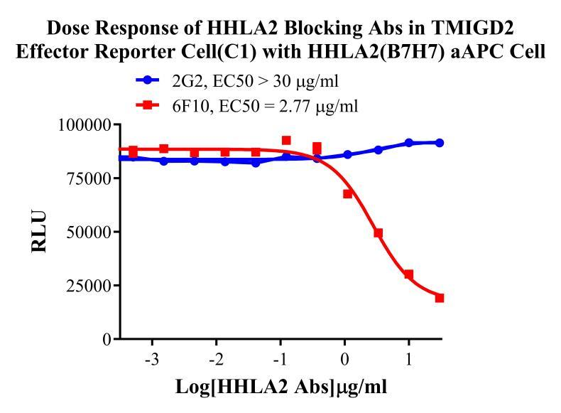
Figure 2. Dose Response of HHLA2 Blocking Abs in TMIGD2 Effector Reporter Cell (C1) with HHLA2(B7H7) aAPC Cell.
Cell Passage Procedures
1.This cell line grows in suspension.
2.Upon receipt, cells should be thawed immediately or stored in liquid nitrogen until use.
3.Before thawing, pre-warm the water bath and culture medium to 37 °C, and prepare a small amount of dry ice.
4.Remove the cryovial from storage and transport it to the cell culture laboratory on dry ice.
5.Rapidly thaw the cells in a 37 °C water bath. Once the cells are completely thawed, spray the cryovial with 70% ethanol for disinfection and transfer it to a biosafety cabinet.
6.Add 10 mL of pre-warmed culture medium into a 15 mL centrifuge tube. Transfer the contents of the cryovial into the tube and centrifuge at 1000 rpm for 5 minutes.
7.Carefully discard the supernatant. Resuspend the cell pellet in 5 mL of pre-warmed culture medium by gentle pipetting. Immediately perform cell counting and adjust the cell density to 3–6 × 10⁵ cells/mL based on the counting results, then transfer the cells into a culture flask.
8.Count the cells every 1–2 days. When the cell density exceeds 1 × 10⁶ cells/mL, passage the cells promptly or add fresh culture medium. Maintain the cell density between 2 × 10⁵ and 1 × 10⁶ cells/mL.
Suspension Cell Cryopreservation Procedure:
1.Collect 8 × 10⁶ cells, centrifuge, and discard the supernatant.
2.Add 1 mL of cell freezing medium (90% FBS + 10% DMSO) and gently pipette to mix thoroughly. Transfer the suspension into a cryovial.
3.Immediately place the cryovial into a controlled-rate freezing container (Nalgene 5100-0001), fill with isopropanol up to the indicated level, and store at −80 °C.
4.After 24 hours, transfer the cryovial to liquid nitrogen for long-term storage.
Related products
CHO-K1 Human CCR4 Cell Line
HEK293 Human NK1R CRE-Luc Cell Line
Raji-Luc-GFP
Jurkat E6.1-Luc
THP-1-GFP
THP-1-Luc
Raji-GFP
Raji-Luc
Jurkat E6.1-GFP
HEK293 Human GAL4-Luc Cell
We Are Pleased to Announce: Global Commercial Licensing Rights for Jurkat E6.1, CHO-K1, and HEK293 Cell Lines Officially Secured.
Explore