

Jurkat E6.1 Human SIRPɑ Effector Reporter Cell
Cat. No: RQP74122
Size: 1 vial of frozen cells (>1E6 per vial in 1 mL)
Unit Price: Contact For Pricing
Product Info
Description
Biological Information
Assay Data
Cell Culture
| Cat. No | RQP74122 |
| Product Name | Jurkat E6.1 Human SIRPɑ Effector Reporter Cell |
| Product Type | Reporter Cell |
| Culture Properties | suspension |
| Stability | 32passages (in-house test, that not means the cell line will be instable beyond the passages we tested.) |
| Mycoplasma Status | Negative |
| Culture Medium | RPMI-1640+10%FBS+1 μg/ml Puromycin+5 μg/ml Blasticidin |
| Freeze Medium | 90% FBS+10% DMSO |
| Storage Conditions | Liquid nitrogen immediately upon delivery |
| Application | Functional(Report Gene) Assay |
For research use only. Not intended for human or animal clinical trials, therapeutic or diagnostic use.
SIRPα (Signal Regulatory Protein α) is a transmembrane protein belonging to the immunoglobulin superfamily. It is primarily expressed on the surface of myeloid cells (such as macrophages and dendritic cells). Its extracellular region contains three immunoglobulin-like domains (D1, D2, and D3), of which the D1 domain specifically binds to CD47. Its intracellular segment contains Immunoreceptor Tyrosine-based Inhibitory Motifs (ITIMs), which recruit SHP-1 and SHP-2 phosphatases to transmit inhibitory signals. By regulating phagocytic activity, SIRPα maintains self-tolerance and prevents macrophages from inadvertently attacking normal cells.
CD47 is a five-pass transmembrane protein widely expressed on the surface of erythrocytes, platelets, and tumor cells. Its extracellular N-terminal IgV-like domain binds to SIRPα; consequently, it is known as a "Don’t eat me" signaling molecule. It plays a critical role in erythrocyte homeostasis (specifically, preventing splenic macrophages from clearing young erythrocytes) and in tumor immune evasion.
Upon binding to CD47, the intracellular ITIM motifs of SIRPα undergo phosphorylation, thereby recruiting SHP-1 and SHP-2 phosphatases to inhibit downstream pro-phagocytic signals through dephosphorylation.
The SIRPα Effector Reporter Cell model accurately simulates the in vivo signal transduction process of SIRPα, as illustrated in the figure below.
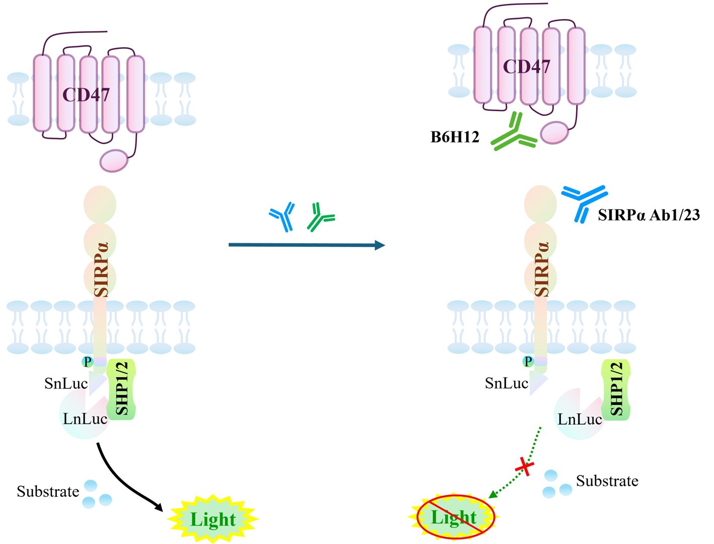
Figure 1. Schematic Diagram of the SIRPα Effector Reporter Cell Model
| Classification | Co-Inhibitory |
| Family | Ig-like cell adhesion molecule family |
| Gene Name | SIRPA |
| Gene Aliases | SHPS1;SIRP;MYD-1;BIT;P84;SHPS-1;SIRPalpha;CD172a;SIRPalpha2; MFR;SIRP-ALPHA-1 |
| Gene ID | 140885 |
| Accession Number | NM_001040023.2 |
| UniProt Number | P78324 |
| Protein Name | SHP substrate 1; SHPS-1 |
| Protein Aliases | Bit;CD172 antigen-like family member A;Inhibitory receptor SHPS-1;Macrophage fusion receptor;MyD-1 antigen |
| Target Species | Human |
| Host cell | Jurkat E6.1 |
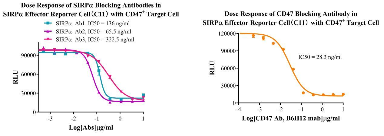
Figure 2. Dose response of SIRPɑ Blocking Antibodies in SIRPɑ Effector Reporter Cell(C11) With CD47+ Target Cell. Dose response of CD47 Blocking Antibody in SIRPɑ Effector Reporter Cell(C11) With CD47+ Target Cell.
Cell Passage Procedures
1.This cell line grows in suspension.
2.Upon receipt, cells should be thawed immediately or stored in liquid nitrogen until use.
3.Before thawing, pre-warm the water bath and culture medium to 37 °C, and prepare a small amount of dry ice.
4.Remove the cryovial from storage and transport it to the cell culture laboratory on dry ice.
5.Rapidly thaw the cells in a 37 °C water bath. Once the cells are completely thawed, spray the cryovial with 70% ethanol for disinfection and transfer it to a biosafety cabinet.
6.Add 10 mL of pre-warmed culture medium into a 15 mL centrifuge tube. Transfer the contents of the cryovial into the tube and centrifuge at 1000 rpm for 5 minutes.
7.Carefully discard the supernatant. Resuspend the cell pellet in 5 mL of pre-warmed culture medium by gentle pipetting. Immediately perform cell counting and adjust the cell density to 3–6 × 10⁵ cells/mL based on the counting results, then transfer the cells into a culture flask.
8.Count the cells every 1–2 days. When the cell density exceeds 1 × 10⁶ cells/mL, passage the cells promptly or add fresh culture medium. Maintain the cell density between 2 × 10⁵ and 1 × 10⁶ cells/mL.
Suspension Cell Cryopreservation Procedure:
1.Collect 8 × 10⁶ cells, centrifuge, and discard the supernatant.
2.Add 1 mL of cell freezing medium (90% FBS + 10% DMSO) and gently pipette to mix thoroughly. Transfer the suspension into a cryovial.
3.Immediately place the cryovial into a controlled-rate freezing container (Nalgene 5100-0001), fill with isopropanol up to the indicated level, and store at −80 °C.
4.After 24 hours, transfer the cryovial to liquid nitrogen for long-term storage.
Related products
CHO-K1 Human CCR4 Cell Line
HEK293 Human NK1R CRE-Luc Cell Line
Raji-Luc-GFP
Jurkat E6.1-Luc
THP-1-GFP
THP-1-Luc
Raji-GFP
Raji-Luc
Jurkat E6.1-GFP
HEK293 Human GAL4-Luc Cell
We Are Pleased to Announce: Global Commercial Licensing Rights for Jurkat E6.1, CHO-K1, and HEK293 Cell Lines Officially Secured.
Explore