

Jurkat E6.1 Human NCR1(NKp46) Effector Reporter Cell
Cat. No: RQP74220
Size: 1 vial of frozen cells (>1E6 per vial in 1 mL)
Unit Price: Contact For Pricing
Product Info
Description
Biological Information
Assay Data
Cell Culture
| Cat. No | RQP74220 |
| Product Name | Jurkat E6.1 Human NCR1(NKp46) Effector Reporter Cell |
| Product Type | Reporter Cell |
| Culture Properties | suspension |
| Stability | 32passages (in-house test, that not means the cell line will be instable beyond the passages we tested.) |
| Mycoplasma Status | Negative |
| Culture Medium | RPMI-1640+10%FBS+1μg/ml puromycin+800μg/ml Hygromycin B+5μg/ml blasticidin |
| Freeze Medium | 90% FBS+10% DMSO |
| Storage Conditions | Liquid nitrogen immediately upon delivery |
| Application | Functional(Report Gene) Assay |
For research use only. Not intended for human or animal clinical trials, therapeutic or diagnostic use.
Natural Cytotoxicity Receptors (NCRs) are specific surface markers of NK cells and serve as their primary activating receptors. The NCR family comprises three members: NKp46 (NCR1, CD335), NKp44 (NCR2, CD336), and NKp30 (NCR3, CD337).
NKp46 (NCR1, CD335) is a 46 kDa type I transmembrane glycoprotein consisting of two extracellular C2-type immunoglobulin (Ig)-like domains; it belongs to the immunoglobulin superfamily. The transmembrane domain of NKp46 contains a single arginine residue, which is responsible for associating with the adaptor subunits FcεRIγ and CD3ζ to transmit activating signals; notably, the intracellular domain of NKp46 lacks ITAM sequences. NKp46 induces NK cell activation—leading to the direct killing of target cells—in a manner independent of HLA class I molecules.
Upon binding to its ligand, NKp46 associates with the adaptor proteins CD3ζ and/or FcεRIγ. The ITAM motifs within these adaptors undergo phosphorylation, a process likely mediated by Src-family kinases (such as LCK and FYN). Through their SH2 domains, these phosphorylated ITAMs recruit and activate tyrosine kinases, such as Syk and/or ZAP70. These tyrosine kinases, in turn, activate transmembrane adaptor proteins—such as LAT—thereby triggering downstream molecules, including phospholipase C (PLCγ), PI3K, as well as Vav1, Vav2, and Vav3. PLCγ subsequently triggers an influx of Ca²⁺ ions; meanwhile, PI3K and Vav1 recruit Rac1 and induce a phosphorylation cascade via the PAK1-MEK-ERK signaling pathway, further activating the MAPK signaling pathway and other downstream responses. Ultimately, these signaling cascades drive actin cytoskeleton rearrangement, degranulation, cytotoxicity, and the transcriptional expression of cytokines or chemokines.
The Jurkat E6.1 Human NCR1(NKp46) Effector Reporter Cell Model—effectively simulates the signal transduction process of NKp46 *in vivo*. The underlying principle is illustrated in the figure below.

Figure 1. Schematic Diagram of the Jurkat E6.1 Human NCR1(NKp46) Effector Reporter Cell Model
| Classification | Co-Stimulatory |
| Family | natural cytotoxicity receptor (NCR) family |
| Gene Name | NCR1 |
| Gene Aliases | LY94;NK-p46;NKP46;CD335 |
| Gene ID | 9437 |
| Accession Number | NM_004829.7 |
| UniProt Number | O76036 |
| Protein Name | Natural cytotoxicity triggering receptor 1 |
| Protein Aliases | Lymphocyte antigen 94 homolog; NK cell-activating receptor; Natural killer cell p46-related protein (NK-p46; NKp46; hNKp46) |
| Target Species | Human |
| Host cell | Jurkat E6.1 |
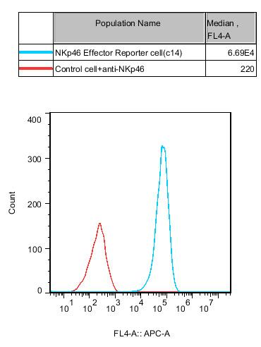
Figure 2. Rcombinant NKp46 Effector Reporter Cell stably expressing NKp46.
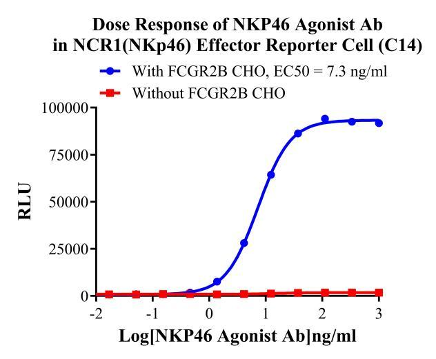
Figure 3. Dose Response of NKp46 Agonist Ab in NCR1(NKp46) Effector Reporter Cell(C14).
Cell Passage Procedures
1.This cell line grows in suspension.
2.Upon receipt, cells should be thawed immediately or stored in liquid nitrogen until use.
3.Before thawing, pre-warm the water bath and culture medium to 37 °C, and prepare a small amount of dry ice.
4.Remove the cryovial from storage and transport it to the cell culture laboratory on dry ice.
5.Rapidly thaw the cells in a 37 °C water bath. Once the cells are completely thawed, spray the cryovial with 70% ethanol for disinfection and transfer it to a biosafety cabinet.
6.Add 10 mL of pre-warmed culture medium into a 15 mL centrifuge tube. Transfer the contents of the cryovial into the tube and centrifuge at 1000 rpm for 5 minutes.
7.Carefully discard the supernatant. Resuspend the cell pellet in 5 mL of pre-warmed culture medium by gentle pipetting. Immediately perform cell counting and adjust the cell density to 3–6 × 10⁵ cells/mL based on the counting results, then transfer the cells into a culture flask.
8.Count the cells every 1–2 days. When the cell density exceeds 1 × 10⁶ cells/mL, passage the cells promptly or add fresh culture medium. Maintain the cell density between 2 × 10⁵ and 1 × 10⁶ cells/mL.
Suspension Cell Cryopreservation Procedure:
1.Collect 8 × 10⁶ cells, centrifuge, and discard the supernatant.
2.Add 1 mL of cell freezing medium (90% FBS + 10% DMSO) and gently pipette to mix thoroughly. Transfer the suspension into a cryovial.
3.Immediately place the cryovial into a controlled-rate freezing container (Nalgene 5100-0001), fill with isopropanol up to the indicated level, and store at −80 °C.
4.After 24 hours, transfer the cryovial to liquid nitrogen for long-term storage.
Related products
CHO-K1 Human CCR4 Cell Line
HEK293 Human NK1R CRE-Luc Cell Line
Raji-Luc-GFP
Jurkat E6.1-Luc
THP-1-GFP
THP-1-Luc
Raji-GFP
Raji-Luc
Jurkat E6.1-GFP
HEK293 Human GAL4-Luc Cell
We Are Pleased to Announce: Global Commercial Licensing Rights for Jurkat E6.1, CHO-K1, and HEK293 Cell Lines Officially Secured.
Explore