

Jurkat E6.1 Human LAG3/NFAT-Luc Cell
Cat. No: RQP74060
Size: 1 vial of frozen cells (>1E6 per vial in 1 mL)
Unit Price: Contact For Pricing
Product Info
Description
Biological Information
Assay Data
Cell Culture
| Cat. No | RQP74060 |
| Product Name | Jurkat E6.1 Human LAG3/NFAT-Luc Cell |
| Product Type | Reporter Cell |
| Culture Properties | suspension |
| Stability | 32passages (in-house test, that not means the cell line will be instable beyond the passages we tested.) |
| Mycoplasma Status | Negative |
| Culture Medium | RPMI-1640+10%FBS+1μg/ml puromycin+400μg/ml Hygromycin B |
| Freeze Medium | 90% FBS+10% DMSO |
| Storage Conditions | Liquid nitrogen immediately upon delivery |
| Application | Functional(Report Gene) Assay |
For research use only. Not intended for human or animal clinical trials, therapeutic or diagnostic use.
Lymphocyte Activation Gene 3 (LAG-3), also known as CD223, is a type I transmembrane protein belonging to the immunoglobulin superfamily (IgSF). The LAG-3 protein shares structural homology with CD4; both possess four Ig-like domains within their extracellular regions. However, LAG-3 contains an additional loop structure within its membrane-distal Ig-like domain, which confers upon it an affinity for MHC Class II molecules that is 100-fold higher than that of CD4. LAG-3 is expressed on activated T cells, B cells, plasmacytoid dendritic cells (pDCs), and NK cells. In addition to binding to MHC Class II (MHC II) molecules, LAG-3 also binds to Fibrinogen-like Protein 1 (FGL-1), alpha-synuclein fibrils (α-syn), the lectin Galectin-3 (Gal-3), and Lymph Node Sinus Endothelial Cell C-type Lectin (LSECtin).
LAG-3 may represent a superior target for immune checkpoint inhibitors compared to CTLA-4 or PD-1. This is because antibodies targeting the latter two checkpoints primarily activate effector T cells without inhibiting Treg activity; in contrast, antagonistic LAG-3 antibodies are capable of both activating effector T cells (by downregulating LAG-3-mediated inhibitory signals) and suppressing the inhibitory activity of induced (i.e., antigen-specific) Tregs.
The Jurkat E6.1 Human LAG3/NFAT-Luc Cell Model—effectively simulates the signal transduction process of LAG3 *in vivo*. The underlying principle is illustrated in the figure below.
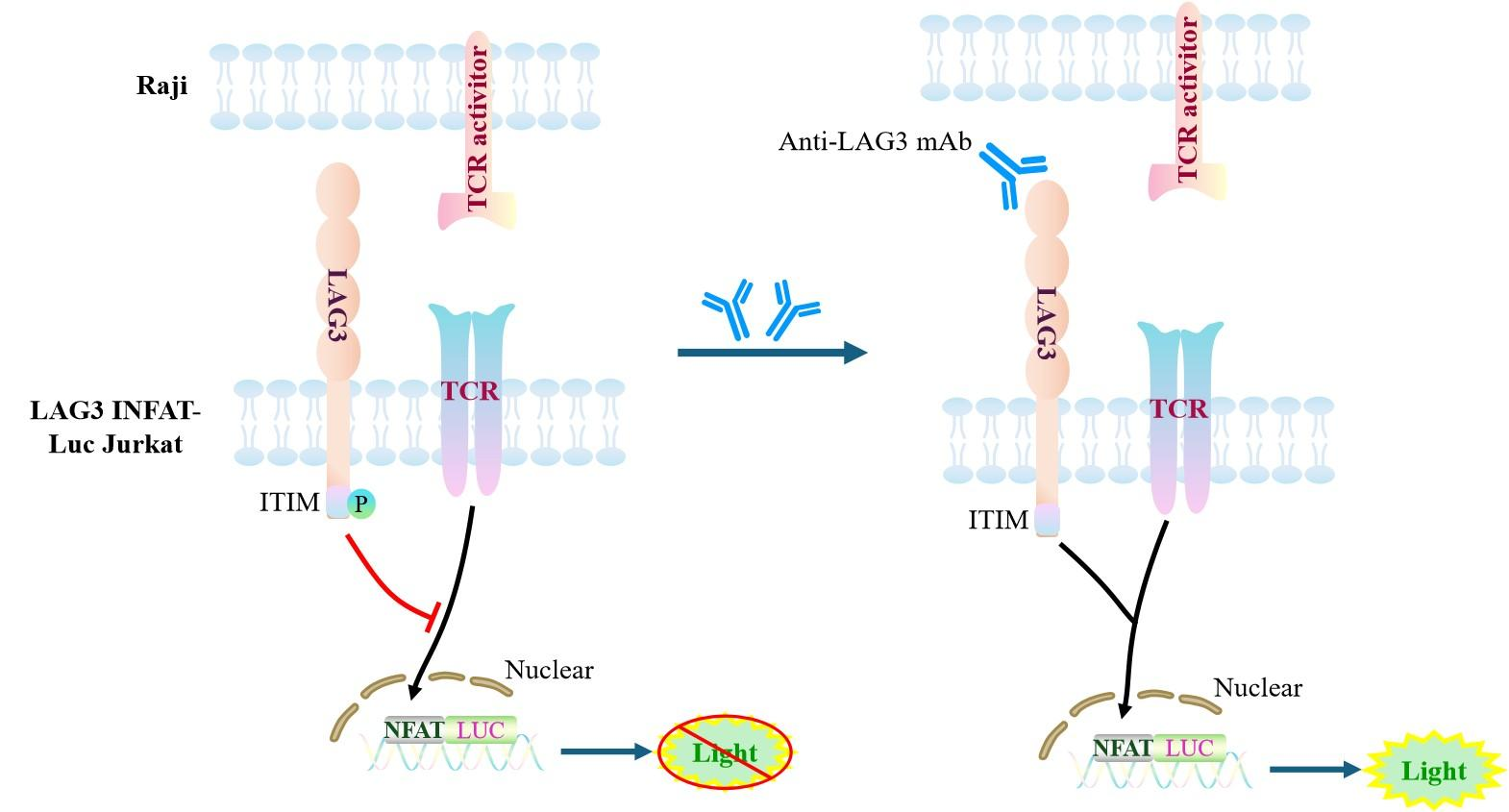
Figure 1. Schematic Diagram of the Jurkat E6.1 Human LAG3/NFAT-Luc Cell Model
| Classification | Co-Inhibitory |
| Family | LAG3 family |
| Gene Name | LAG3 |
| Gene Aliases | CD223 |
| Gene ID | 3902 |
| Accession Number | NM_002286.6 |
| UniProt Number | P18627 |
| Protein Name | LAG-3 |
| Protein Aliases | sLAG-3 |
| Target Species | Human |
| Host cell | Jurkat E6.1 |
Jurkat E6.1 cell line expressing full length human LAG3. Expression is confirmed by flow cytometry.
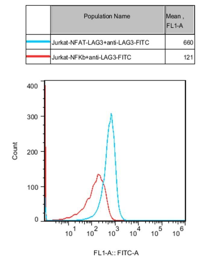
Figure 2. Recombinant LAG3/NFAT-Luc/Jurkat stably expressing LAG3.
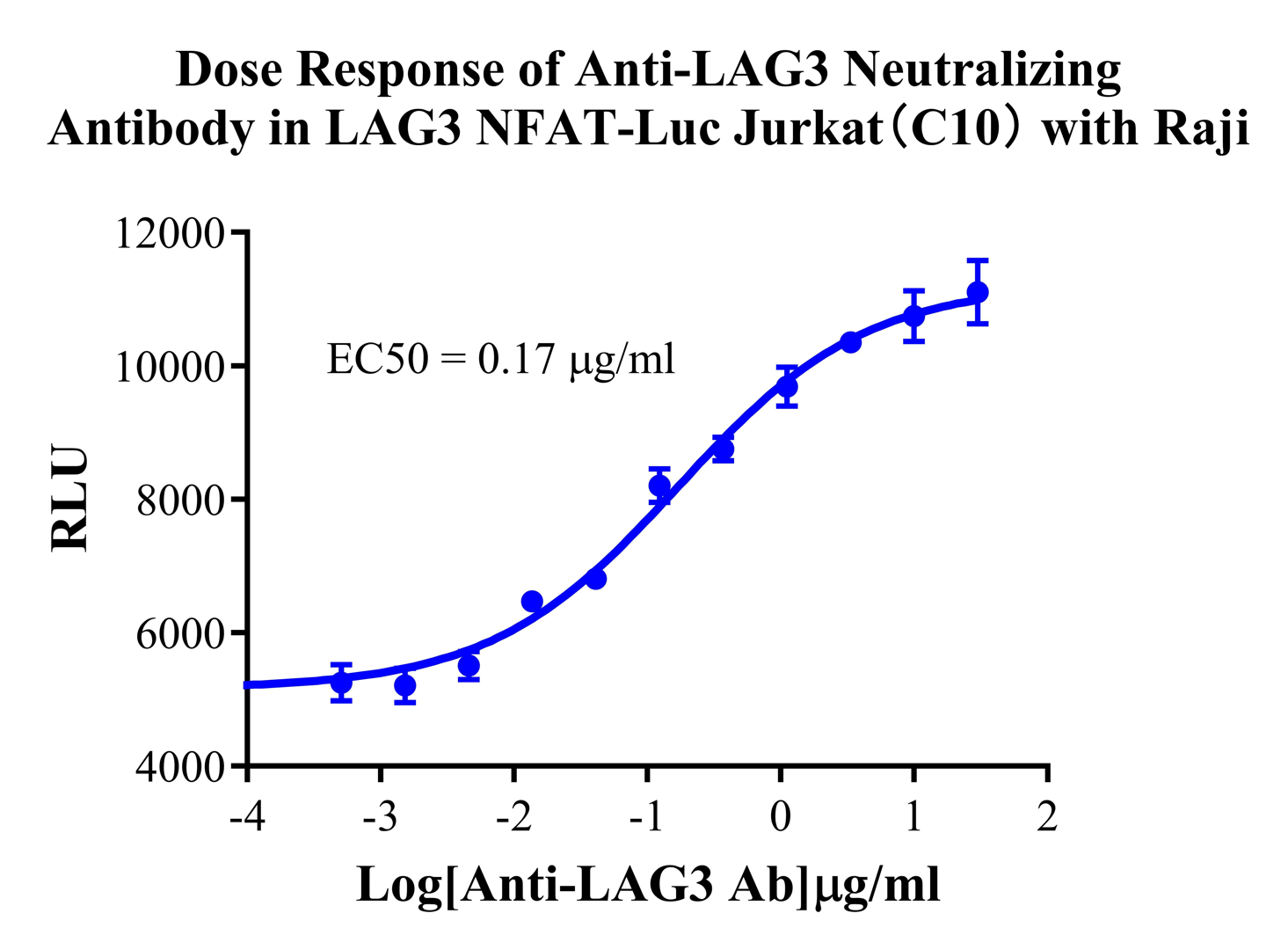
Figure 3. Dose Response of Anti-LAG3 Neutralizing Antibody in LAG3 NFAT-Luc Jurkat(C10) with Raji.
Cell Passage Procedures
1.This cell line grows in suspension.
2.Upon receipt, cells should be thawed immediately or stored in liquid nitrogen until use.
3.Before thawing, pre-warm the water bath and culture medium to 37 °C, and prepare a small amount of dry ice.
4.Remove the cryovial from storage and transport it to the cell culture laboratory on dry ice.
5.Rapidly thaw the cells in a 37 °C water bath. Once the cells are completely thawed, spray the cryovial with 70% ethanol for disinfection and transfer it to a biosafety cabinet.
6.Add 10 mL of pre-warmed culture medium into a 15 mL centrifuge tube. Transfer the contents of the cryovial into the tube and centrifuge at 1000 rpm for 5 minutes.
7.Carefully discard the supernatant. Resuspend the cell pellet in 5 mL of pre-warmed culture medium by gentle pipetting. Immediately perform cell counting and adjust the cell density to 3–6 × 10⁵ cells/mL based on the counting results, then transfer the cells into a culture flask.
8.Count the cells every 1–2 days. When the cell density exceeds 1 × 10⁶ cells/mL, passage the cells promptly or add fresh culture medium. Maintain the cell density between 2 × 10⁵ and 1 × 10⁶ cells/mL.
Suspension Cell Cryopreservation Procedure:
1.Collect 8 × 10⁶ cells, centrifuge, and discard the supernatant.
2.Add 1 mL of cell freezing medium (90% FBS + 10% DMSO) and gently pipette to mix thoroughly. Transfer the suspension into a cryovial.
3.Immediately place the cryovial into a controlled-rate freezing container (Nalgene 5100-0001), fill with isopropanol up to the indicated level, and store at −80 °C.
4.After 24 hours, transfer the cryovial to liquid nitrogen for long-term storage.
Related products
CHO-K1 Human CCR4 Cell Line
HEK293 Human NK1R CRE-Luc Cell Line
Raji-Luc-GFP
Jurkat E6.1-Luc
THP-1-GFP
THP-1-Luc
Raji-GFP
Raji-Luc
Jurkat E6.1-GFP
HEK293 Human GAL4-Luc Cell
We Are Pleased to Announce: Global Commercial Licensing Rights for Jurkat E6.1, CHO-K1, and HEK293 Cell Lines Officially Secured.
Explore