

Jurkat E6.1 Human LAG3/IL-2-Luc Cell
Cat. No: RQP74079
Size: 1 vial of frozen cells (>1E6 per vial in 1 mL)
Unit Price: Contact For Pricing
Product Info
Description
Biological Information
Assay Data
Cell Culture
| Cat. No | RQP74079 |
| Product Name | Jurkat E6.1 Human LAG3/IL-2-Luc Cell |
| Product Type | Reporter Cell |
| Culture Properties | suspension |
| Stability | 32passages (in-house test, that not means the cell line will be instable beyond the passages we tested.) |
| Mycoplasma Status | Negative |
| Culture Medium | RPMI-1640+10%FBS+1μg/ml puromycin+10μg/ml blasticidin |
| Freeze Medium | 90% FBS+10% DMSO |
| Storage Conditions | Liquid nitrogen immediately upon delivery |
| Application | Functional(Report Gene) Assay |
For research use only. Not intended for human or animal clinical trials, therapeutic or diagnostic use.
Lymphocyte Activation Gene 3 (LAG-3), also known as CD223, is a type I transmembrane protein belonging to the immunoglobulin superfamily (IgSF). The LAG-3 protein shares structural homology with CD4; both possess four Ig-like domains within their extracellular regions. However, LAG-3 contains an additional loop structure within its membrane-distal Ig-like domain, which confers upon it an affinity for MHC Class II molecules that is 100-fold higher than that of CD4. LAG-3 is expressed on activated T cells, B cells, plasmacytoid dendritic cells (pDCs), and NK cells. In addition to binding to MHC Class II (MHC II) molecules, LAG-3 also binds to Fibrinogen-like Protein 1 (FGL-1), alpha-synuclein fibrils (α-syn), the lectin Galectin-3 (Gal-3), and Lymph Node Sinus Endothelial Cell C-type Lectin (LSECtin).
LAG-3 may represent a superior target for immune checkpoint inhibitors compared to CTLA-4 or PD-1. This is because antibodies targeting the latter two checkpoints primarily activate effector T cells without inhibiting Treg activity; in contrast, antagonistic LAG-3 antibodies are capable of both activating effector T cells (by downregulating LAG-3-mediated inhibitory signals) and suppressing the inhibitory activity of induced (i.e., antigen-specific) Tregs.
The Jurkat E6.1 Human LAG3/IL-2-Luc Cell Model—effectively simulates the signal transduction process of LAG3 *in vivo*. The underlying principle is illustrated in the figure below.
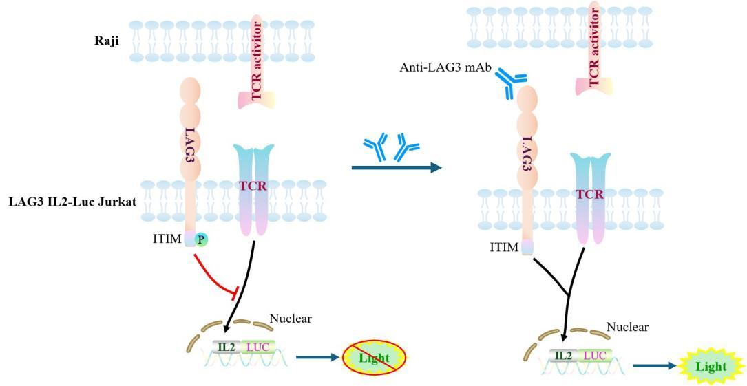
Figure 1. Schematic Diagram of the Jurkat E6.1 Human LAG3/IL-2-Luc Cell Model
| Classification | Co-Inhibitory |
| Family | LAG3 family |
| Gene Name | LAG3 |
| Gene Aliases | CD223 |
| Gene ID | 3902 |
| Accession Number | NM_002286.6 |
| UniProt Number | P18627 |
| Protein Name | LAG-3 |
| Protein Aliases | sLAG-3 |
| Target Species | Human |
| Host cell | Jurkat E6.1 |
Jurkat E6.1 cell line expressing full length human LAG3/IL-2. Expression is confirmed by flow cytometry.
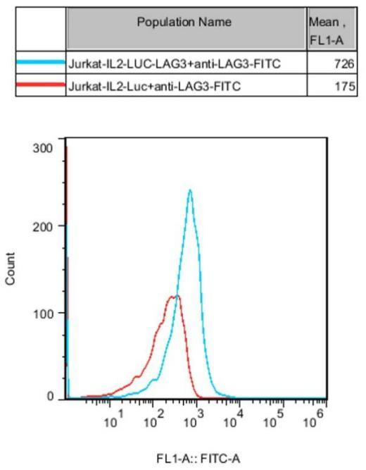
Figure 2. Recombinant LAG3/IL2-Luc/Jurkat stably expressing LAG3.
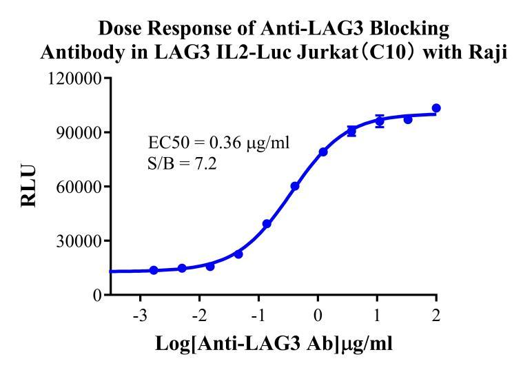
Figure 3. Dose Response of Anti-LAG3 Blocking Antibody in LAG3 IL2-Luc Jurkat (C10) with Raji.
Cell Passage Procedures
1.This cell line grows in suspension.
2.Upon receipt, cells should be thawed immediately or stored in liquid nitrogen until use.
3.Before thawing, pre-warm the water bath and culture medium to 37 °C, and prepare a small amount of dry ice.
4.Remove the cryovial from storage and transport it to the cell culture laboratory on dry ice.
5.Rapidly thaw the cells in a 37 °C water bath. Once the cells are completely thawed, spray the cryovial with 70% ethanol for disinfection and transfer it to a biosafety cabinet.
6.Add 10 mL of pre-warmed culture medium into a 15 mL centrifuge tube. Transfer the contents of the cryovial into the tube and centrifuge at 1000 rpm for 5 minutes.
7.Carefully discard the supernatant. Resuspend the cell pellet in 5 mL of pre-warmed culture medium by gentle pipetting. Immediately perform cell counting and adjust the cell density to 3–6 × 10⁵ cells/mL based on the counting results, then transfer the cells into a culture flask.
8.Count the cells every 1–2 days. When the cell density exceeds 1 × 10⁶ cells/mL, passage the cells promptly or add fresh culture medium. Maintain the cell density between 2 × 10⁵ and 1 × 10⁶ cells/mL.
Suspension Cell Cryopreservation Procedure:
1.Collect 8 × 10⁶ cells, centrifuge, and discard the supernatant.
2.Add 1 mL of cell freezing medium (90% FBS + 10% DMSO) and gently pipette to mix thoroughly. Transfer the suspension into a cryovial.
3.Immediately place the cryovial into a controlled-rate freezing container (Nalgene 5100-0001), fill with isopropanol up to the indicated level, and store at −80 °C.
4.After 24 hours, transfer the cryovial to liquid nitrogen for long-term storage.
Related products
CHO-K1 Human CCR4 Cell Line
HEK293 Human NK1R CRE-Luc Cell Line
Raji-Luc-GFP
Jurkat E6.1-Luc
THP-1-GFP
THP-1-Luc
Raji-GFP
Raji-Luc
Jurkat E6.1-GFP
HEK293 Human GAL4-Luc Cell
We Are Pleased to Announce: Global Commercial Licensing Rights for Jurkat E6.1, CHO-K1, and HEK293 Cell Lines Officially Secured.
Explore