

Jurkat E6.1 Human IL33 Effector Reporter Cell
Cat. No: RQP74176
Size: 1 vial of frozen cells (>1E6 per vial in 1 mL)
Unit Price: Contact For Pricing
Product Info
Description
Biological Information
Assay Data
Cell Culture
| Cat. No | RQP74176 |
| Product Name | Jurkat E6.1 Human IL33 Effector Reporter Cell |
| Product Type | Reporter Cell |
| Culture Properties | suspension |
| Stability | 32passages (in-house test, that not means the cell line will be instable beyond the passages we tested.) |
| Mycoplasma Status | Negative |
| Culture Medium | RPMI-1640+10%FBS+1μg/ml puromycin+800μg/ml Hygromycin B+10μg/ml blasticidin |
| Freeze Medium | 90% FBS+10% DMSO |
| Storage Conditions | Liquid nitrogen immediately upon delivery |
| Application | Functional(Report Gene) Assay |
For research use only. Not intended for human or animal clinical trials, therapeutic or diagnostic use.
Interleukin-33 (IL-33) is a member of the IL-1 cytokine family. IL-33 binds to a heterodimer formed by IL-33R (also known as ST2) and the IL-1 Receptor Accessory Protein (IL-1RAcP), thereby initiating signal transduction and triggering a local immune response. IL-33R is highly expressed in T helper 2 (Th2) cells, mast cells, activated leukocytes—such as regulatory T (Treg) cells—Group 2 innate lymphoid cells (ILC2s), CD8+ T cells, and natural killer (NK) cells; this expression induces the transcription of Th2-associated cytokine genes and facilitates host defense against pathogens. The IL-1 Receptor Accessory Protein (IL-1RAcP) serves as a co-receptor for IL-1Rα, IL-33R, and IL-36R, respectively.
IL-1RAcP does not directly interact with IL-33. Instead, it is recruited to IL-33R following IL-33 stimulation to form an activated heterodimeric receptor complex. This receptor complex subsequently recruits downstream signaling components via its TIR domain. The activated receptor complex then recruits downstream signaling proteins—such as Myeloid Differentiation Primary Response 88 (MyD88) and TNF Receptor-Associated Factor 6 (TRAF6)—which subsequently induce the activation of IL1R-associated kinases (e.g., IRAK-1/4). This process activates Mitogen-Activated Protein Kinases (MAPKs)—specifically ERK, p38, and JNK—as well as the downstream IKK complex, ultimately leading to the activation of the transcription factors AP-1 and NF-κB. IL-33 has been identified as a key mediator in various inflammatory conditions, including asthma, cardiovascular diseases, and allergic disorders.
The Jurkat E6.1 Human IL33 Effector Reporter Cell Model—effectively simulates the signal transduction process of IL33 *in vivo*. The underlying principle is illustrated in the figure below.
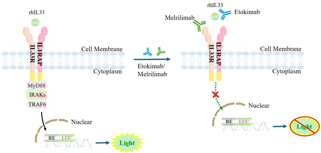
Figure 1. Schematic Diagram of the Jurkat E6.1 Human IL33 Effector Reporter Cell Model
| Classification | Cytokine&Growth Factor |
| Family | Interleukin-1 receptor family |
| Gene Name | IL1RL1 |
| Gene Aliases | ST2;FIT-1;ST2L;ST2V;DER4;T1;IL33R |
| Gene ID | 9173 |
| Accession Number | NM_016232.5 |
| UniProt Number | Q01638 |
| Protein Name | Interleukin-1 receptor-like 1 |
| Protein Aliases | Protein ST2 |
| Family-2 | Interleukin-1 receptor family |
| Gene Name-2 | IL1RAP |
| Gene Aliases-2 | IL-1RAcP;IL1R3;C3orf13 |
| Gene ID-2 | 3556 |
| Accession Number-2 | NM_002182.4 |
| UniProt Number-2 | Q9NPH3 |
| Protein Name-2 | IL-1 receptor accessory protein; IL-1RAcP |
| Protein Aliases-2 | N/A |
| Target Species | Human |
| Host cell | Jurkat E6.1 |
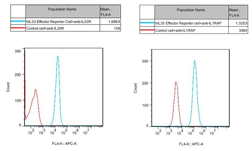
Figure 2. Recombinant hIL33 Effector Reporter Cell constitutively expressing IL33R&IL1RAcP.
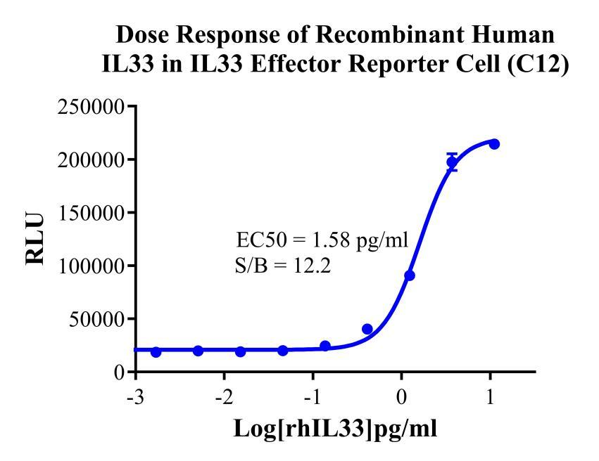
Figure 3. Dose Response of Recombinant Human lL33 in IL33 Effector Reporter Cell (C12).
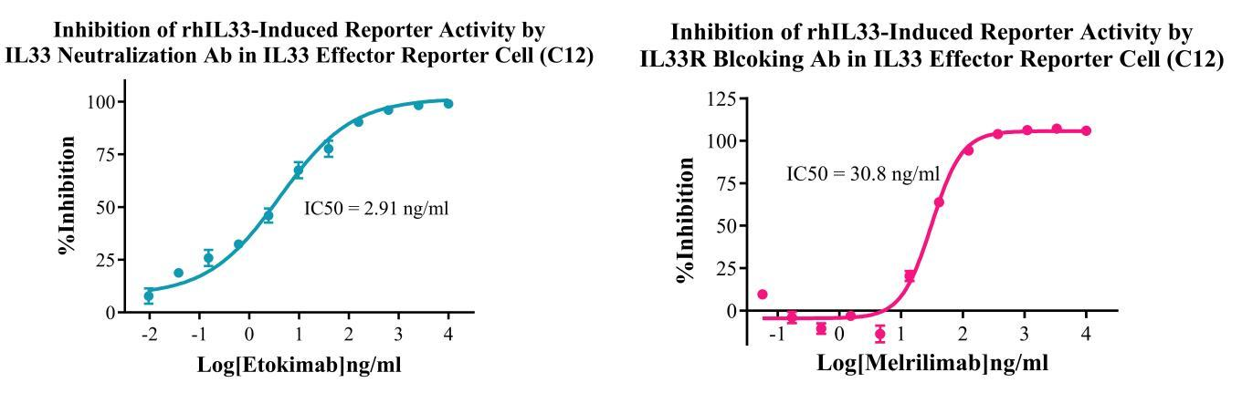
Figure 4. Inhibition of rhlL33-Induced Reporter Activity by IL33 Neutralization/Blocking Ab in IL33 Effector Reporter Cell(C12).
Cell Passage Procedures
1.This cell line grows in suspension.
2.Upon receipt, cells should be thawed immediately or stored in liquid nitrogen until use.
3.Before thawing, pre-warm the water bath and culture medium to 37 °C, and prepare a small amount of dry ice.
4.Remove the cryovial from storage and transport it to the cell culture laboratory on dry ice.
5.Rapidly thaw the cells in a 37 °C water bath. Once the cells are completely thawed, spray the cryovial with 70% ethanol for disinfection and transfer it to a biosafety cabinet.
6.Add 10 mL of pre-warmed culture medium into a 15 mL centrifuge tube. Transfer the contents of the cryovial into the tube and centrifuge at 1000 rpm for 5 minutes.
7.Carefully discard the supernatant. Resuspend the cell pellet in 5 mL of pre-warmed culture medium by gentle pipetting. Immediately perform cell counting and adjust the cell density to 3–6 × 10⁵ cells/mL based on the counting results, then transfer the cells into a culture flask.
8.Count the cells every 1–2 days. When the cell density exceeds 1 × 10⁶ cells/mL, passage the cells promptly or add fresh culture medium. Maintain the cell density between 2 × 10⁵ and 1 × 10⁶ cells/mL.
Suspension Cell Cryopreservation Procedure:
1.Collect 8 × 10⁶ cells, centrifuge, and discard the supernatant.
2.Add 1 mL of cell freezing medium (90% FBS + 10% DMSO) and gently pipette to mix thoroughly. Transfer the suspension into a cryovial.
3.Immediately place the cryovial into a controlled-rate freezing container (Nalgene 5100-0001), fill with isopropanol up to the indicated level, and store at −80 °C.
4.After 24 hours, transfer the cryovial to liquid nitrogen for long-term storage.
Related products
CHO-K1 Human CCR4 Cell Line
HEK293 Human NK1R CRE-Luc Cell Line
Raji-Luc-GFP
Jurkat E6.1-Luc
THP-1-GFP
THP-1-Luc
Raji-GFP
Raji-Luc
Jurkat E6.1-GFP
HEK293 Human GAL4-Luc Cell
We Are Pleased to Announce: Global Commercial Licensing Rights for Jurkat E6.1, CHO-K1, and HEK293 Cell Lines Officially Secured.
Explore