


Jurkat E6.1 Human IL1 Effector Reporter Cell
Cat. No: RQP74253
Size: 1 vial of frozen cells (>1E6 per vial in 1 mL)
Unit Price: Contact For Pricing
Product Info
Description
Biological Information
Assay Data
Cell Culture
| Cat. No | RQP74253 |
| Product Name | Jurkat E6.1 Human IL1 Effector Reporter Cell |
| Product Type | Reporter Cell |
| Culture Properties | suspension |
| Stability | 32passages (in-house test, that not means the cell line will be instable beyond the passages we tested.) |
| Mycoplasma Status | Negative |
| Culture Medium | RPMI-1640+10%FBS+1μg/ml puromycin+800μg/ml Hygromycin B+10μg/ml blasticidin |
| Freeze Medium | 90% FBS+10% DMSO |
| Storage Conditions | Liquid nitrogen immediately upon delivery |
| Application | Functional(Report Gene) Assay |
For research use only. Not intended for human or animal clinical trials, therapeutic or diagnostic use.
The IL-1 family comprises 11 cytokines, including 7 ligands with agonist activity (IL-1α, IL-1β, IL-18, IL-33, IL-36α, IL-36β, IL-36γ), and 4 members with antagonistic activity [IL-1 receptor antagonist (IL-1Rα), IL-36Ra, IL-37, IL-38]. IL-1α, IL-1β, IL-18, IL-33, IL-36α, IL-36β, and IL-36γ activate MPKK and NF-κB, leading to inflammatory responses. Other members of the IL-1 family, however, exert anti-inflammatory effects. IL-1 is a signature cytokine of this family; it jointly stimulates the maturation and proliferation of T helper cells and B cells, activates NK cells, and is associated with inflammation. IL-1 is produced and secreted by epithelial cells, endothelial cells, platelets, fibroblasts, and immune cells (such as macrophages and neutrophils) as part of innate and adaptive immune responses, protecting the body from injury and infection.
Binding of IL-1α or IL-1β to the IL-1R1 receptor induces the dimerization of IL-1R1 with the IL-1R1 co-receptor (IL-1RAcP); the resulting complex recruits MyD88, which in turn recruits IL-1 receptor-associated kinase 1 (IRAK1). IRAK1 activates the TNF receptor-associated factor 6 (TRAF6)/TGFβ-activated kinase 1 (TAK1) signaling cascade, thereby activating the transcription of inflammatory and immune genes.
The IL-1 Effector Reporter Cell model effectively mimics the in vivo signaling pathway of IL-1; the mechanism is illustrated in the figure below.
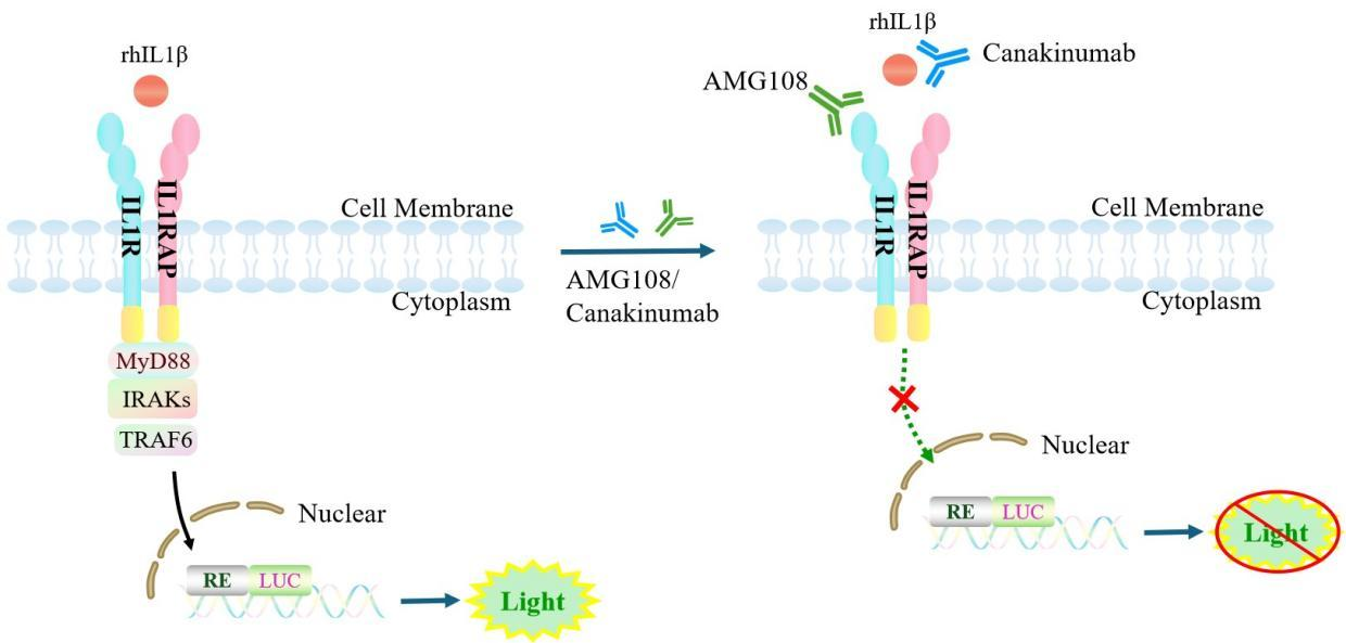
Figure 1. Schematic diagram of the IL-1 effector reporter cell model
| Classification | Cytokine&Growth Factor |
| Family | IL‑1 cytokine family |
| Gene Name | IL1B |
| Gene Aliases | IL1F2;IL-1B;IL1-BETA |
| Gene ID | 3553 |
| Accession Number | NM_000576.3 |
| UniProt Number | P01584 |
| Protein Name | IL-1 beta |
| Protein Aliases | N/A |
| Family-2 | IL-1 family |
| Gene Name-2 | IL1RN |
| Gene Aliases-2 | IL1RA;ICIL-1RA;IL1F3;IRAP;IL-1RN;MGC10430 |
| Gene ID-2 | 3557 |
| Accession Number-2 | NM_173842.3 |
| UniProt Number-2 | P18510 |
| Protein Name-2 | IL-1RN; IL-1ra; IRAP |
| Protein Aliases-2 | ICIL-1RA;IL1 inhibitor |
| Family-3 | interleukin-1 receptor family |
| Gene Name-3 | IL1R1 |
| Gene Aliases-3 | IL1R;IL1RA;D2S1473;CD121A |
| Gene ID-3 | 3554 |
| Accession Number-3 | NM_000877.4 |
| UniProt Number-3 | P14778 |
| Protein Name-3 | IL-1R-1; IL-1RT-1; IL-1RT1 |
| Protein Aliases-3 | CD121 antigen-like family member A;IL-1R-alpha;Interleukin-1 receptor type I |
| Target Species | Human |
| Host cell | Jurkat E6.1 |
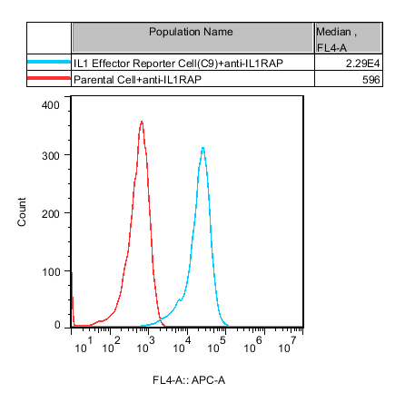
Figure 2. Recombinant IL1 Effector Reporter Cell constitutively expressing IL1RAcP.

Figure 3. Dose Response of Recombinant Human IL1β in IL1 Effector Reporter Cell (C9).
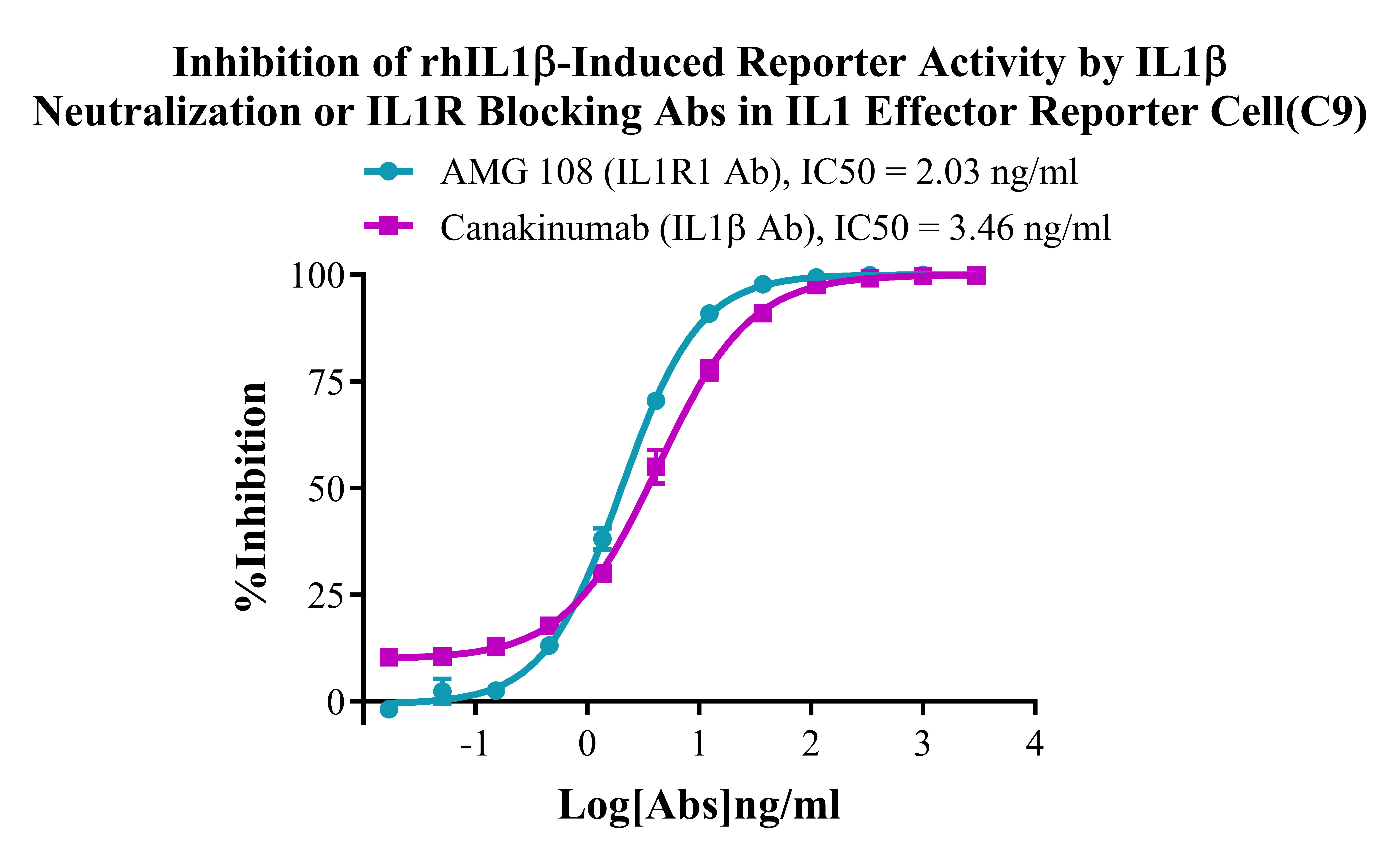
Figure 4. Inhibition of rhIL1β-Induced Reporter Activity by IL1β Neutralization or ILlR Blocking Abs in IL1 Effector Reporter Cell(C9).
Cell Passage Procedures
1.This cell line grows in suspension.
2.Upon receipt, cells should be thawed immediately or stored in liquid nitrogen until use.
3.Before thawing, pre-warm the water bath and culture medium to 37 °C, and prepare a small amount of dry ice.
4.Remove the cryovial from storage and transport it to the cell culture laboratory on dry ice.
5.Rapidly thaw the cells in a 37 °C water bath. Once the cells are completely thawed, spray the cryovial with 70% ethanol for disinfection and transfer it to a biosafety cabinet.
6.Add 10 mL of pre-warmed culture medium into a 15 mL centrifuge tube. Transfer the contents of the cryovial into the tube and centrifuge at 1000 rpm for 5 minutes.
7.Carefully discard the supernatant. Resuspend the cell pellet in 5 mL of pre-warmed culture medium by gentle pipetting. Immediately perform cell counting and adjust the cell density to 3–6 × 10⁵ cells/mL based on the counting results, then transfer the cells into a culture flask.
8.Count the cells every 1–2 days. When the cell density exceeds 1 × 10⁶ cells/mL, passage the cells promptly or add fresh culture medium. Maintain the cell density between 2 × 10⁵ and 1 × 10⁶ cells/mL.
Suspension Cell Cryopreservation Procedure:
1.Collect 8 × 10⁶ cells, centrifuge, and discard the supernatant.
2.Add 1 mL of cell freezing medium (90% FBS + 10% DMSO) and gently pipette to mix thoroughly. Transfer the suspension into a cryovial.
3.Immediately place the cryovial into a controlled-rate freezing container (Nalgene 5100-0001), fill with isopropanol up to the indicated level, and store at −80 °C.
4.After 24 hours, transfer the cryovial to liquid nitrogen for long-term storage.
Related products
CHO-K1 Human CCR4 Cell Line
HEK293 Human NK1R CRE-Luc Cell Line
Raji-Luc-GFP
Jurkat E6.1-Luc
THP-1-GFP
THP-1-Luc
Raji-GFP
Raji-Luc
Jurkat E6.1-GFP
HEK293 Human GAL4-Luc Cell
We Are Pleased to Announce: Global Commercial Licensing Rights for Jurkat E6.1, CHO-K1, and HEK293 Cell Lines Officially Secured.
Explore