

Jurkat E6.1 Human FcεRI Effector Reporter Cell
Cat. No: RQP74285
Size: 1 vial of frozen cells (>1E6 per vial in 1 mL)
Unit Price: Contact For Pricing
Product Info
Description
Biological Information
Assay Data
Cell Culture
| Cat. No | RQP74285 |
| Product Name | Jurkat E6.1 Human FcεRI Effector Reporter Cell |
| Product Type | Reporter Cell |
| Culture Properties | suspension |
| Stability | 32passages (in-house test, that not means the cell line will be instable beyond the passages we tested.) |
| Mycoplasma Status | Negative |
| Culture Medium | RPMI-1640+10%FBS+ 1 μg/ml Puromycin+ 10 μg/ml Blasticidin + 800 μg/ml Hygromycin B+ 1000 μg/ml G418 |
| Freeze Medium | 90% FBS+10% DMSO |
| Storage Conditions | Liquid nitrogen immediately upon delivery |
| Application | Functional(Report Gene) Assay |
For research use only. Not intended for human or animal clinical trials, therapeutic or diagnostic use.
IgE consists of two heavy chains (ε chains) and two light chains; its Fc region contains a binding site for the high-affinity receptor (FcεRI), and its half-life is only 2–3 days (compared to 21 days for IgG). As a “sentinel” of the immune system, IgE can bind to surface antigens of parasites, activating eosinophils to release toxic granules (such as major alkaline protein) and eliminate parasites (such as schistosomes and hookworms) through ADCC (antibody-dependent cell-mediated cytotoxicity) ; it can also trigger life-threatening allergic reactions due to hypersensitivity; IgE can also target tumor antigens (such as HER2) to activate the ADCC effect of macrophages; and recent studies have indicated that meningeal mast cells can exacerbate Alzheimer’s disease pathology via IgE-MRGPRX2.
IgE has two surface receptors: FcεRI and FcεRII. The high-affinity immunoglobulin E receptor (FcεRI) is a tetrameric membrane protein complex expressed on mast cells and basophils, belonging to the family of immune receptors involved in antigen recognition. Upon cross-linking, it activates the Syk kinase, triggering the MAPK/NF-κB pathway, leading to degranulation and the release of inflammatory cytokines. FcεRII (CD23) is a low-affinity receptor expressed on B cells, dendritic cells, and epithelial cells. It enhances antigen presentation through the internalization of the IgE-CD23 complex; soluble CD23 (sCD23) competitively binds to IgE, thereby inhibiting allergic reactions.
The FcεRI Effector Reporter Cell drug target model effectively mimics the FcεRI signaling pathway; the mechanism is illustrated in the figure below.
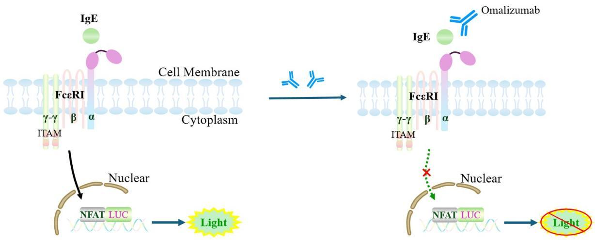
Figure 1. Schematic diagram of the FcεRI Effector Reporter Cell model
| Classification | Fc Effector |
| Family | Fc receptor family (FcγR family) |
| Gene Name | FCER1A |
| Gene Aliases | FCE1A; Fc fragment of IgE receptor Ia |
| Gene ID | 2205 |
| Accession Number | NM_001387280.1 |
| UniProt Number | P12319 |
| Protein Name | Fc-epsilon RI-alpha (FcERI) |
| Protein Aliases | IgE Fc receptor subunit alpha |
| Target Species | Human |
| Host cell | Jurkat E6.1 |
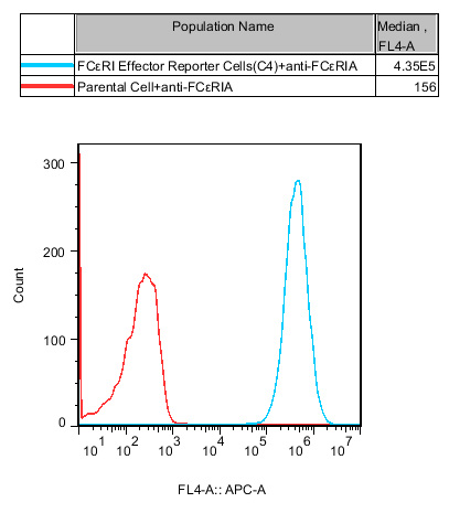
Figure 2. Recombinant FcεRI Effector Reporter Cell stably expressing FcεRI.
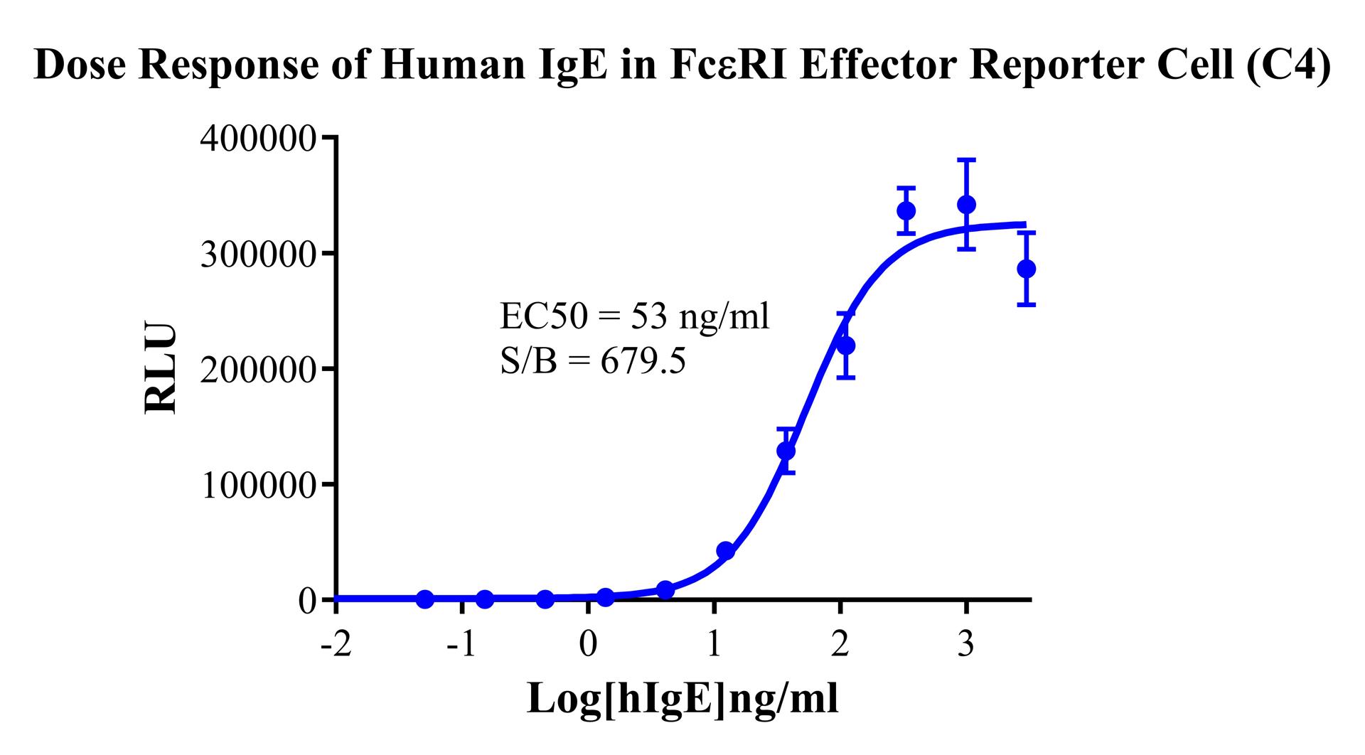
Figure 3. Dose Response of Human IgE in FcεRI Effector Reporter Cell(C4).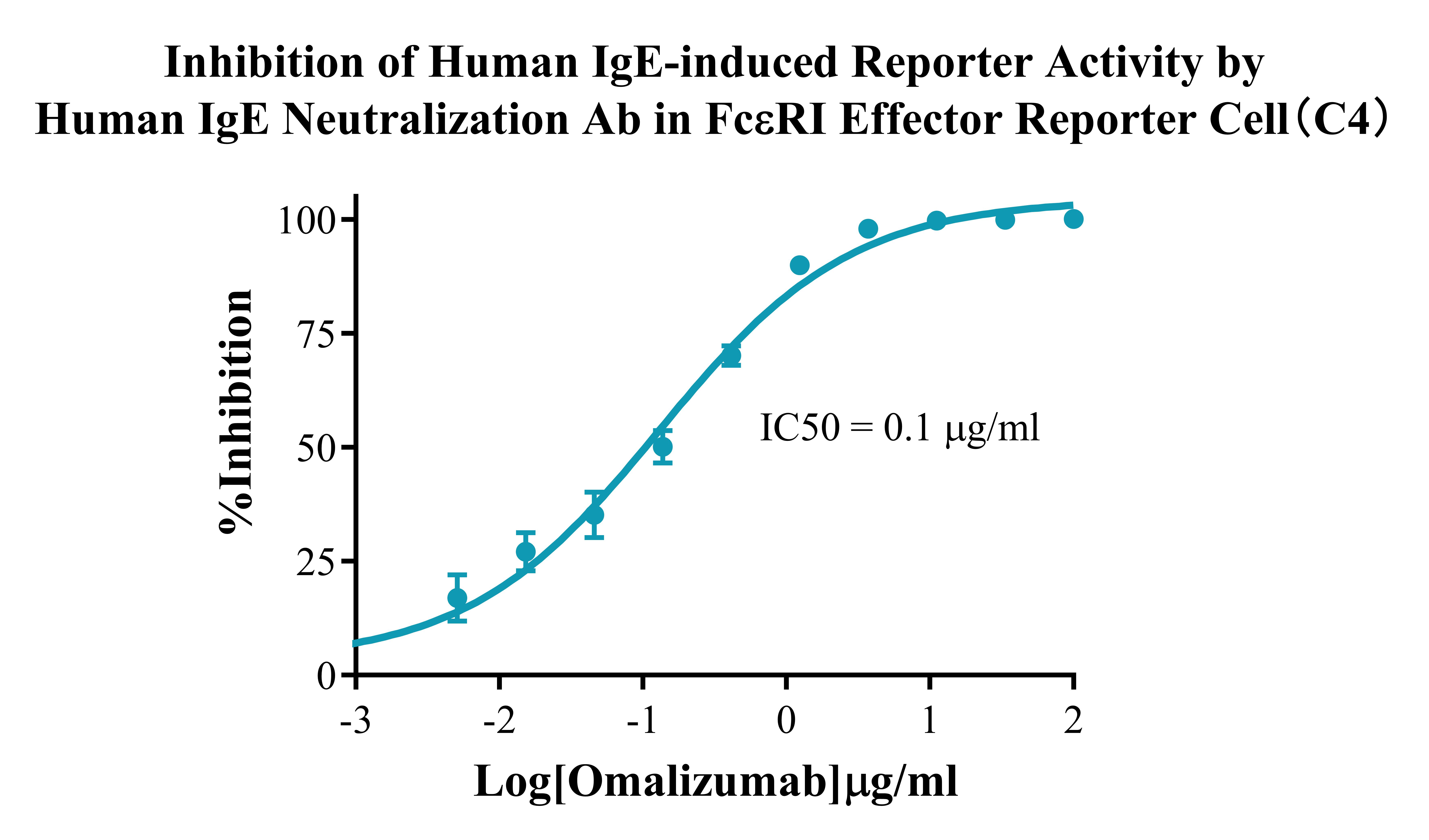
Figure 4. Inhibition of Human IgE-induced Reporter Activity by Human IgE Neutralization Ab in FcεRI Effector Reporter Cell(C4).
Cell Passage Procedures
1.This cell line grows in suspension.
2.Upon receipt, cells should be thawed immediately or stored in liquid nitrogen until use.
3.Before thawing, pre-warm the water bath and culture medium to 37 °C, and prepare a small amount of dry ice.
4.Remove the cryovial from storage and transport it to the cell culture laboratory on dry ice.
5.Rapidly thaw the cells in a 37 °C water bath. Once the cells are completely thawed, spray the cryovial with 70% ethanol for disinfection and transfer it to a biosafety cabinet.
6.Add 10 mL of pre-warmed culture medium into a 15 mL centrifuge tube. Transfer the contents of the cryovial into the tube and centrifuge at 1000 rpm for 5 minutes.
7.Carefully discard the supernatant. Resuspend the cell pellet in 5 mL of pre-warmed culture medium by gentle pipetting. Immediately perform cell counting and adjust the cell density to 3–6 × 10⁵ cells/mL based on the counting results, then transfer the cells into a culture flask.
8.Count the cells every 1–2 days. When the cell density exceeds 1 × 10⁶ cells/mL, passage the cells promptly or add fresh culture medium. Maintain the cell density between 2 × 10⁵ and 1 × 10⁶ cells/mL.
Suspension Cell Cryopreservation Procedure:
1.Collect 8 × 10⁶ cells, centrifuge, and discard the supernatant.
2.Add 1 mL of cell freezing medium (90% FBS + 10% DMSO) and gently pipette to mix thoroughly. Transfer the suspension into a cryovial.
3.Immediately place the cryovial into a controlled-rate freezing container (Nalgene 5100-0001), fill with isopropanol up to the indicated level, and store at −80 °C.
4.After 24 hours, transfer the cryovial to liquid nitrogen for long-term storage.
Related products
CHO-K1 Human CCR4 Cell Line
HEK293 Human NK1R CRE-Luc Cell Line
Raji-Luc-GFP
Jurkat E6.1-Luc
THP-1-GFP
THP-1-Luc
Raji-GFP
Raji-Luc
Jurkat E6.1-GFP
HEK293 Human GAL4-Luc Cell
We Are Pleased to Announce: Global Commercial Licensing Rights for Jurkat E6.1, CHO-K1, and HEK293 Cell Lines Officially Secured.
Explore