

Jurkat E6.1 Human TCRαβ KO NFAT-Luc Cell
Cat. No: RQP74467
Size: 1 vial of frozen cells (>1E6 per vial in 1 mL)
Unit Price: Contact For Pricing
Product Info
Description
Biological Information
Assay Data
Cell Culture
| Cat. No | RQP74467 |
| Product Name | Jurkat E6.1 Human TCRαβ KO NFAT-Luc Cell |
| Product Type | Reporter Cell |
| Culture Properties | suspension |
| Stability | 32passages (in-house test, that not means the cell line will be instable beyond the passages we tested.) |
| Mycoplasma Status | Negative |
| Culture Medium | RPMI-1640+10%FBS+800 μg/ml Hygromycin B |
| Freeze Medium | 90% FBS+10% DMSO |
| Storage Conditions | Liquid nitrogen immediately upon delivery |
| Application | Functional(Report Gene) Assay |
For research use only. Not intended for human or animal clinical trials, therapeutic or diagnostic use.
T cells play a central role in cell-mediated immunity, mediating long-term, antigen-specific, effector, and memory responses. T cells are targets for many immunotherapies, including checkpoint inhibitors, bispecific T-cell engagers, and immune agonists. Furthermore, T cells themselves have been studied as therapeutic agents using chimeric antigen receptor (CAR) addition or autologous adoptive transfer in hematological malignancies. In recent years, various immunotherapeutic strategies aimed at inducing, enhancing, and/or engineering T cell responses have emerged as promising approaches for treating diseases such as cancer and autoimmune disorders.
The T cell receptor (TCR) is the molecular structure that enables T cells to specifically recognize and bind antigen peptide-MHC molecules. The activation of T cells mediated by the TCR plays a crucial role in thymocyte T cell development, T cell subset differentiation, and the functional exertion of effector T cells. The TCR can specifically recognize antigen peptides presented on the surface of antigen-presenting cells via MHC molecules, converting extracellular recognition into signals transmissible to the interior of the cell. This process activates tyrosine kinases adjacent to the TCR, promotes the assembly of signal transduction complexes, activates downstream signaling pathways such as MAPK, PKC, and calcium ions, ultimately activating corresponding transcription factors, regulating the expression of effector protein molecules, and completing T cell activation.
The TCRαβ KO NFAT-Luc Jurkat reporter gene drug target model effectively simulates the signal transduction process of TCR in vivo, as illustrated in the figure below.
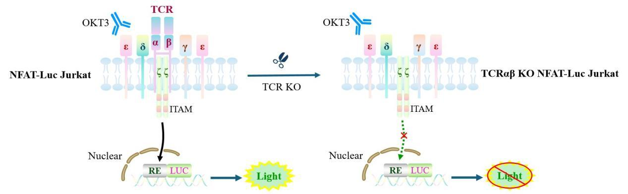
Figure 1. Schematic diagram of the TCRαβ KO NFAT-Luc Jurkat cell model.
| Classification | T Cell Activation |
| Family | Immunoglobulin Superfamily (IgSF) |
| Gene Name | TRA |
| Gene Aliases | TCRA; |
| Gene ID | 6955 |
| Accession Number | NG_001332 |
| UniProt Number | P0DSE1 |
| Protein Name | M1-specific T cell receptor alpha chain |
| Protein Aliases | TR alpha chain TRAV27*01J42*01C*01 |
| Family-2 | Immunoglobulin Superfamily (IgSF) |
| Gene Name-2 | TRB |
| Gene Aliases-2 | TCRB; |
| Gene ID-2 | 6957 |
| Accession Number-2 | NG_001333 |
| UniProt Number-2 | P0DSE2 |
| Protein Name-2 | M1-specific T cell receptor beta chain |
| Protein Aliases-2 | TR beta chain TRBV19*01J2S7*01C*02 |
| Target Species | Human |
| Host cell | Jurkat E6.1 |
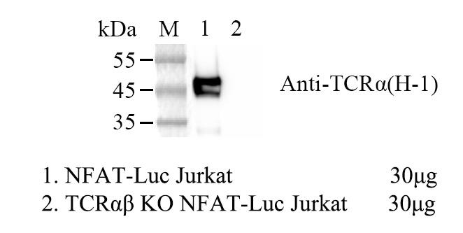
Figure 2. WB of TCRαβ KO NFAT-Luc Jurkat.
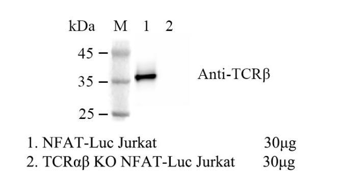
Figure 3. WB of TCRαβ KO NFAT-Luc Jurkat.
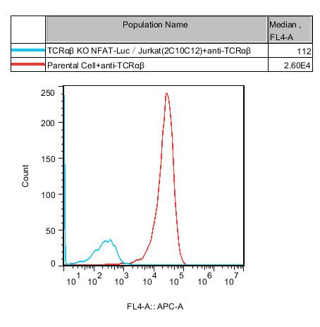
Figure 4. TCRαβ KO NFAT-Luc Jurkat, which refers to the knockout of TCRαβ in NFAT-Luc Jurkat cells.

Figure 5. Sequencing Results of TRAC KO
A: chr14-22547665--CC (TRAC:p.M54Lfs*57) TRAC(ENST00000611116.2):c.158_159insCC
B: chr14-22547665--TC (TRAC:p.S53Ffs*58) TRAC(ENST00000611116.2):c.157_158insTC
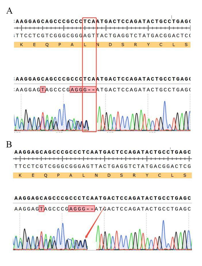
Figure 6. Sequencing Results of TRBC1 Knockout
A: chr7-142791898-TCA-C (TRBC1:p.L68Pfs*2)
B: chr7-142791900--GCCCAGGG (8bp) (TRBC1:p.N69Afs*10)
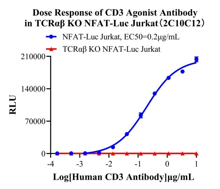
Figure 7. Dose Response of CD3 Agonist Antibody in TCRαβ KO NFAT-Luc Jurkat(2C10C12).
Cell Passage Procedures
1.This cell line grows in suspension.
2.Upon receipt, cells should be thawed immediately or stored in liquid nitrogen until use.
3.Before thawing, pre-warm the water bath and culture medium to 37 °C, and prepare a small amount of dry ice.
4.Remove the cryovial from storage and transport it to the cell culture laboratory on dry ice.
5.Rapidly thaw the cells in a 37 °C water bath. Once the cells are completely thawed, spray the cryovial with 70% ethanol for disinfection and transfer it to a biosafety cabinet.
6.Add 10 mL of pre-warmed culture medium into a 15 mL centrifuge tube. Transfer the contents of the cryovial into the tube and centrifuge at 1000 rpm for 5 minutes.
7.Carefully discard the supernatant. Resuspend the cell pellet in 5 mL of pre-warmed culture medium by gentle pipetting. Immediately perform cell counting and adjust the cell density to 3–6 × 10⁵ cells/mL based on the counting results, then transfer the cells into a culture flask.
8.Count the cells every 1–2 days. When the cell density exceeds 1 × 10⁶ cells/mL, passage the cells promptly or add fresh culture medium. Maintain the cell density between 2 × 10⁵ and 1 × 10⁶ cells/mL.
Suspension Cell Cryopreservation Procedure:
1.Collect 8 × 10⁶ cells, centrifuge, and discard the supernatant.
2.Add 1 mL of cell freezing medium (90% FBS + 10% DMSO) and gently pipette to mix thoroughly. Transfer the suspension into a cryovial.
3.Immediately place the cryovial into a controlled-rate freezing container (Nalgene 5100-0001), fill with isopropanol up to the indicated level, and store at −80 °C.
4.After 24 hours, transfer the cryovial to liquid nitrogen for long-term storage.
Related products
CHO-K1 Human CCR4 Cell Line
HEK293 Human NK1R CRE-Luc Cell Line
Raji-Luc-GFP
Jurkat E6.1-Luc
THP-1-GFP
THP-1-Luc
Raji-GFP
Raji-Luc
Jurkat E6.1-GFP
HEK293 Human GAL4-Luc Cell
We Are Pleased to Announce: Global Commercial Licensing Rights for Jurkat E6.1, CHO-K1, and HEK293 Cell Lines Officially Secured.
Explore