

HEK293 Human VEGFC/VEGFR3 Effector Reporter Cell
Cat. No: RQP74347
Size: 1 vial of frozen cells (>1E6 per vial in 1 mL)
Unit Price: Contact For Pricing
Product Info
Description
Biological Information
Assay Data
Cell Culture
| Cat. No | RQP74347 |
| Product Name | HEK293 Human VEGFC/VEGFR3 Effector Reporter Cell |
| Product Type | Reporter Cell |
| Culture Properties | Adherent |
| Stability | 32passages (in-house test, that not means the cell line will be instable beyond the passages we tested.) |
| Mycoplasma Status | Negative |
| Culture Medium | DMEM+10%FBS+2 μg/ml Puromycin+200 μg/ml Hygromycin B |
| Freeze Medium | 90% FBS+10% DMSO |
| Storage Conditions | Liquid nitrogen immediately upon delivery |
| Application | Functional(Report Gene) Assay |
For research use only. Not intended for human or animal clinical trials, therapeutic or diagnostic use.
VEGF, also known as vascular permeability factor (VPF), is a dimeric glycoprotein that binds to cell-surface VEGF receptors (VEGFRs), thereby activating intracellular tyrosine kinases and initiating a cascade of signaling events involved in angiogenesis and vasculogenesis. In mammals, the VEGF family comprises five members: VEGFA, VEGFB, VEGFC, VEGFD, and placental growth factor (PGF). VEGFC is a secreted dimeric glycoprotein (full-length 58 kDa) that requires proteolytic cleavage—by enzymes such as ADAMTS3—to be activated into a 21 kDa mature peptide, thereby enhancing its biological activity. Its primary function is to orchestrate lymphangiogenesis; during development, it is widely expressed in the mesenchyme of the heart, kidneys, and brain, while in adulthood, it is predominantly enriched in lymphatic endothelial cells, pericytes, macrophages, and the tumor microenvironment, playing a pivotal role by driving embryonic lymphatic system development and adult lymphatic remodeling.
Members of the VEGF family bind in an overlapping manner to three receptor tyrosine kinases: VEGFR1, VEGFR2, and VEGFR3. All of these receptors are transmembrane tyrosine kinase receptors, each comprising three main domains: an extracellular domain, a transmembrane domain, and an intracellular C-terminal domain. Upon binding to its receptor, VEGF induces the formation of a homodimeric complex, triggering a conformational change in the protein structure that leads to the autophosphorylation of tyrosine residues and the subsequent transduction of signals downstream.
The VEGFC/VEGFR3 Effector Reporter Cell model accurately simulates the *in vivo* signal transduction process of the VEGFC/VEGFR3 pathway; the underlying principle is illustrated in the figure below.
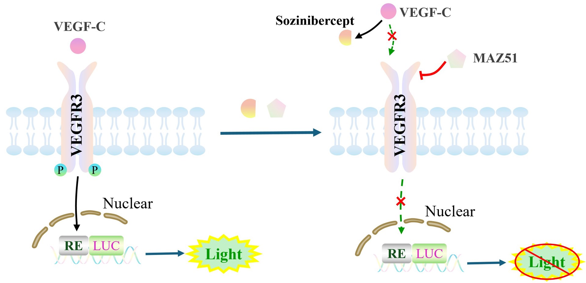
Figure 1. Schematic Diagram of the HEK293 Human VEGFC/VEGFR3 Effector Reporter Cell Model
| Classification | Cytokine&Growth Factor |
| Family-1 | PDGF/VEGF growth factor family |
| Gene Name-1 | VEGFC |
| Gene Aliases-1 | VRP;VEGF-C |
| Gene ID-1 | 7424 |
| Accession Number-1 | NM_005429.5 |
| UniProt Number-1 | P49767 |
| Protein Name-1 | VEGF-C |
| Protein Aliases-1 | Flt4 ligand (Flt4-L);Vascular endothelial growth factor-related protein (VRP) |
| Family-2 | PDGF receptor subfamily |
| Gene Name-2 | FLT4 |
| Gene Aliases-2 | VEGFR3;PCL;VEGFR-3 |
| Gene ID-2 | 2324 |
| Accession Number-2 | NM_182925.5 |
| UniProt Number-2 | P35916 |
| Protein Name-2 | VEGFR-3 |
| Protein Aliases-2 | Fms-like tyrosine kinase 4 (FLT-4);Tyrosine-protein kinase receptor FLT4 |
| Target Species | Human |
| Host cell | HEK293 |
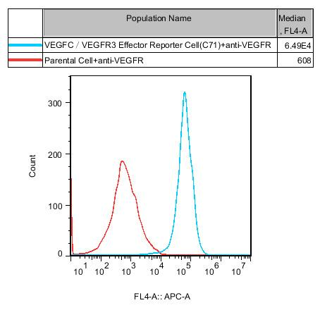
Figure 2. Recombinant VEGFC/VEGFR3 Effector Reporter Cell stably expressing VEGFR3.
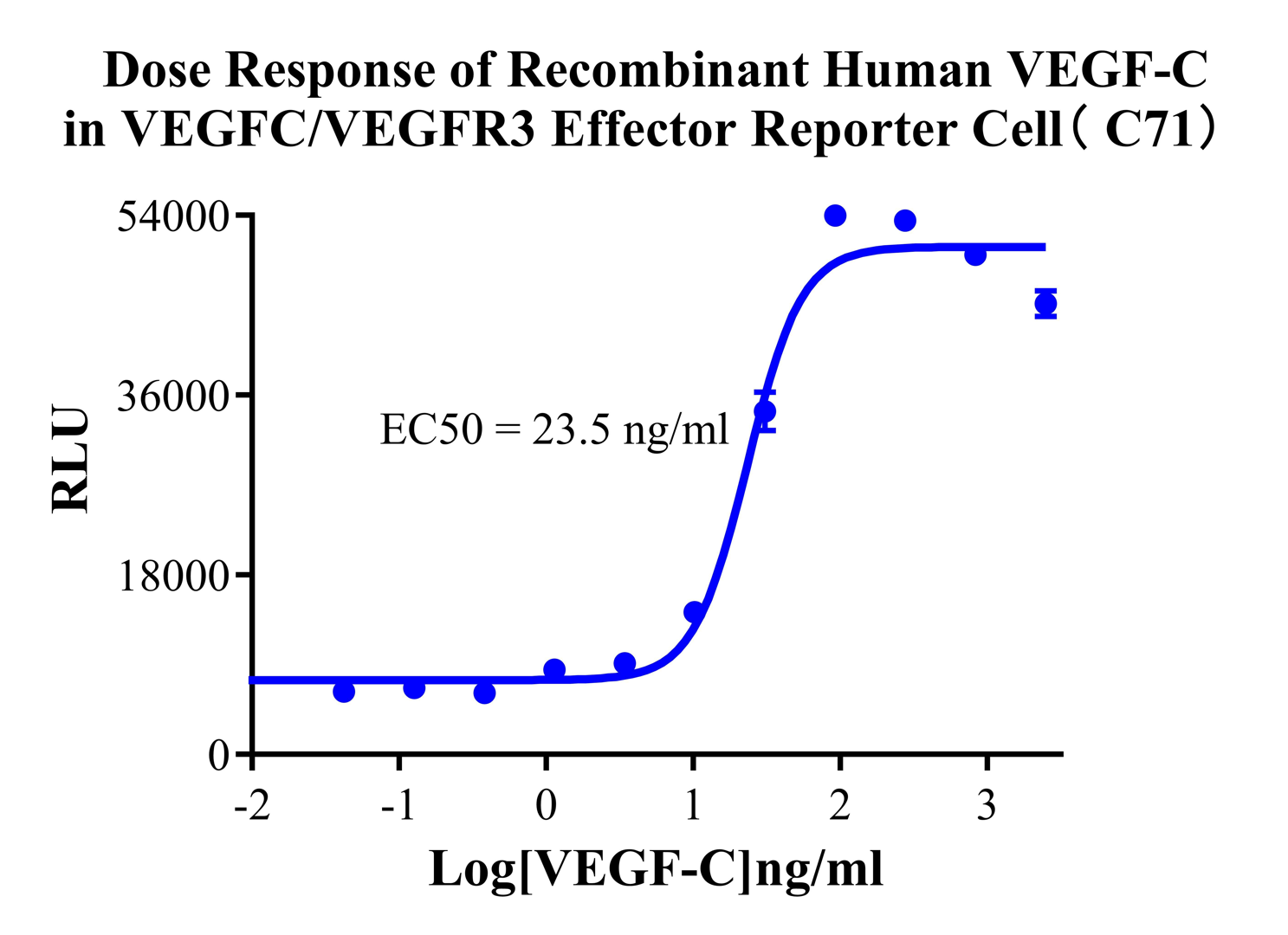
Figure 3. Dose Response of Recombinant Human VEGF-C in VEGFC/VEGFR3 Effector Reporter Cell (C71).
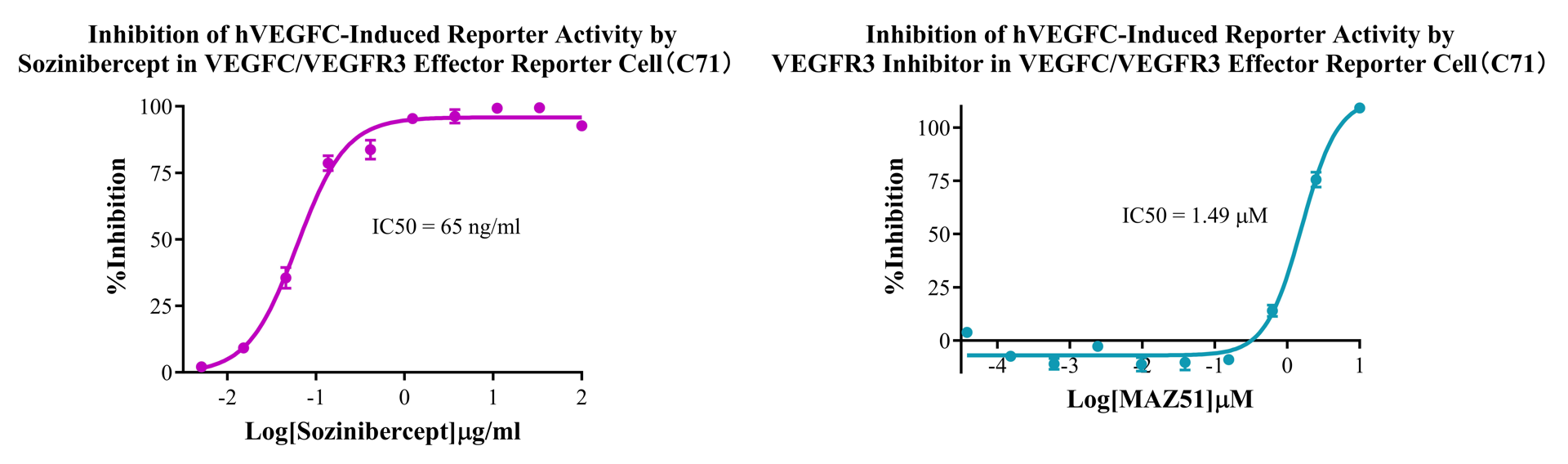
Figure 4&5. Inhibition of hVEGFC-Induced Reporter Activity by Sozinibercept in VEGFC/EGFR3 Effector Reporter Cell (C71). Inhibition of hVEGFC-Induced Reporter Activity by VEGFR3 Inhibitor in VEGFC/EGFR3 Effector Reporter Cell (C71).
Cell Resuscitation
1)Rapidly thaw the frozen cells in a 37 °C water bath for approximately 60 seconds. Once thawed (which may take slightly less or more than 60 seconds), immediately transfer the cell suspension from the cryovial into a 15 mL centrifuge tube containing 10 mL of pre-warmed HEK293 Human VEGFC/VEGFR3 Effector Reporter Cell complete culture medium.
2)Centrifuge cells at 1000 rpm for 5 min to remove medium, then resuspend cells in 5 mL of pre-warmed complete medium.
3)Transfer the cell suspension into a T25 culture flask and incubate at 37 °C with 5% CO₂.
4)After approximately 24–36 hours, replace the medium or passage the cells to remove non-adherent dead cells.
Subculturing procedure
1)When the cell density reaches the appropriate confluency for passaging, wash the cells with PBS, then add 1 mL trypsin to detach the cells. When more than 80% of the cells detach upon gently tapping the culture flask, add complete culture medium to terminate digestion. Gently pipette to obtain a single-cell suspension, transfer to a 15 mL centrifuge tube, and centrifuge at 1000 rpm for 5 minutes.
2)Discard supernatant after centrifugation. Resuspend cells in fresh medium to a single-cell suspension and transfer to a new culture flask for continued growth.
Cell Freezing
After trypsinization and centrifugation of cells from each T75 flask or 10 cm culture dish, discard the supernatant. Add 2 mL of cryopreservation medium (90% FBS + 10% DMSO), gently resuspend thoroughly, and aliquot into two cryovials. Immediately place the cryovials into a controlled-rate freezing container (e.g., Nalgene 5100-0001), fill with isopropanol to the indicated level, and store at −80 °C. After 24 hours, transfer the cryovials to liquid nitrogen for long-term storage.
Related products
CHO-K1 Human CCR4 Cell Line
HEK293 Human NK1R CRE-Luc Cell Line
Raji-Luc-GFP
Jurkat E6.1-Luc
THP-1-GFP
THP-1-Luc
Raji-GFP
Raji-Luc
Jurkat E6.1-GFP
HEK293 Human GAL4-Luc Cell
We Are Pleased to Announce: Global Commercial Licensing Rights for Jurkat E6.1, CHO-K1, and HEK293 Cell Lines Officially Secured.
Explore