

HEK293 Human PDGF/PDGFRA Effector Reporter Cell
Cat. No: RQP74284
Size: 1 vial of frozen cells (>1E6 per vial in 1 mL)
Unit Price: Contact For Pricing
Product Info
Description
Biological Information
Assay Data
Cell Culture
| Cat. No | RQP74284 |
| Product Name | HEK293 Human PDGF/PDGFRA Effector Reporter Cell |
| Product Type | Reporter Cell |
| Culture Properties | Adherent |
| Stability | 32passages (in-house test, that not means the cell line will be instable beyond the passages we tested.) |
| Mycoplasma Status | Negative |
| Culture Medium | DMEM+10%FBS+ 2 μg/ml Puromycin+ 200 μg/ml Hygromycin B |
| Freeze Medium | 90% FBS+10% DMSO |
| Storage Conditions | Liquid nitrogen immediately upon delivery |
| Application | Functional(Report Gene) Assay |
For research use only. Not intended for human or animal clinical trials, therapeutic or diagnostic use.
Platelet-derived growth factor (PDGF) is a class of important growth factors primarily secreted by platelets, endothelial cells, and macrophages; it plays a pivotal role in various biological processes, including cell proliferation, migration, differentiation, and angiogenesis. It is instrumental not only in normal physiological processes—such as wound healing and tissue repair—but also in a variety of pathological conditions, including cancer, fibrosis, and atherosclerosis. The PDGF family comprises four distinct subtypes: PDGF-A, PDGF-B, PDGF-C, and PDGF-D. By binding to its cognate receptors (PDGFRs), PDGF activates downstream signaling pathways, thereby regulating cellular behavior.
Platelet-derived growth factor receptor alpha (PDGFRA)—also known as CD140A or PDGFR2—is a cell-surface tyrosine kinase receptor belonging to the PDGF receptor family. Upon binding to its ligand, PDGF, PDGFRA undergoes dimerization and phosphorylation, thereby activating downstream signaling pathways such as JAK-STAT3, PI3K-AKT-mTOR, and RAS-MAPK; this activation plays a critical role in regulating key cellular functions, including proliferation and apoptosis. Conversely, the aberrant activation of the PDGFRA protein can lead to tumorigenesis and promote tumor angiogenesis.
The HEK293 Human PDGF/PDGFRA Effector Reporter Cell Model—effectively simulates the signal transduction process of PDGFRA *in vivo*. The underlying principle is illustrated in the figure below.
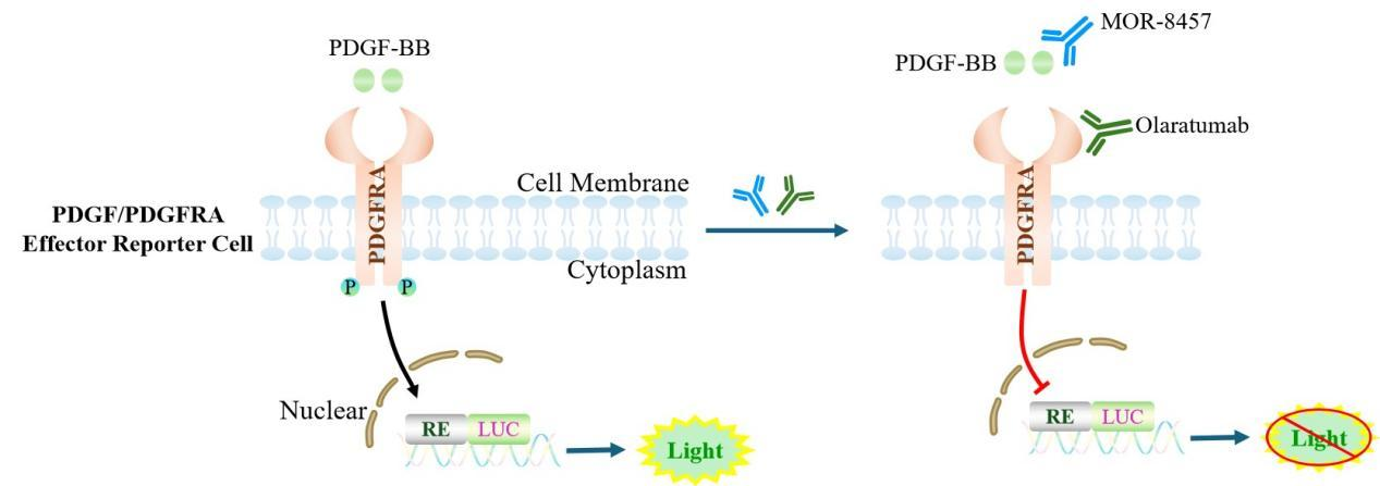
Figure 1. Schematic Diagram of the HEK293 Human PDGF/PDGFRA Effector Reporter Cell Model
| Classification | Cytokine&Growth Factor |
| Family | Receptor tyrosine kinase (RTK) family |
| Gene Name | PDGFRA |
| Gene Aliases | CD140a;PDGFR2;GAS9;RHEPDGFRA |
| Gene ID | 5156 |
| Accession Number | NM_006206.6 |
| UniProt Number | P16234 |
| Protein Name | PDGF-R-alpha; PDGFR-alpha |
| Protein Aliases | |
| Target Species | Human |
| Host cell | HEK293 |
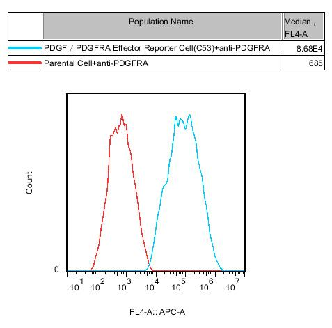
Figure 2. Recombinant PDGF/PDGFRA Effector Reporter Cell stably expressing PDGFRA.
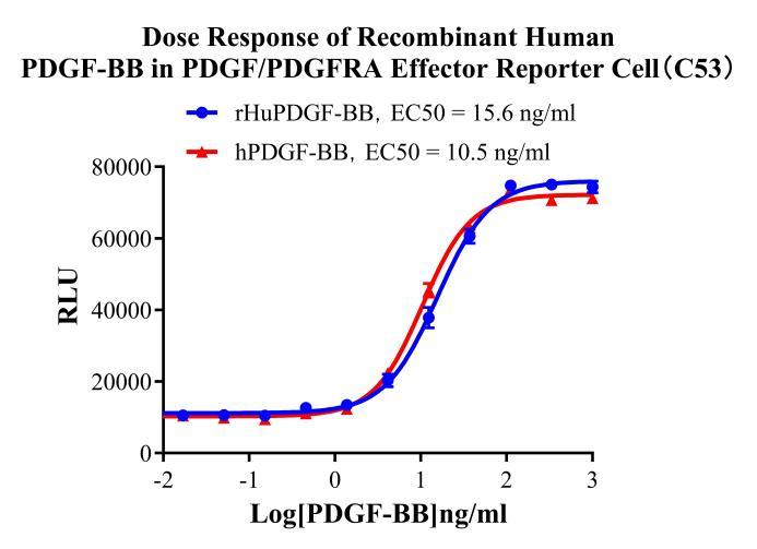
Figure 3. Dose Response of Recombinant Human PDGF-BB in PDGF/PDGFRA Effector Reporter Cell(C53).
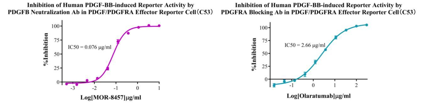
Figure 4. Inhibition of Human PDGF-BB-induced Reporter Activity by PDGFB Neutralization Ab in PDGF/PDGFRA Effector Reporter Cell(C53). Inhibition of Human PDGF-BB-induced Reporter Activity by PDGFRA Blocking Ab in PDGF/DGFRA Effector Reporter Cel(C53).
Cell Resuscitation
1)Rapidly thaw the frozen cells in a 37 °C water bath for approximately 60 seconds. Once thawed (which may take slightly less or more than 60 seconds), immediately transfer the cell suspension from the cryovial into a 15 mL centrifuge tube containing 10 mL of pre-warmed HEK293 Human PDGF/PDGFRA Effector Reporter Cell complete culture medium.
2)Centrifuge cells at 1000 rpm for 5 min to remove medium, then resuspend cells in 5 mL of pre-warmed complete medium.
3)Transfer the cell suspension into a T25 culture flask and incubate at 37 °C with 5% CO₂.
4)After approximately 24–36 hours, replace the medium or passage the cells to remove non-adherent dead cells.
Subculturing procedure
1)When the cell density reaches the appropriate confluency for passaging, wash the cells with PBS, then add 1 mL trypsin to detach the cells. When more than 80% of the cells detach upon gently tapping the culture flask, add complete culture medium to terminate digestion. Gently pipette to obtain a single-cell suspension, transfer to a 15 mL centrifuge tube, and centrifuge at 1000 rpm for 5 minutes.
2)Discard supernatant after centrifugation. Resuspend cells in fresh medium to a single-cell suspension and transfer to a new culture flask for continued growth.
Cell Freezing
After trypsinization and centrifugation of cells from each T75 flask or 10 cm culture dish, discard the supernatant. Add 2 mL of cryopreservation medium (90% FBS + 10% DMSO), gently resuspend thoroughly, and aliquot into two cryovials. Immediately place the cryovials into a controlled-rate freezing container (e.g., Nalgene 5100-0001), fill with isopropanol to the indicated level, and store at −80 °C. After 24 hours, transfer the cryovials to liquid nitrogen for long-term storage.
Related products
CHO-K1 Human CCR4 Cell Line
HEK293 Human NK1R CRE-Luc Cell Line
Raji-Luc-GFP
Jurkat E6.1-Luc
THP-1-GFP
THP-1-Luc
Raji-GFP
Raji-Luc
Jurkat E6.1-GFP
HEK293 Human GAL4-Luc Cell
We Are Pleased to Announce: Global Commercial Licensing Rights for Jurkat E6.1, CHO-K1, and HEK293 Cell Lines Officially Secured.
Explore