

HEK293 Human OX40/NFκB-Luc Cell
Cat. No: RQP74017
Size: 1 vial of frozen cells (>1E6 per vial in 1 mL)
Unit Price: Contact For Pricing
Product Info
Description
Biological Information
Assay Data
Cell Culture
| Cat. No | RQP74017 |
| Product Name | HEK293 Human OX40/NFκB-Luc Cell |
| Product Type | Reporter Cell |
| Culture Properties | Adherent |
| Stability | 32passages (in-house test, that not means the cell line will be instable beyond the passages we tested.) |
| Mycoplasma Status | Negative |
| Culture Medium | MEM + 10% Foetal Bovine Serum (FBS)+ 1% Non Essential Amino Acids (NEAA) + 1mM Sodium Pyruvate (NaP) +1μg/ml puromycin+100μg/ml Hygromycin B |
| Freeze Medium | 90% FBS+10% DMSO |
| Storage Conditions | Liquid nitrogen immediately upon delivery |
| Application | Functional(Report Gene) Assay |
For research use only. Not intended for human or animal clinical trials, therapeutic or diagnostic use.
OX40 (also known as ACT35, CD134, or TNFRSF4) belongs to the TNFR superfamily and is a type I transmembrane protein. OX40 is primarily expressed on activated CD4+ and CD8+ T cells, while its expression levels on NK and NKT cells are relatively low. OX40L (also known as CD252, TNFSF4, CD134L, or GP34) is the ligand for OX40 and is a type II glycoprotein. OX40L is predominantly expressed on antigen-presenting cells, such as B cells and dendritic cells.
Similar to other members of the TNF family, OX40 signaling is mediated via TNF receptor-associated factors (TRAFs). In vivo, OX40 signals are transduced into T cells through TRAF2 and TRAF5; in vitro, this transduction occurs via TRAF1, TRAF3, and TRAF5. The expression of OX40 and OX40L is regulated by a variety of factors. On T cells, OX40 expression is induced by signals such as those from the TCR, CD28/CD80, and CD40/CD40L pathways, reaching peak expression levels 48 to 72 hours after T cell activation. While TCR signaling can initiate OX40 expression on various cell types, CD28 and other cytokines can further enhance its expression on activated T cells. IL-2, IL-4, and TNF can augment or prolong OX40 expression. The OX40/OX40L axis plays a pivotal role in enhancing effector T cell function, maintaining effector T cell survival, and inhibiting effector T cell apoptosis.
HEK293 Human OX40/NFκB-Luc Cell Model—effectively simulates the signal transduction process of OX40 *in vivo*. The underlying principle is illustrated in the figure below.
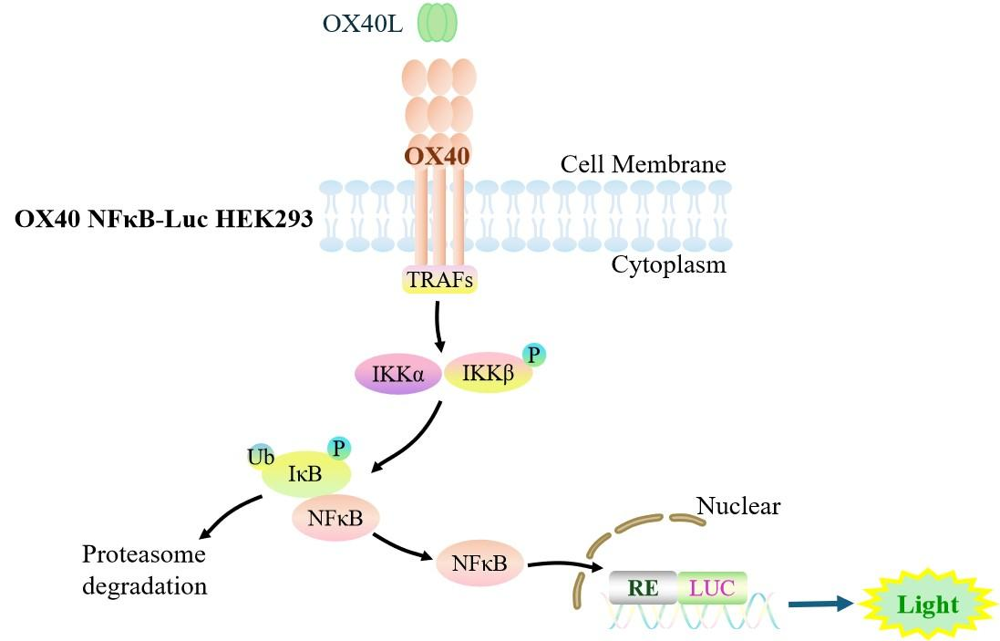
Figure 1. Schematic Diagram of the HEK293 Human OX40/NFκB-Luc Cell Model
| Classification | Co-Stimulatory |
| Family | Tumor necrosis factor receptor superfamily |
| Gene Name | TNFRSF4 |
| Gene Aliases | ACT35;OX40;CD134 |
| Gene ID | 7293 |
| Accession Number | NM_003327.4 |
| UniProt Number | P43489 |
| Protein Name | Tumor necrosis factor receptor superfamily member 4 |
| Protein Aliases | ACT35 antigen;OX40L receptor;TAX transcriptionally-activated glycoprotein 1 receptor |
| Target Species | Human |
| Host cell | HEK293 |
HEK293 cell line expressing full length human OX40. Expression is confirmed by flow cytometry.
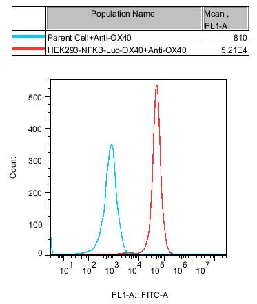
Figure 2. Recombinant OX40/NFκB-Luc/HEK293 stably expressing OX40.
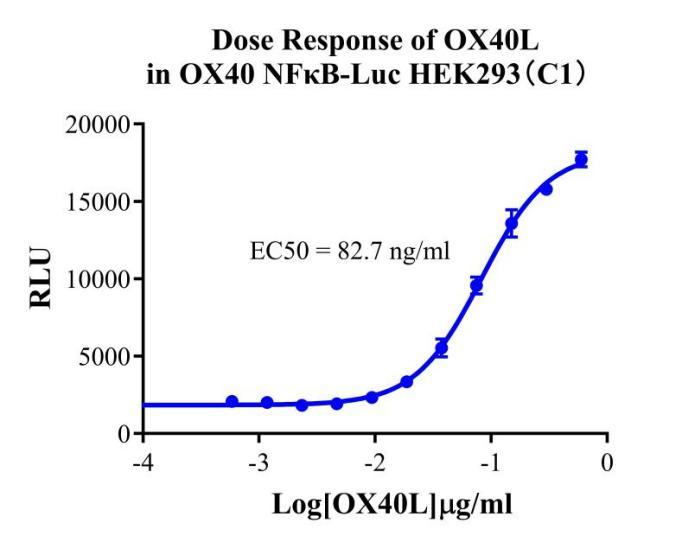
Figure 3. Dose Response of OX40L in OX40 NFκB-Luc HEK293(C1).
Cell Resuscitation
1)Rapidly thaw the frozen cells in a 37 °C water bath for approximately 60 seconds. Once thawed (which may take slightly less or more than 60 seconds), immediately transfer the cell suspension from the cryovial into a 15 mL centrifuge tube containing 10 mL of pre-warmed HEK293 Human OX40/NFκB-Luc Cell complete culture medium.
2)Centrifuge cells at 1000 rpm for 5 min to remove medium, then resuspend cells in 5 mL of pre-warmed complete medium.
3)Transfer the cell suspension into a T25 culture flask and incubate at 37 °C with 5% CO₂.
4)After approximately 24–36 hours, replace the medium or passage the cells to remove non-adherent dead cells.
Subculturing procedure
1)When the cell density reaches the appropriate confluency for passaging, wash the cells with PBS, then add 1 mL trypsin to detach the cells. When more than 80% of the cells detach upon gently tapping the culture flask, add complete culture medium to terminate digestion. Gently pipette to obtain a single-cell suspension, transfer to a 15 mL centrifuge tube, and centrifuge at 1000 rpm for 5 minutes.
2)Discard supernatant after centrifugation. Resuspend cells in fresh medium to a single-cell suspension and transfer to a new culture flask for continued growth.
Cell Freezing
After trypsinization and centrifugation of cells from each T75 flask or 10 cm culture dish, discard the supernatant. Add 2 mL of cryopreservation medium (90% FBS + 10% DMSO), gently resuspend thoroughly, and aliquot into two cryovials. Immediately place the cryovials into a controlled-rate freezing container (e.g., Nalgene 5100-0001), fill with isopropanol to the indicated level, and store at −80 °C. After 24 hours, transfer the cryovials to liquid nitrogen for long-term storage.
Related products
CHO-K1 Human CCR4 Cell Line
HEK293 Human NK1R CRE-Luc Cell Line
Raji-Luc-GFP
Jurkat E6.1-Luc
THP-1-GFP
THP-1-Luc
Raji-GFP
Raji-Luc
Jurkat E6.1-GFP
HEK293 Human GAL4-Luc Cell
We Are Pleased to Announce: Global Commercial Licensing Rights for Jurkat E6.1, CHO-K1, and HEK293 Cell Lines Officially Secured.
Explore