

HEK293 Human NOD1 Effector Reporter Cell
Cat. No: RQP74531
Size: 1 vial of frozen cells (>1E6 per vial in 1 mL)
Unit Price: Contact For Pricing
Product Info
Description
Biological Information
Assay Data
Cell Culture
| Cat. No | RQP74531 |
| Product Name | HEK293 Human NOD1 Effector Reporter Cell |
| Product Type | Reporter Cell |
| Culture Properties | Adherent |
| Stability | 32passages (in-house test, that not means the cell line will be instable beyond the passages we tested.) |
| Mycoplasma Status | Negative |
| Culture Medium | MEM + 10%FBS +1% NEAA + 1mM NaP + 1μg/ml Puromycin + 100 μg/ml Hygromycin B |
| Freeze Medium | 90% FBS+10% DMSO |
| Storage Conditions | Liquid nitrogen immediately upon delivery |
| Application | Functional(Report Gene) Assay |
For research use only. Not intended for human or animal clinical trials, therapeutic or diagnostic use.
NOD-like receptors (NLRs) constitute a critical class of intracellular pattern recognition receptors within the innate immune system; their core function is to sense pathogen-associated molecular patterns (PAMPs) or danger-associated molecular patterns (DAMPs), thereby initiating an immune response. Based on their distinct N-terminal domains, the human NLR family can be broadly classified into four major subfamilies: NLRA (e.g., CIITA), NLRB (e.g., NAIP), NLRC (e.g., NOD1, NOD2, NLRC3/4/5), and NLRP (containing a PYD domain; e.g., NLRP1, NLRP3). Among these, NOD1 and NOD2 both belong to the NLRC subfamily and primarily recognize fragments of bacterial cell wall peptidoglycans, serving as classic sensors in antibacterial innate immunity.
NOD1 (full name: Nucleotide-binding oligomerization domain-containing protein 1) was one of the first members of the NLR family to be identified. It is expressed in a wide variety of tissue cells, including epithelial cells, endothelial cells, fibroblasts, and monocytes/macrophages. Unlike NOD2—which primarily recognizes muramyl dipeptide—NOD1 specifically recognizes the structural unit γ-D-glutamyl-meso-diaminopimelic acid (iE-DAP) found in the peptidoglycans of Gram-negative bacteria and certain Gram-positive bacteria. Upon pathogen invasion or stimulation by cellular stress signals, NOD1 undergoes a conformational change after binding to its ligand via its LRR domain; this exposes the NACHT domain and triggers ATP hydrolysis, leading to the formation of oligomers (typically considered to be dimers or higher-order complexes). Aberrant activation of NOD1 has been implicated in various diseases, such as inflammatory bowel disease, sarcoidosis, asthma, and *Helicobacter pylori*-induced gastric cancer; conversely, loss-of-function mutations may increase susceptibility to infections by certain intracellular bacteria.
The HEK293 Human NOD1 Effector Reporter Cell model effectively mimics the in vivo MET signaling transduction process. The principle is illustrated in the figure below.
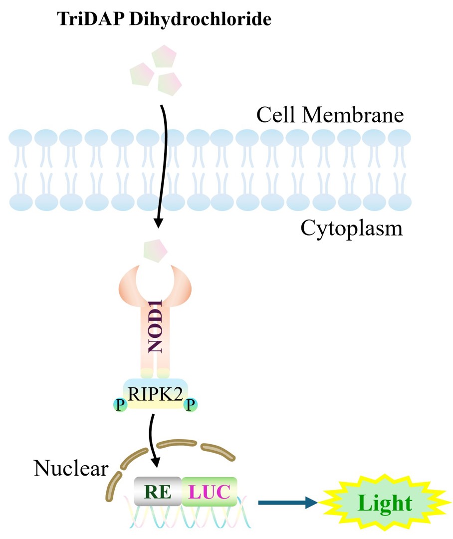
Figure 1. Schematic diagram of the HEK293 Human NOD1 Effector Reporter Cell model
| Classification | TLR |
| Family | NOD1-NOD2 family |
| Gene Name | NOD1 |
| Gene Aliases | CARD4;NLRC1;CLR7.1 |
| Gene ID | 10392 |
| Accession Number | NM_006092.4 |
| UniProt Number | Q9Y239 |
| Protein Name | hNod1 |
| Protein Aliases | Caspase recruitment domain-containing protein 4 |
| Target Species | Human |
| Host cell | HEK293 |
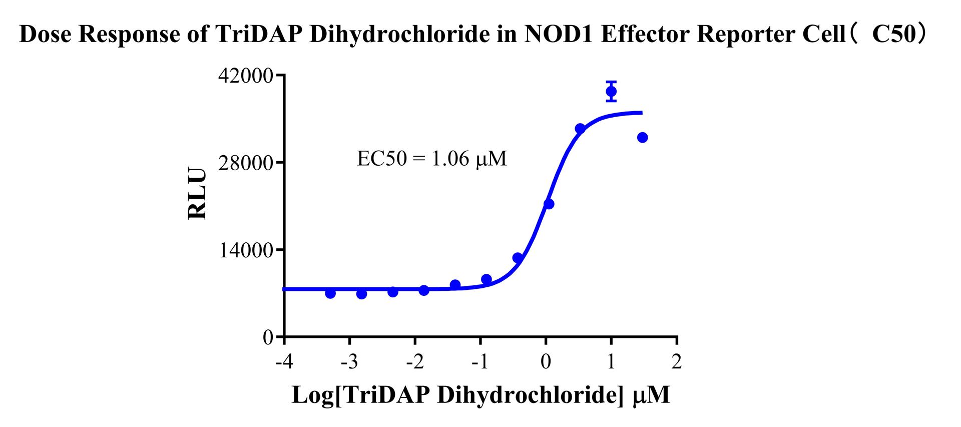
Figure 1. Dose Response of TriDAP Dihydrochloride in NOD1 Effector Reporter Cell(C50).
Cell Resuscitation
1)Rapidly thaw the frozen cells in a 37 °C water bath for approximately 60 seconds. Once thawed (which may take slightly less or more than 60 seconds), immediately transfer the cell suspension from the cryovial into a 15 mL centrifuge tube containing 10 mL of pre-warmed HEK293 Human NOD1 Effector Reporter Cell complete culture medium.
2)Centrifuge cells at 1000 rpm for 5 min to remove medium, then resuspend cells in 5 mL of pre-warmed complete medium.
3)Transfer the cell suspension into a T25 culture flask and incubate at 37 °C with 5% CO₂.
4)After approximately 24–36 hours, replace the medium or passage the cells to remove non-adherent dead cells.
Subculturing procedure
1)When the cell density reaches the appropriate confluency for passaging, wash the cells with PBS, then add 1 mL trypsin to detach the cells. When more than 80% of the cells detach upon gently tapping the culture flask, add complete culture medium to terminate digestion. Gently pipette to obtain a single-cell suspension, transfer to a 15 mL centrifuge tube, and centrifuge at 1000 rpm for 5 minutes.
2)Discard supernatant after centrifugation. Resuspend cells in fresh medium to a single-cell suspension and transfer to a new culture flask for continued growth.
Cell Freezing
After trypsinization and centrifugation of cells from each T75 flask or 10 cm culture dish, discard the supernatant. Add 2 mL of cryopreservation medium (90% FBS + 10% DMSO), gently resuspend thoroughly, and aliquot into two cryovials. Immediately place the cryovials into a controlled-rate freezing container (e.g., Nalgene 5100-0001), fill with isopropanol to the indicated level, and store at −80 °C. After 24 hours, transfer the cryovials to liquid nitrogen for long-term storage.
Related products
CHO-K1 Human CCR4 Cell Line
HEK293 Human NK1R CRE-Luc Cell Line
Raji-Luc-GFP
Jurkat E6.1-Luc
THP-1-GFP
THP-1-Luc
Raji-GFP
Raji-Luc
Jurkat E6.1-GFP
HEK293 Human GAL4-Luc Cell
We Are Pleased to Announce: Global Commercial Licensing Rights for Jurkat E6.1, CHO-K1, and HEK293 Cell Lines Officially Secured.
Explore