

HEK293 Human ISRE-Luc Cell
Cat. No: RQPB0005
Size: 1 vial of frozen cells (>1E6 per vial in 1 mL)
Unit Price: Contact For Pricing
Product Info
Description
Biological Information
Assay Data
Cell Culture
| Cat. No | RQPB0005 |
| Product Name | HEK293 Human ISRE-Luc Cell |
| Product Type | Reporter Cell |
| Culture Properties | Adherent |
| Stability | 32passages (in-house test, that not means the cell line will be instable beyond the passages we tested.) |
| Mycoplasma Status | Negative |
| Culture Medium | DMEM +10%FBS+200μg/ml Hygromycin B |
| Freeze Medium | 90% FBS+10% DMSO |
| Storage Conditions | Liquid nitrogen immediately upon delivery |
| Application | Functional(Report Gene) Assay |
For research use only. Not intended for human or animal clinical trials, therapeutic or diagnostic use.
Interferon (IFN) is a multifunctional soluble glycoprotein produced by monocytes and lymphocytes, and serves as a key effector molecule in both innate and adaptive immunity. IFN cytokines are classified into three types—Type I, Type II, and Type III—based on the specific interferon receptors they bind to. Type I interferons comprise a diverse group, including IFN-α, IFN-β, IFN-δ, IFN-κ, IFN-ω, IFN-τ, IFN-ε, and IFN-ξ; Type II interferon is IFN-γ; Type III interferon is IFN-λ, which includes IFN-λ1, IFN-λ2, and IFN-λ3. The primary functions of type I interferons are antiviral and antitumor effects, while type II interferons primarily induce the expression of major histocompatibility complex (MHC) antigens and exert immunomodulatory effects; however, their antiviral activity is weaker than that of type I interferons. Type III interferons, discovered in 2003, are a novel class of interferons with broad-spectrum antiviral and immunomodulatory activities.
Upon ligand binding, both type I and type II receptors are activated, leading to the dimerization and rearrangement of receptor subunits. This triggers the activation of associated JAK kinases through autophosphorylation, which in turn activates STAT proteins. Upon phosphorylation, the activated STAT forms homodimers and subsequently translocates to the cell nucleus, where it initiates the transcription of interferon-stimulated genes.
Type I interferon also induces the formation of the ISGF3 complex, which consists of STAT1, STAT2, and IRF9. The ISGF3 complex binds to the ISRE (interferon-stimulated response element), further inducing the transcription of interferon-stimulated genes containing ISREs within their promoters.
Binding of IFN-γ to the IFN-γ receptor leads to tyrosine phosphorylation of STAT1 at the Tyr701 site. The phosphorylated STAT1 homodimer translocates to the cell nucleus and binds to the GAS (IFN-γ activation site) element, thereby inducing the expression of IFN-γ-regulated genes.
ISRE-Luc HEK293 reporter cells are HEK293 cells in which the ISRE regulates the expression of the Luc luciferase reporter gene. The mechanism by which IFN binds to its receptor and activates ISRE is illustrated in the figure below.
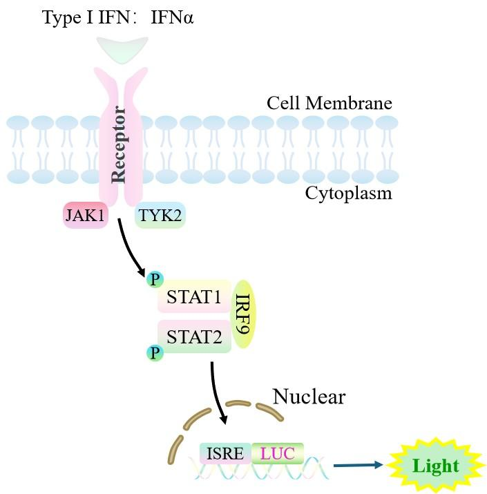
Figure 1. Schematic diagram of the ISRE-Luc HEK293 cell model
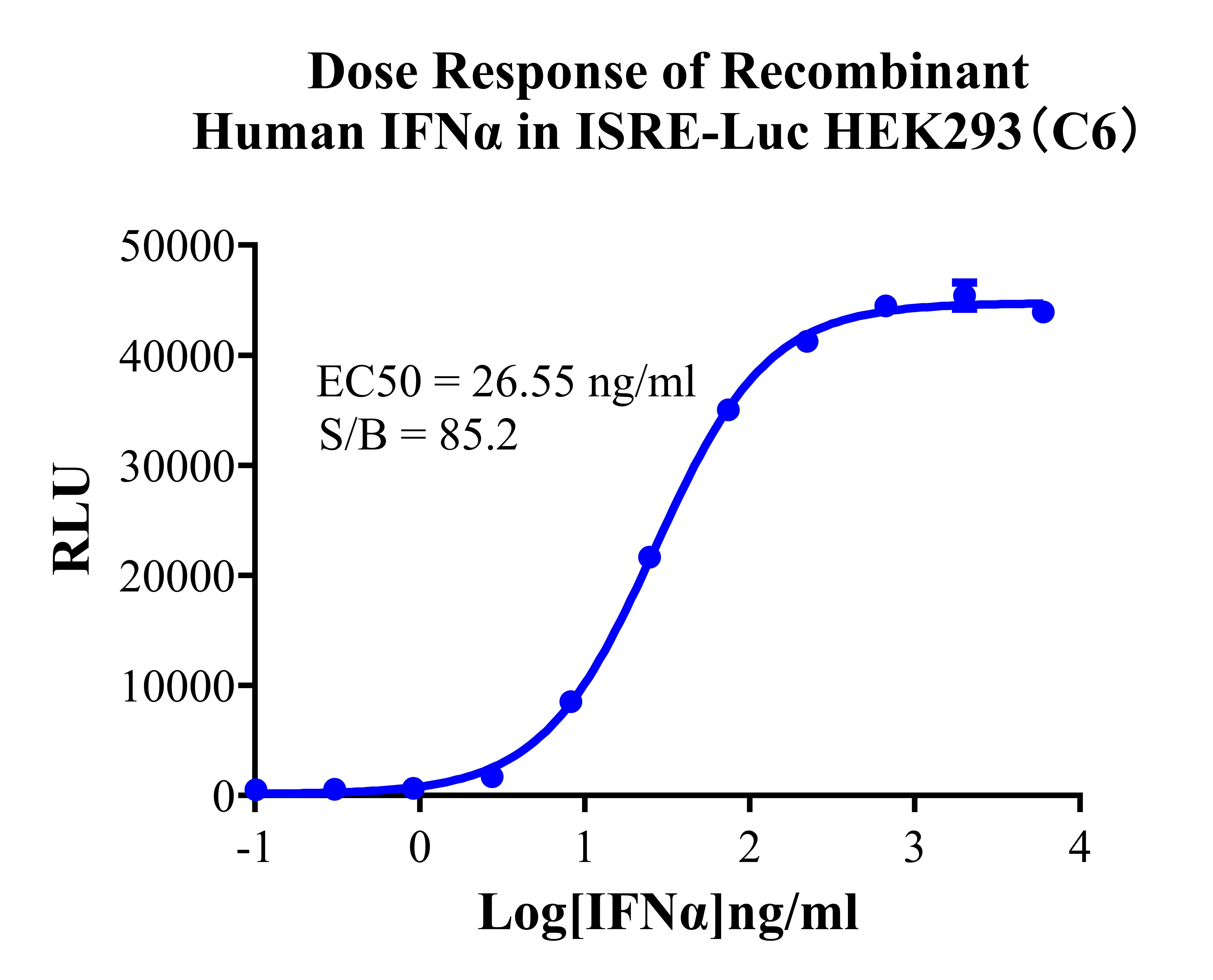
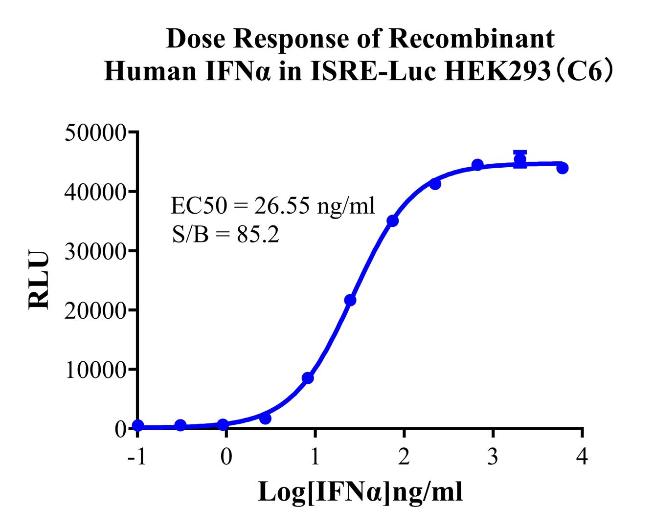
Figure 2. Dose Response of Recombinant Human IFNα in ISRE-Luc HEK293(C6).
Cell Resuscitation
1)Rapidly thaw the frozen cells in a 37 °C water bath for approximately 60 seconds. Once thawed (which may take slightly less or more than 60 seconds), immediately transfer the cell suspension from the cryovial into a 15 mL centrifuge tube containing 10 mL of pre-warmed HEK293 Human ISRE-Luc Cell complete culture medium.
2)Centrifuge cells at 1000 rpm for 5 min to remove medium, then resuspend cells in 5 mL of pre-warmed complete medium.
3)Transfer the cell suspension into a T25 culture flask and incubate at 37 °C with 5% CO₂.
4)After approximately 24–36 hours, replace the medium or passage the cells to remove non-adherent dead cells.
Subculturing procedure
1)When the cell density reaches the appropriate confluency for passaging, wash the cells with PBS, then add 1 mL trypsin to detach the cells. When more than 80% of the cells detach upon gently tapping the culture flask, add complete culture medium to terminate digestion. Gently pipette to obtain a single-cell suspension, transfer to a 15 mL centrifuge tube, and centrifuge at 1000 rpm for 5 minutes.
2)Discard supernatant after centrifugation. Resuspend cells in fresh medium to a single-cell suspension and transfer to a new culture flask for continued growth.
Cell Freezing
After trypsinization and centrifugation of cells from each T75 flask or 10 cm culture dish, discard the supernatant. Add 2 mL of cryopreservation medium (90% FBS + 10% DMSO), gently resuspend thoroughly, and aliquot into two cryovials. Immediately place the cryovials into a controlled-rate freezing container (e.g., Nalgene 5100-0001), fill with isopropanol to the indicated level, and store at −80 °C. After 24 hours, transfer the cryovials to liquid nitrogen for long-term storage.
Related products
CHO-K1 Human CCR4 Cell Line
HEK293 Human NK1R CRE-Luc Cell Line
Raji-Luc-GFP
Jurkat E6.1-Luc
THP-1-GFP
THP-1-Luc
Raji-GFP
Raji-Luc
Jurkat E6.1-GFP
HEK293 Human GAL4-Luc Cell
We Are Pleased to Announce: Global Commercial Licensing Rights for Jurkat E6.1, CHO-K1, and HEK293 Cell Lines Officially Secured.
Explore