

HEK293 Human IL3 Effector Reporter Cell
Cat. No: RQP74433
Size: 1 vial of frozen cells (>1E6 per vial in 1 mL)
Unit Price: Contact For Pricing
Product Info
Description
Biological Information
Assay Data
Cell Culture
| Cat. No | RQP74433 |
| Product Name | HEK293 Human IL3 Effector Reporter Cell |
| Product Type | Reporter Cell |
| Culture Properties | Adherent |
| Stability | 32passages (in-house test, that not means the cell line will be instable beyond the passages we tested.) |
| Mycoplasma Status | Negative |
| Culture Medium | DMEM+10%FBS+2 μg/ml Puromycin+ 5 μg/ml Blasticidin+ 200 μg/ml Hygromycin B |
| Freeze Medium | 90% FBS+10% DMSO |
| Storage Conditions | Liquid nitrogen immediately upon delivery |
| Application | Functional(Report Gene) Assay |
For research use only. Not intended for human or animal clinical trials, therapeutic or diagnostic use.
IL-3 (Interleukin-3) is a pleiotropic hematopoietic growth factor produced by activated T cells, NK cells, mast cells, and other cell types. Its primary function is to promote the survival, proliferation, and differentiation of multi-lineage hematopoietic progenitor cells; specifically, it regulates the development of myeloid and lymphoid cells—such as basophils, mast cells, and macrophages—while also participating in inflammation, allergic responses, and the remodeling of the tumor immune microenvironment. The receptor encoded by the *IL3R* (*CD123/CD131*) gene serves as the key receptor for Interleukin-3 (IL-3). Composed of two subunits—α (CD123) and β (CD131)—this receptor belongs to the cytokine receptor superfamily. The α subunit (CD123) specifically binds to IL-3, thereby determining receptor specificity; conversely, the β subunit (CD131, also known as the common β chain) is shared with the IL-5 and GM-CSF receptors, forming heterodimers or oligomers to transduce signals. By activating signaling pathways such as JAK-STAT, PI3K-Akt, and Ras-MAPK, this receptor regulates the proliferation, differentiation, and survival of hematopoietic cells. It plays a pivotal role in the regulation of early hematopoiesis, facilitating the colony formation of multi-lineage progenitor cells, including those of the myeloid and erythroid lineages.
The IL3 Effector Reporter Cell model serves as an excellent *in vitro* mimic of the signal transduction processes mediated by IL-3 *in vivo*. The underlying principle is illustrated in the figure below.
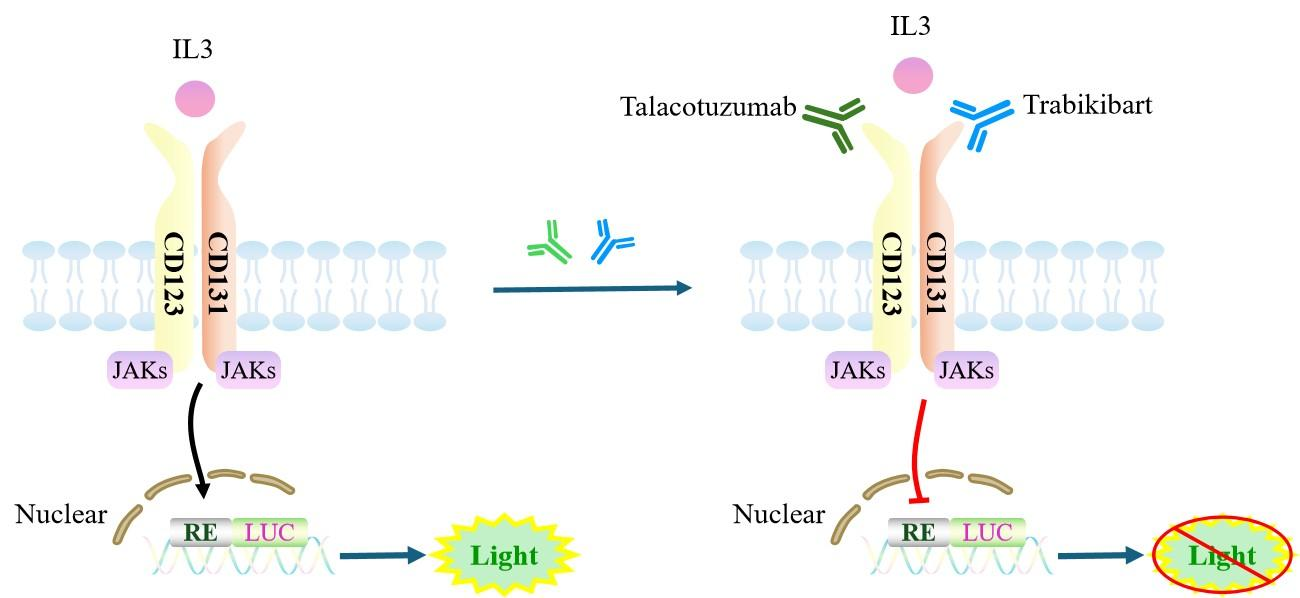
Figure 1. Schematic Diagram of the IL3 Effector Reporter Cell Model
| Classification | Cytokine&Growth Factor |
| Family | type I cytokine receptor family |
| Gene Name | IL3RA |
| Gene Aliases | CD123;IL3R |
| Gene ID | 3563 |
| Accession Number | NM_002183.4 |
| UniProt Number | P26951 |
| Protein Name | IL-3 receptor subunit alpha; IL-3R subunit alpha; IL-3R-alpha; IL-3RA |
| Protein Aliases | N/A |
| Target Species | Human |
| Host cell | HEK293 |
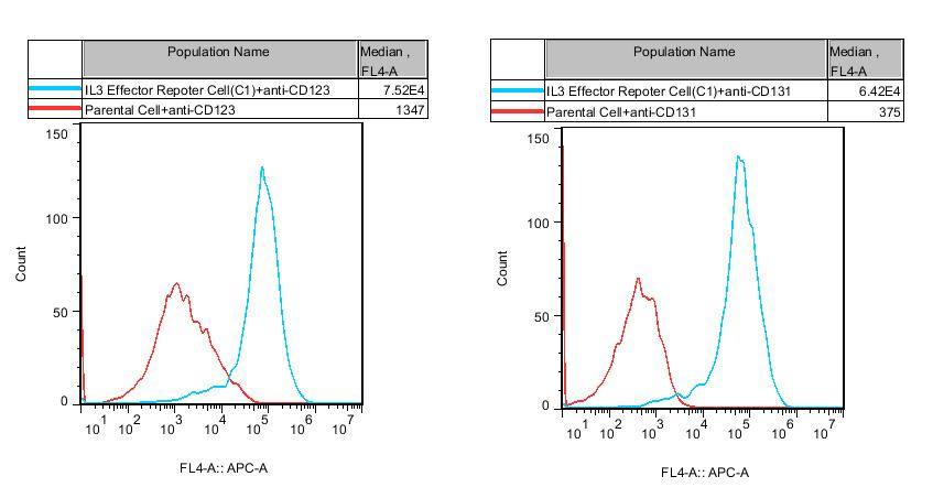
Figure 2. Recombinant IL3 Effector Reporter Cell stably expressing CD123 &CD131.
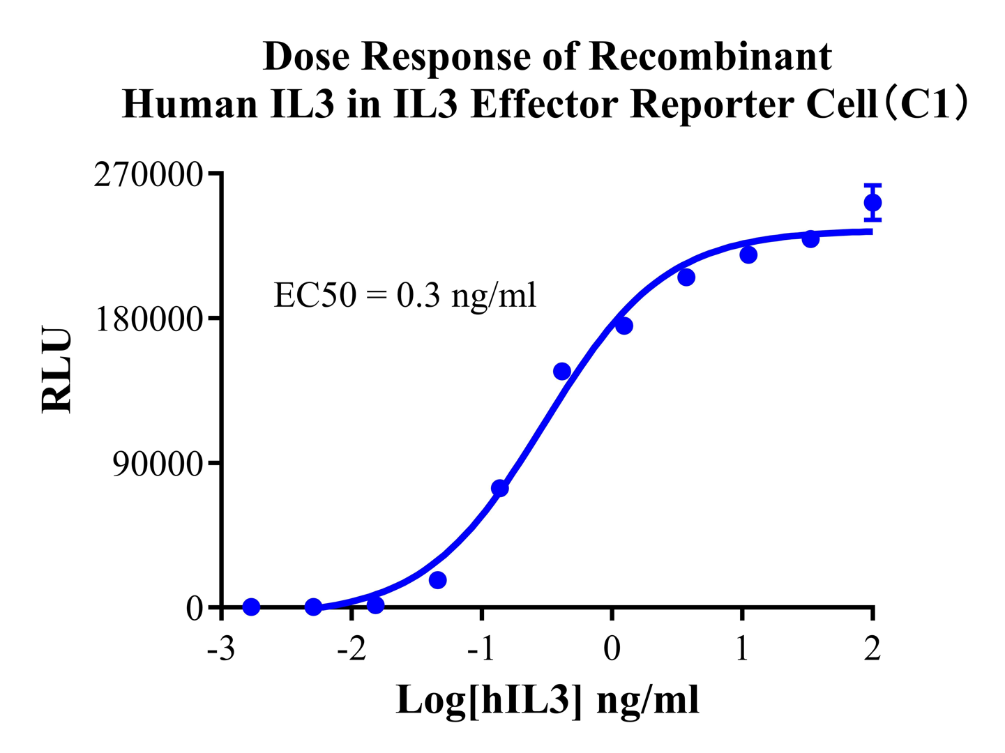
Figure 3. Dose Response of Recombinant Human IL3 in IL3 Effector Reporter Cell(C1).
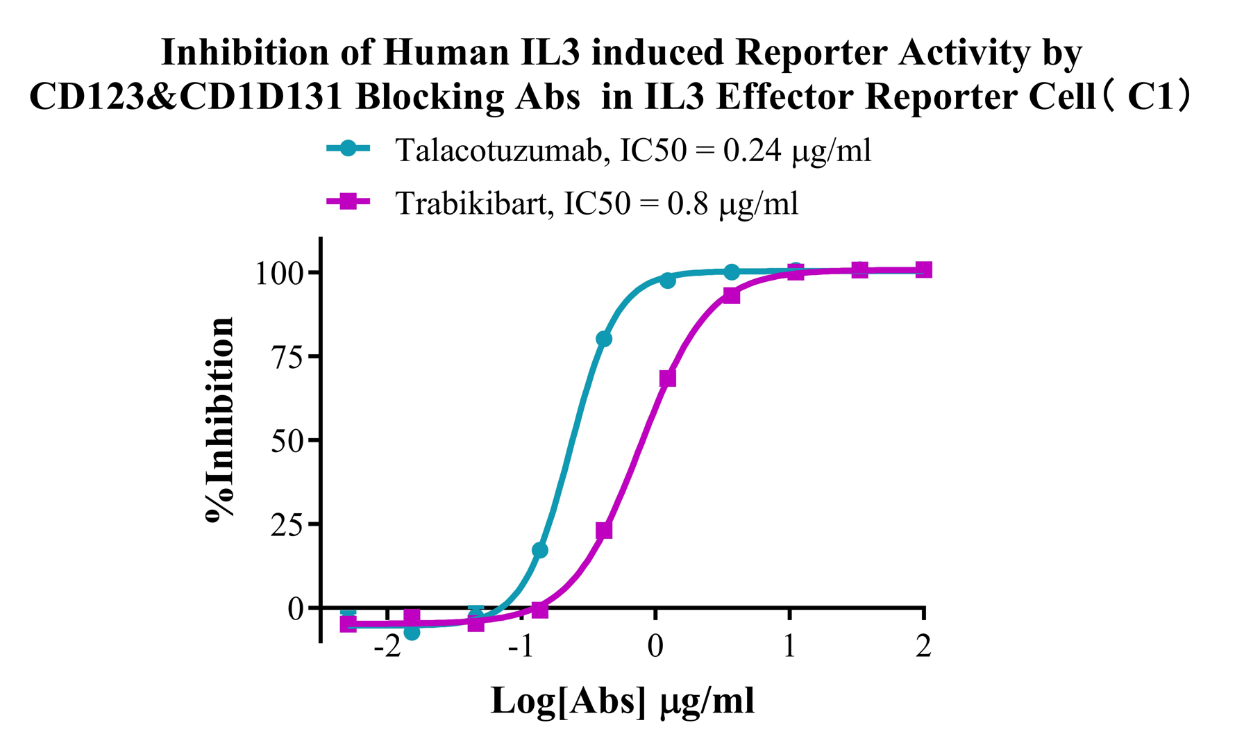
Figure 4. Inhibition of Human IL3 induced Reporter Activity by CD123&CD1D131 Blocking Abs in IL3 Effector Reporter Cell(C1).
Cell Resuscitation
1)Rapidly thaw the frozen cells in a 37 °C water bath for approximately 60 seconds. Once thawed (which may take slightly less or more than 60 seconds), immediately transfer the cell suspension from the cryovial into a 15 mL centrifuge tube containing 10 mL of pre-warmed HEK293 Human IL3 Effector Reporter Cell complete culture medium.
2)Centrifuge cells at 1000 rpm for 5 min to remove medium, then resuspend cells in 5 mL of pre-warmed complete medium.
3)Transfer the cell suspension into a T25 culture flask and incubate at 37 °C with 5% CO₂.
4)After approximately 24–36 hours, replace the medium or passage the cells to remove non-adherent dead cells.
Subculturing procedure
1)When the cell density reaches the appropriate confluency for passaging, wash the cells with PBS, then add 1 mL trypsin to detach the cells. When more than 80% of the cells detach upon gently tapping the culture flask, add complete culture medium to terminate digestion. Gently pipette to obtain a single-cell suspension, transfer to a 15 mL centrifuge tube, and centrifuge at 1000 rpm for 5 minutes.
2)Discard supernatant after centrifugation. Resuspend cells in fresh medium to a single-cell suspension and transfer to a new culture flask for continued growth.
Cell Freezing
After trypsinization and centrifugation of cells from each T75 flask or 10 cm culture dish, discard the supernatant. Add 2 mL of cryopreservation medium (90% FBS + 10% DMSO), gently resuspend thoroughly, and aliquot into two cryovials. Immediately place the cryovials into a controlled-rate freezing container (e.g., Nalgene 5100-0001), fill with isopropanol to the indicated level, and store at −80 °C. After 24 hours, transfer the cryovials to liquid nitrogen for long-term storage.
Related products
CHO-K1 Human CCR4 Cell Line
HEK293 Human NK1R CRE-Luc Cell Line
Raji-Luc-GFP
Jurkat E6.1-Luc
THP-1-GFP
THP-1-Luc
Raji-GFP
Raji-Luc
Jurkat E6.1-GFP
HEK293 Human GAL4-Luc Cell
We Are Pleased to Announce: Global Commercial Licensing Rights for Jurkat E6.1, CHO-K1, and HEK293 Cell Lines Officially Secured.
Explore