


HEK293 Human CSF1R/SRE-Luc Cell
Cat. No: RQP74010
Size: 1 vial of frozen cells (>1E6 per vial in 1 mL)
Unit Price: Contact For Pricing
Product Info
Description
Biological Information
Assay Data
Cell Culture
| Cat. No | RQP74010 |
| Product Name | HEK293 Human CSF1R/SRE-Luc Cell |
| Product Type | Reporter Cell |
| Culture Properties | Adherent |
| Stability | 32passages (in-house test, that not means the cell line will be instable beyond the passages we tested.) |
| Mycoplasma Status | Negative |
| Culture Medium | DMEM +10%FBS+200μg/ml Hygromycin B+1μg/ml puromycin |
| Freeze Medium | 90% FBS+10% DMSO |
| Storage Conditions | Liquid nitrogen immediately upon delivery |
| Application | Functional(Report Gene) Assay |
For research use only. Not intended for human or animal clinical trials, therapeutic or diagnostic use.
Colony-stimulating factor 1 (CSF1), also known as macrophage colony-stimulating factor (M-CSF), is a homodimer formed by two identical polypeptide chains linked by disulfide bonds. It is one of the most common pro-inflammatory cytokines involved in various inflammatory diseases. It plays a significant role in the development and progression of osteoarthritis, cancer, and other autoimmune diseases.
CSF1 regulates the proliferation, differentiation, and survival of the monocyte-macrophage lineage by binding to and activating its sole receptor, CSF1R (c-FMS, CD115). CSF1R belongs to the type III receptor tyrosine kinase (RTK) family and consists of an extracellular ligand-binding domain, transmembrane helices, and an intracellular kinase domain; its abnormal activation is closely associated with tumorigenesis, chronic inflammation, and bone remodeling disorders. Under physiological conditions, the CSF1/CSF1R axis is critical for embryonic development (e.g., placental formation), tissue homeostasis (e.g., osteoclast differentiation), and tissue repair. CSF1 binding induces CSF1R dimerization and autophosphorylation, activating signaling pathways such as Ras/MAPK, PI3K/AKT/mTOR, and JAK/STAT.
The CSF1R SRE-Luc HEK293 reporter gene drug target model effectively mimics the in vivo CSF1 signaling pathway; the mechanism is illustrated in the figure below.
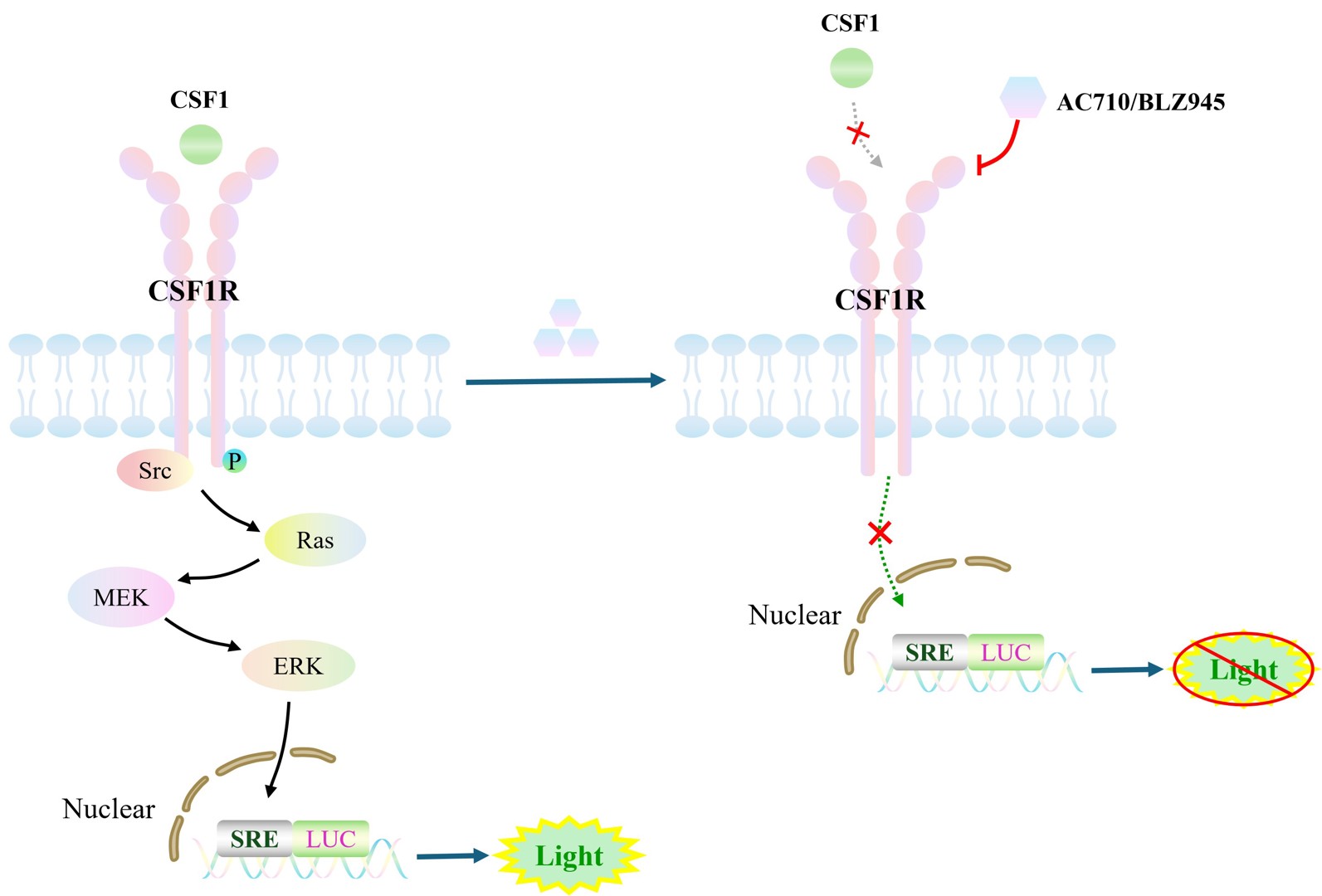
Figure 1. Schematic diagram of the CSF1R SRE-Luc HEK293 cell model
| Classification | Cytokine&Growth Factor |
| Family | Receptor tyrosine kinase (RTK) family |
| Gene Name | CSF1R |
| Gene Aliases | FMS;C-FMS;CSFR;CD115 |
| Gene ID | 1436 |
| Accession Number | NM_001288705.3 |
| UniProt Number | P07333 |
| Protein Name | Macrophage colony-stimulating factor 1 receptor |
| Protein Aliases | CSF-1-R; CSF-1R; M-CSF-R; |
| Target Species | Human |
| Host cell | HEK293 |
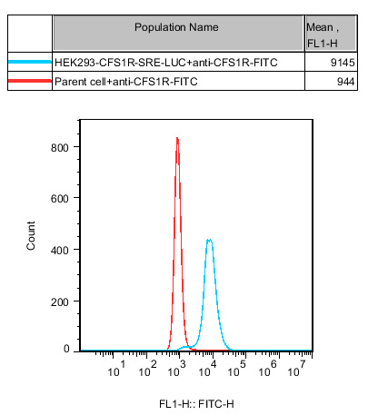
Figure 2. Recombinant CSF1R/SRE-Luc/HEK293 stably expressing CSF1R.
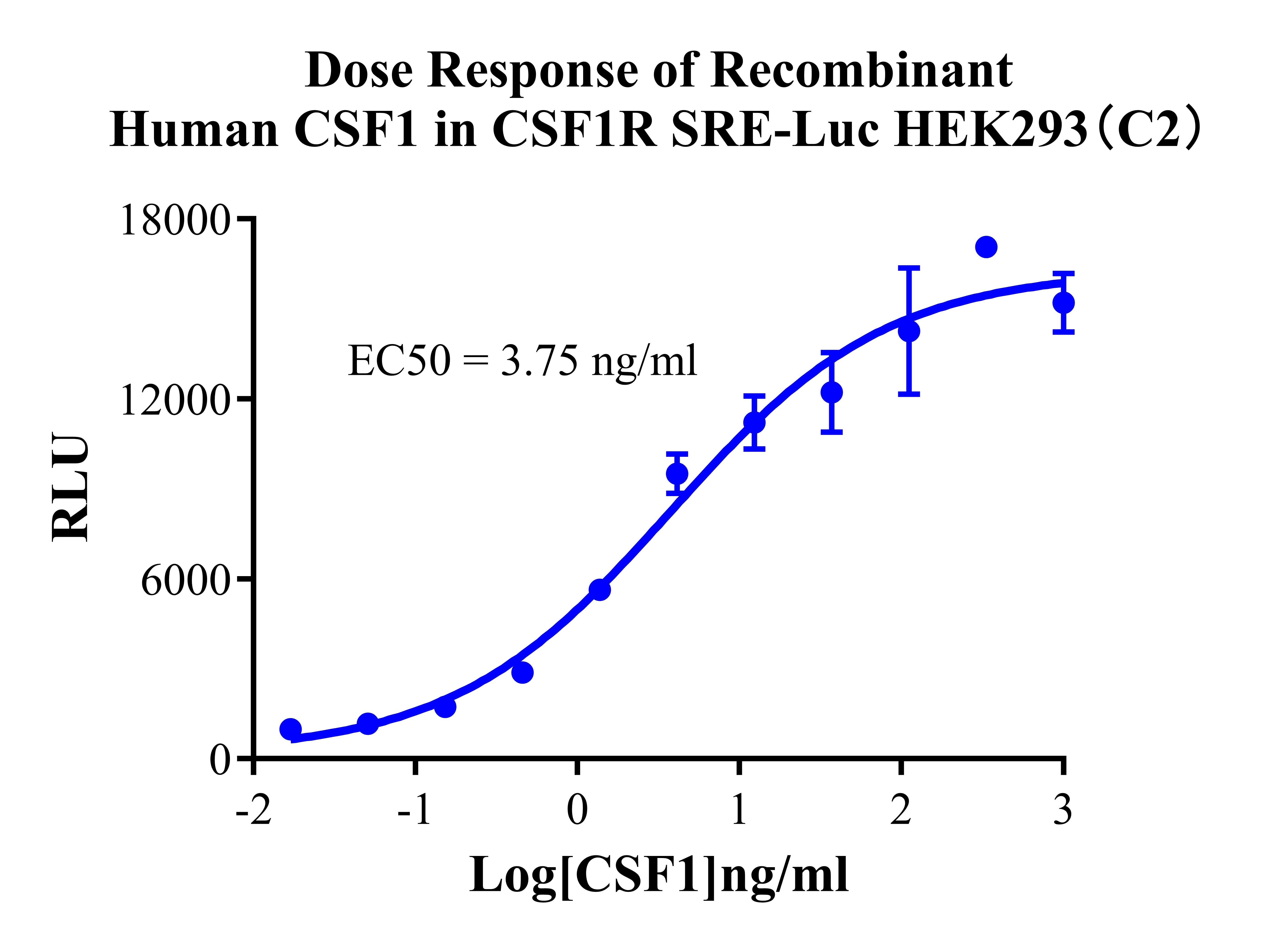
Figure 3. Detect Luciferase assay by Ultra Luciferase Detection Kit CBPH0001(we strongly suggest to purchase from Cobioer). Dose Response of Recombinant Human CSF1 in CSF1R SRE-Luc HEK293(C2).
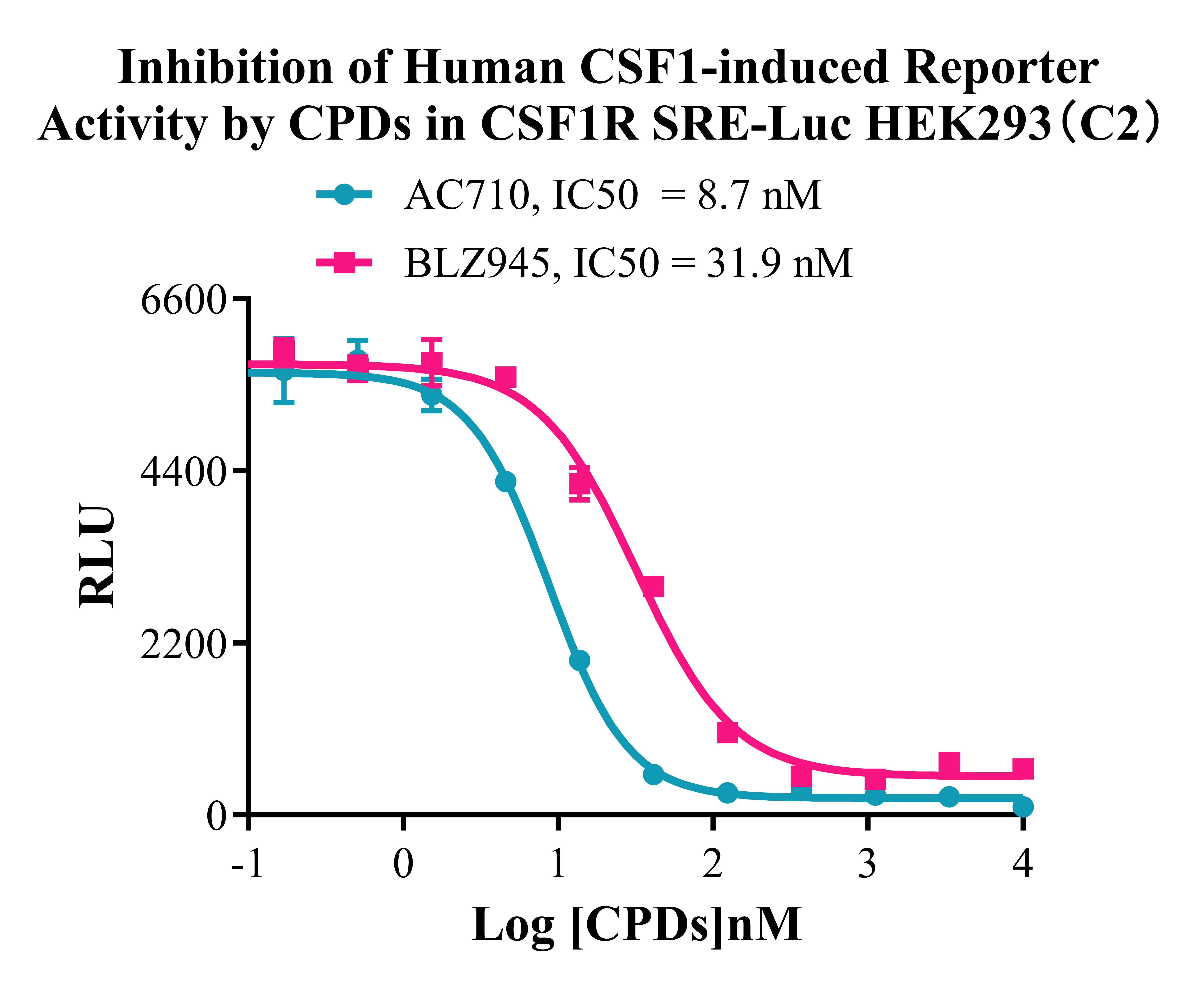
Figure 4. Inhibition of Human CSF1-induced Reporter Activity by CPDs in CSF1R SRE-Luc HEK293(C2).
Cell Resuscitation
1)Rapidly thaw the frozen cells in a 37 °C water bath for approximately 60 seconds. Once thawed (which may take slightly less or more than 60 seconds), immediately transfer the cell suspension from the cryovial into a 15 mL centrifuge tube containing 10 mL of pre-warmed HEK293 Human CSF1R/SRE-Luc Cell complete culture medium.
2)Centrifuge cells at 1000 rpm for 5 min to remove medium, then resuspend cells in 5 mL of pre-warmed complete medium.
3)Transfer the cell suspension into a T25 culture flask and incubate at 37 °C with 5% CO₂.
4)After approximately 24–36 hours, replace the medium or passage the cells to remove non-adherent dead cells.
Subculturing procedure
1)When the cell density reaches the appropriate confluency for passaging, wash the cells with PBS, then add 1 mL trypsin to detach the cells. When more than 80% of the cells detach upon gently tapping the culture flask, add complete culture medium to terminate digestion. Gently pipette to obtain a single-cell suspension, transfer to a 15 mL centrifuge tube, and centrifuge at 1000 rpm for 5 minutes.
2)Discard supernatant after centrifugation. Resuspend cells in fresh medium to a single-cell suspension and transfer to a new culture flask for continued growth.
Cell Freezing
After trypsinization and centrifugation of cells from each T75 flask or 10 cm culture dish, discard the supernatant. Add 2 mL of cryopreservation medium (90% FBS + 10% DMSO), gently resuspend thoroughly, and aliquot into two cryovials. Immediately place the cryovials into a controlled-rate freezing container (e.g., Nalgene 5100-0001), fill with isopropanol to the indicated level, and store at −80 °C. After 24 hours, transfer the cryovials to liquid nitrogen for long-term storage.
Related products
CHO-K1 Human CCR4 Cell Line
HEK293 Human NK1R CRE-Luc Cell Line
Raji-Luc-GFP
Jurkat E6.1-Luc
THP-1-GFP
THP-1-Luc
Raji-GFP
Raji-Luc
Jurkat E6.1-GFP
HEK293 Human GAL4-Luc Cell
We Are Pleased to Announce: Global Commercial Licensing Rights for Jurkat E6.1, CHO-K1, and HEK293 Cell Lines Officially Secured.
Explore