

HEK293 Human BCMA/NFκB-Luc Cell
Cat. No: RQP74072
Size: 1 vial of frozen cells (>1E6 per vial in 1 mL)
Unit Price: Contact For Pricing
Product Info
Description
Biological Information
Assay Data
Cell Culture
| Cat. No | RQP74072 |
| Product Name | HEK293 Human BCMA/NFκB-Luc Cell |
| Product Type | Reporter Cell |
| Culture Properties | Adherent |
| Stability | 32passages (in-house test, that not means the cell line will be instable beyond the passages we tested.) |
| Mycoplasma Status | Negative |
| Culture Medium | MEM + 10% Foetal Bovine Serum (FBS)+ 1% Non Essential Amino Acids (NEAA) + 1mM Sodium Pyruvate (NaP) +100μg/ml Hygromycin B+1μg/ml puromycin |
| Freeze Medium | 90% FBS+10% DMSO |
| Storage Conditions | Liquid nitrogen immediately upon delivery |
| Application | Functional(Report Gene) Assay |
For research use only. Not intended for human or animal clinical trials, therapeutic or diagnostic use.
B-cell maturation antigen (BCMA) is a transmembrane glycoprotein belonging to the tumor necrosis factor receptor (TNFR) superfamily. It is expressed on the surface of normal, malignant plasma cells, and mature B cells, with minimal expression in other tissues. BCMA, along with two other functionally related TNFR superfamily members—B-cell activating factor receptor (BAFF-R) and TACI—synergistically regulates B-cell proliferation, maturation, survival, and differentiation into plasma cells (PC). BCMA has a soluble form, sBCMA, which is shed directly from membrane-bound BCMA via γ-secretase activity. sBCMA retains the extracellular domain and part of the transmembrane region. sBCMA is a potential biomarker for B-cell involvement in human autoimmune diseases such as systemic lupus erythematosus, rheumatoid arthritis, and multiple sclerosis.
BCMA has two agonistic ligands: a proliferation-inducing ligand (APRIL) and BAFF. APRIL binds to BCMA with much higher affinity than BAFF and can also bind to TACI, whereas BAFF shows higher selectivity for BAFF-R. Upon ligand binding to BCMA, multiple growth and survival signaling cascades are activated in multiple myeloma cells, most commonly the nuclear factor κ light chain enhancer of activated B cells (NF-κB), but also including the RAS/MAPK and PI3K-Akt signaling pathways. These pathways stimulate proliferation by regulating cell cycle checkpoints, enhance survival by upregulating anti-apoptotic proteins (such as Mcl-1, BCL-2, BCL-XL), and promote the production of cell adhesion molecules (e.g., ICAM-I), angiogenic factors (e.g., VEGF, IL-8), and immunosuppressive molecules (e.g., IL-10, PD-L1, TGF-β).
The HEK293 Human BCMA/NFκB-Luc Cell model effectively mimics the in vivo BCMA signaling transduction process. The principle is illustrated in the figure below.
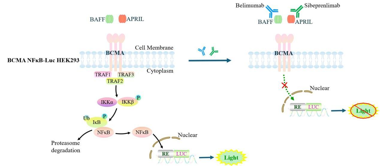
Figure 1. Schematic diagram of the HEK293 Human BCMA/NFκB-Luc Cell model.
| Classification | Co-Stimulatory |
| Family | Tumor necrosis factor receptor superfamily |
| Gene Name | BCMA |
| Gene Aliases | TNFRSF17;BCM;CD269;TNFRSF13A |
| Gene ID | 608 |
| Accession Number | NM_001192.3 |
| UniProt Number | Q02223 |
| Protein Name | B-cell maturation protein |
| Protein Aliases | Tumor necrosis factor receptor superfamily member 17 |
| Target Species | Human |
| Host cell | HEK293 |
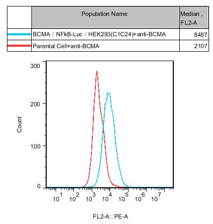
Figure 2. Recombinant BCMA/NFκB-Luc/HEK293 stably expressing BCMA.
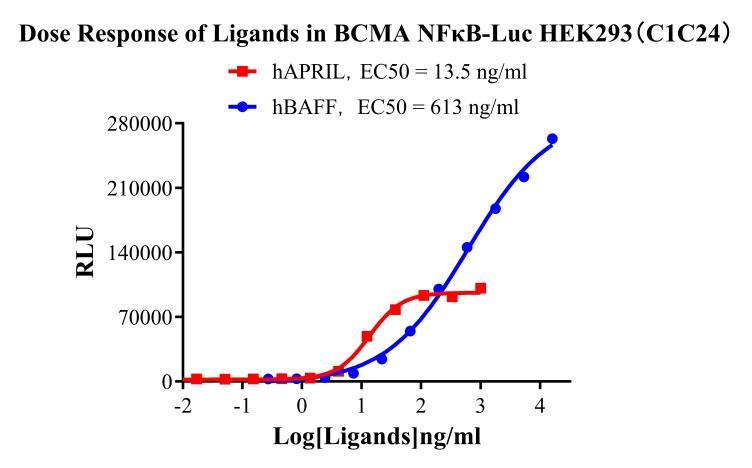
Figure 3. Dose Response of Ligands in BCMA NFκB-Luc/HEK293 (C1C4).
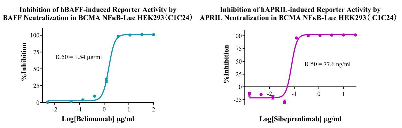
Figure 4. Inhibition of Human hBAFF-induced Reporter Activity by BAFF Neutralization in BCMA NFκB Luc HEK293 (C1C4).Inhibition of hAPRIL-induced Reporter Activity by APRIL Neutralization in BCMA NFκB Luc HEK293(C1C4).
Cell Resuscitation
1)Rapidly thaw the frozen cells in a 37 °C water bath for approximately 60 seconds. Once thawed (which may take slightly less or more than 60 seconds), immediately transfer the cell suspension from the cryovial into a 15 mL centrifuge tube containing 10 mL of pre-warmed HEK293 Human BCMA/NFκB-Luc Cell complete culture medium.
2)Centrifuge cells at 1000 rpm for 5 min to remove medium, then resuspend cells in 5 mL of pre-warmed complete medium.
3)Transfer the cell suspension into a T25 culture flask and incubate at 37 °C with 5% CO₂.
4)After approximately 24–36 hours, replace the medium or passage the cells to remove non-adherent dead cells.
Subculturing procedure
1)When the cell density reaches the appropriate confluency for passaging, wash the cells with PBS, then add 1 mL trypsin to detach the cells. When more than 80% of the cells detach upon gently tapping the culture flask, add complete culture medium to terminate digestion. Gently pipette to obtain a single-cell suspension, transfer to a 15 mL centrifuge tube, and centrifuge at 1000 rpm for 5 minutes.
2)Discard supernatant after centrifugation. Resuspend cells in fresh medium to a single-cell suspension and transfer to a new culture flask for continued growth.
Cell Freezing
After trypsinization and centrifugation of cells from each T75 flask or 10 cm culture dish, discard the supernatant. Add 2 mL of cryopreservation medium (90% FBS + 10% DMSO), gently resuspend thoroughly, and aliquot into two cryovials. Immediately place the cryovials into a controlled-rate freezing container (e.g., Nalgene 5100-0001), fill with isopropanol to the indicated level, and store at −80 °C. After 24 hours, transfer the cryovials to liquid nitrogen for long-term storage.
Related products
CHO-K1 Human CCR4 Cell Line
HEK293 Human NK1R CRE-Luc Cell Line
Raji-Luc-GFP
Jurkat E6.1-Luc
THP-1-GFP
THP-1-Luc
Raji-GFP
Raji-Luc
Jurkat E6.1-GFP
HEK293 Human GAL4-Luc Cell
We Are Pleased to Announce: Global Commercial Licensing Rights for Jurkat E6.1, CHO-K1, and HEK293 Cell Lines Officially Secured.
Explore