
CHO-K1 Human Lnluc-LDLR Cell
Cat. No: RQP75087
Size: 1 vial of frozen cells (>1E6 per vial in 1 mL)
Unit Price: Contact For Pricing
Product Info
Description
Assay Data
Cell Culture
| Cat. No | RQP75087 |
| Product Name | CHO-K1 Human Lnluc-LDLR Cell |
| Culture Properties | Adherent |
| Stability | 32passages (in-house test, that not means the cell line will be instable beyond the passages we tested.) |
| Mycoplasma Status | Negative |
| Culture Medium | F12K+ 10% FBS+ 5 μg/ml Puromycin |
| Freeze Medium | 90% FBS+10% DMSO |
| Storage Conditions | Liquid nitrogen immediately upon delivery |
| Application | Binding Assay,FACS |
For research use only. Not intended for human or animal clinical trials, therapeutic or diagnostic use.
The low-density lipoprotein receptor (LDLR) is a transmembrane glycoprotein primarily located on the surface of hepatocytes; its core function is to clear low-density lipoprotein (LDL) from the blood and maintain cholesterol homeostasis. Its structure is highly modular: the ligand-binding domain recognizes apolipoprotein B-100 on LDL, the EGF-homology domain responds to pH changes, and the cytoplasmic tail mediates endocytic signaling. Upon binding LDL, the LDLR enters the cell via clathrin-coated pits; within acidic endosomes, it releases the LDL—allowing the empty receptor to be recycled to the cell membrane—while the LDL is degraded by lysosomes for cellular use. This efficient cycle enables the LDLR to repeatedly clear cholesterol, making it a central molecule in lipid regulation.
PCSK9 (proprotein convertase subtilisin/kexin type 9) is primarily synthesized by the liver and secreted into the bloodstream, serving as a key negative regulator of cholesterol metabolism. Following autocatalytic cleavage of its precursor, the pro-domain binds tightly to the catalytic domain to form the mature protein. Rather than degrading other substrates, PCSK9 acts as a molecular chaperone that specifically binds to the LDLR on the hepatocyte surface, targeting the receptor's EGF-A domain.
PCSK9 binds to the LDLR either on the cell surface or during endocytosis; this binding affinity increases significantly within acidic endosomes, resulting in a tight complex. Under normal conditions, after releasing LDL in the acidic endosome, the LDLR is recognized by recycling proteins (such as SNX17) and directed back to the cell membrane; however, PCSK9 binding locks the LDLR conformation, preventing interaction with recycling proteins and thereby blocking the recycling pathway. The LDLR-PCSK9 complex is subsequently trafficked to lysosomes as the endosome matures and undergoes co-degradation, leading to a reduction in surface LDLR levels and impaired blood LDL clearance. PCSK9 inhibitors lower cholesterol levels significantly by blocking this interaction and protecting the LDLR, a mechanism that establishes PCSK9 as a therapeutic target for lipid-lowering therapy.
The CHO-K1 Human Lnluc-LDLR Cell model effectively simulates the in vivo LDLR signal transduction process, the principle is illustrated in the figure below.
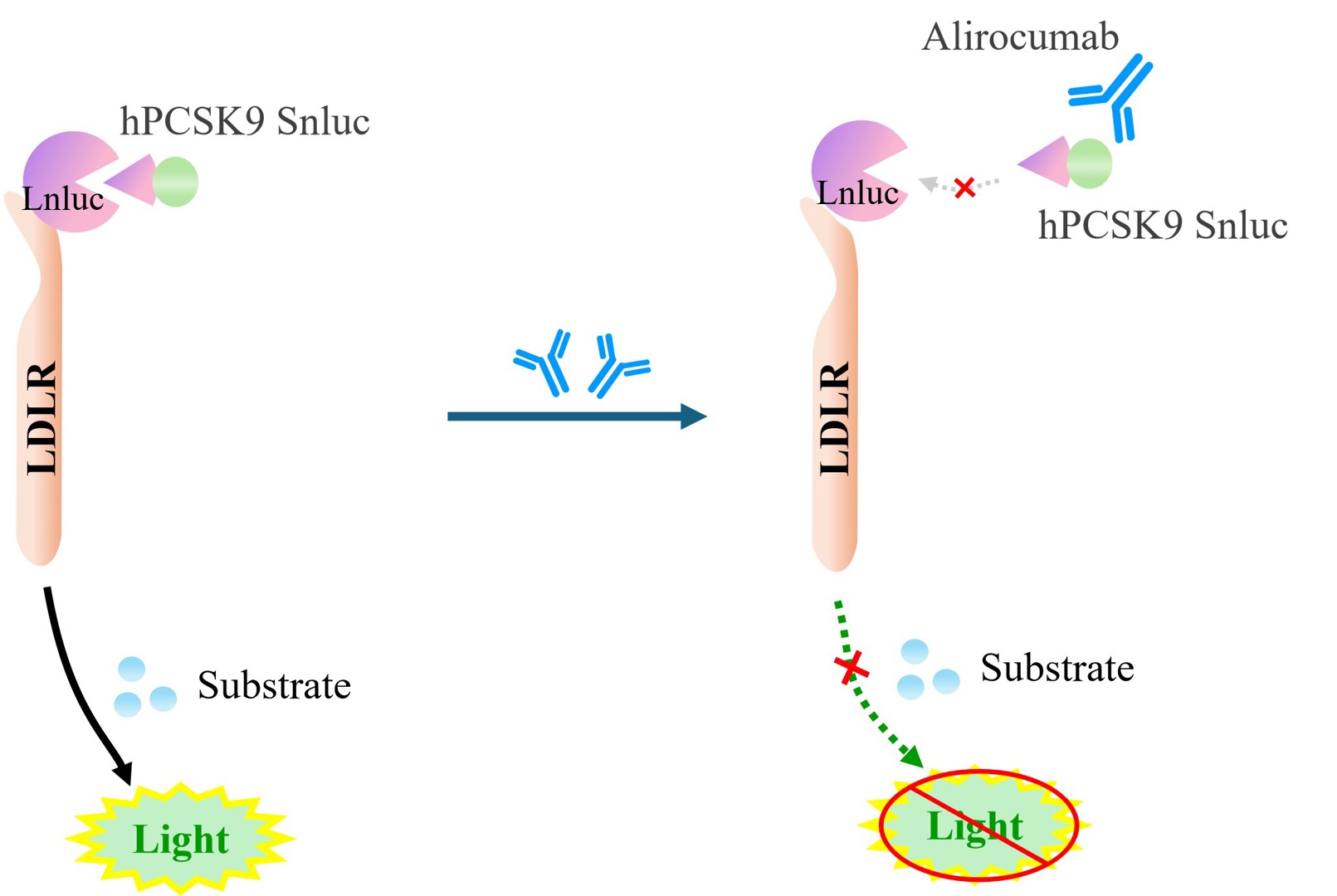
Figure 1. Schematic diagram of the CHO-K1 Human Lnluc-LDLR Cell model
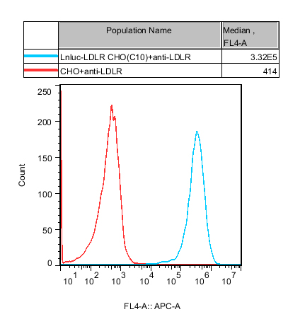
Figure 2. Recombinant Lnluc-LDLR CHO stably expressing LDLR.
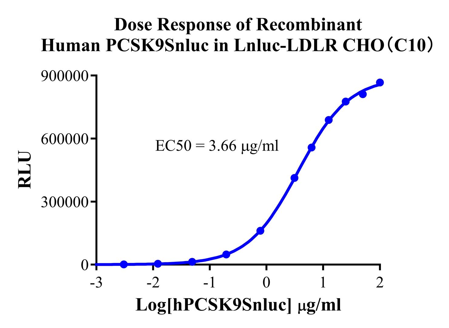
Figure 3. Dose Response of Recombinant Human PCSK9Snluc in Lnluc-LDLR CHO(C10).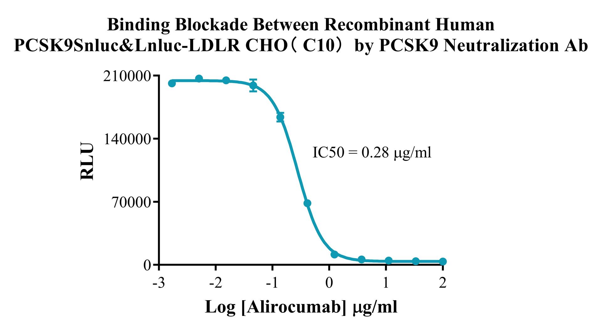
Figure 4. Binding Blockade Between Recombinant Human PCSK9Snluc&Lnluc-LDLR CHO(C10) by PCSK9 Neutralization Ab.
Cell Resuscitation
1)Rapidly thaw the frozen cells in a 37 °C water bath for approximately 60 seconds. Once thawed (which may take slightly less or more than 60 seconds), immediately transfer the cell suspension from the cryovial into a 15 mL centrifuge tube containing 10 mL of pre-warmed CHO-K1 Human Lnluc-LDLR Cell complete culture medium.
2)Centrifuge cells at 1000 rpm for 5 min to remove medium, then resuspend cells in 5 mL of pre-warmed complete medium.
3)Transfer the cell suspension into a T25 culture flask and incubate at 37 °C with 5% CO₂.
4)After approximately 24–36 hours, replace the medium or passage the cells to remove non-adherent dead cells.
Subculturing procedure
1)When the cell density reaches the appropriate confluency for passaging, wash the cells with PBS, then add 1 mL trypsin to detach the cells. When more than 80% of the cells detach upon gently tapping the culture flask, add complete culture medium to terminate digestion. Gently pipette to obtain a single-cell suspension, transfer to a 15 mL centrifuge tube, and centrifuge at 1000 rpm for 5 minutes.
2)Discard supernatant after centrifugation. Resuspend cells in fresh medium to a single-cell suspension and transfer to a new culture flask for continued growth.
Cell Freezing
After trypsinization and centrifugation of cells from each T75 flask or 10 cm culture dish, discard the supernatant. Add 2 mL of cryopreservation medium (90% FBS + 10% DMSO), gently resuspend thoroughly, and aliquot into two cryovials. Immediately place the cryovials into a controlled-rate freezing container (e.g., Nalgene 5100-0001), fill with isopropanol to the indicated level, and store at −80 °C. After 24 hours, transfer the cryovials to liquid nitrogen for long-term storage.
Related products
CHO-K1 Human NTSR2 Cell Line
CHO-K1 Human CCR4 Cell Line
HEK293 Human NK1R CRE-Luc Cell Line
Raji-Luc-GFP
Jurkat E6.1-Luc
THP-1-GFP
Raji-GFP
Raji-Luc
Jurkat E6.1-GFP
HEK293 Human GAL4-Luc Cell
We Are Pleased to Announce: Global Commercial Licensing Rights for Jurkat E6.1, CHO-K1, HEK293, THP-1 and RAJI Cell Lines Officially Secured.
Explore