

CHO-K1 Human ERBB4 Dimerization Cell
Cat. No: RQP74450
Size: 1 vial of frozen cells (>1E6 per vial in 1 mL)
Unit Price: Contact For Pricing
Product Info
Description
Biological Information
Assay Data
Cell Culture
| Cat. No | RQP74450 |
| Product Name | CHO-K1 Human ERBB4 Dimerization Cell |
| Product Type | Reporter Cell |
| Culture Properties | Adherent |
| Stability | 32passages (in-house test, that not means the cell line will be instable beyond the passages we tested.) |
| Mycoplasma Status | Negative |
| Culture Medium | F12K+10%FBS+5 μg/ml Puromycin+ 5 μg/ml Blasticidin |
| Freeze Medium | 90% FBS+10% DMSO |
| Storage Conditions | Liquid nitrogen immediately upon delivery |
| Application | Functional(Report Gene) Assay |
For research use only. Not intended for human or animal clinical trials, therapeutic or diagnostic use.
ERBB4 (HER4) is one of the core members of the human epidermal growth factor receptor (HER/ERBB) family. It is a transmembrane receptor with tyrosine kinase activity that is widely distributed in cardiomyocytes, central nervous system neurons, mammary glands, and epithelial tissues, and is localized at the subcellular level to the plasma membrane, cell nucleus, and even mitochondria.
The specific ligands for HER4 are the neurotrophic proteins NRG1, NRG2, and heterotrophin (HRG). Upon binding, these ligands trigger homodimerization of ERBB4 or the formation of heterodimers with ERBB2/3. This activates classic downstream signaling pathways, such as PI3K/AKT, RAS/RAF/MAPK, and PLCγ/PKC, to regulate cell growth and metabolism. Simultaneously, it releases the intracellular domain (4ICD) via a γ-secretase-mediated topological cleavage mechanism; upon nuclear entry, the 4ICD binds to STAT5, HIF-1α, and other transcription factors to drive the expression of target genes such as Cyclin D1 and MYC, or directly inhibits BAX-induced apoptosis in mitochondria, thereby playing a central role in key biological processes including cardiac development, mammary duct morphogenesis, oligodendrocyte differentiation, and neuronal synaptic plasticity. Due to its dual roles (oncogenic/tumor-suppressive), this target is widely associated with various diseases: overexpression or mutations (such as the extracellular domain V236L) and NRG1 fusion genes can drive the progression of breast cancer, lung adenocarcinoma, and melanoma, while signal attenuation leads to schizophrenia (impaired ERBB4-EphB2 interaction causing synaptic dysfunction), Alzheimer’s disease (abnormal cleavage of γ-secretase exacerbates Aβ toxicity), and dilated cardiomyopathy (loss of myocardial anti-apoptotic capacity), and has thus become a hotspot for targeted therapy.
The ERBB4 (HER4) Dimerization CHO Reporter Gene Drug Target Model effectively mimics the in vivo ERBB4 signaling pathway; the mechanism is illustrated in the figure below.
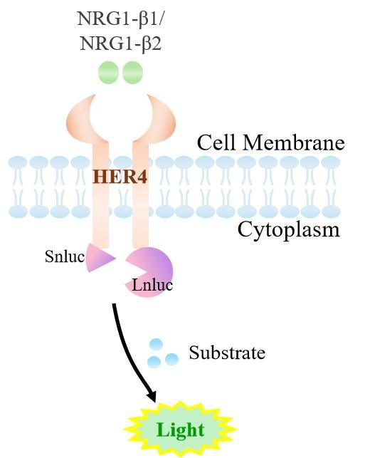
Figure 1. Schematic diagram of the CHO cell model for ERBB4 (HER4) dimerization
| Classification | Cytokine&Growth Factor |
| Family | Epidermal growth factor receptor (EGFR/ERBB) family |
| Gene Name | ERBB4 |
| Gene Aliases | ALS19;HER4 |
| Gene ID | 2066 |
| Accession Number | NM_005235.3 |
| UniProt Number | Q15303 |
| Protein Name | Receptor tyrosine-protein kinase erbB-4 |
| Protein Aliases | |
| Target Species | Human |
| Host cell | CHO-K1 |
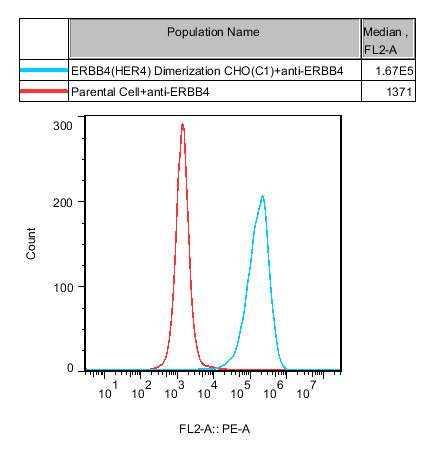
Figure 2. Recombinant HER4 Dimerization CHO stably expressing ERBB4.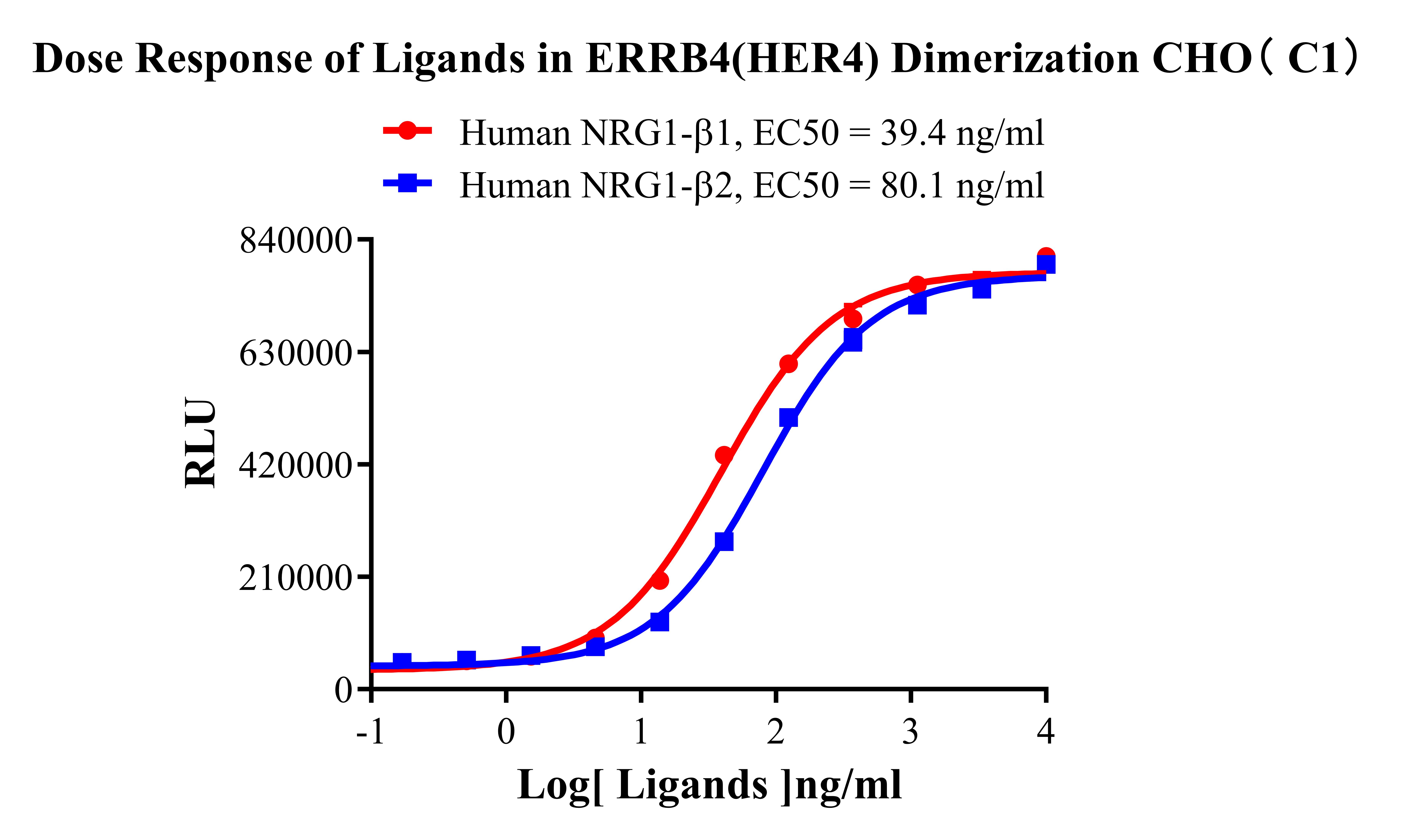
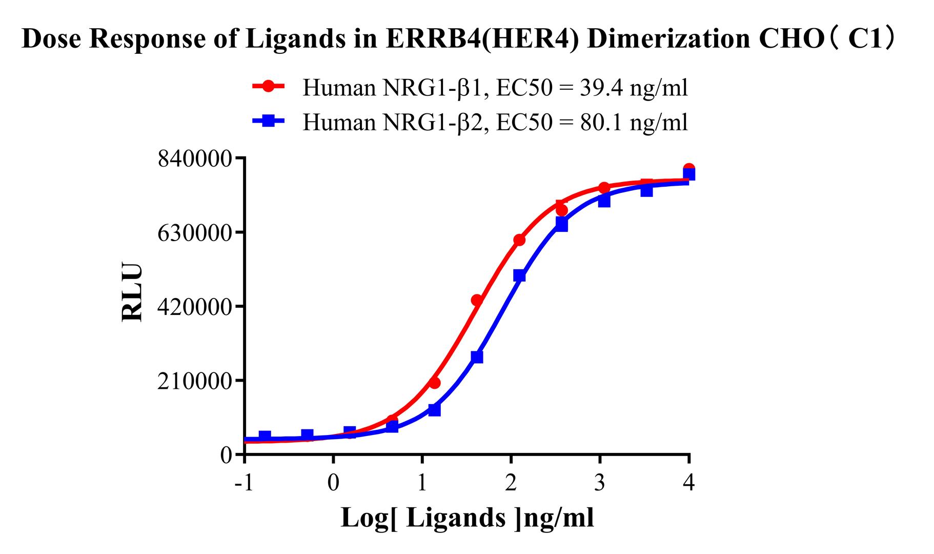
Figure 3. Dose Response of Ligands in ERRB4(HER4) Dimerization CHO(C1).
Cell Resuscitation
1)Rapidly thaw the frozen cells in a 37 °C water bath for approximately 60 seconds. Once thawed (which may take slightly less or more than 60 seconds), immediately transfer the cell suspension from the cryovial into a 15 mL centrifuge tube containing 10 mL of pre-warmed CHO-K1 Human ERBB4 Dimerization Cell complete culture medium.
2)Centrifuge cells at 1000 rpm for 5 min to remove medium, then resuspend cells in 5 mL of pre-warmed complete medium.
3)Transfer the cell suspension into a T25 culture flask and incubate at 37 °C with 5% CO₂.
4)After approximately 24–36 hours, replace the medium or passage the cells to remove non-adherent dead cells.
Subculturing procedure
1)When the cell density reaches the appropriate confluency for passaging, wash the cells with PBS, then add 1 mL trypsin to detach the cells. When more than 80% of the cells detach upon gently tapping the culture flask, add complete culture medium to terminate digestion. Gently pipette to obtain a single-cell suspension, transfer to a 15 mL centrifuge tube, and centrifuge at 1000 rpm for 5 minutes.
2)Discard supernatant after centrifugation. Resuspend cells in fresh medium to a single-cell suspension and transfer to a new culture flask for continued growth.
Cell Freezing
After trypsinization and centrifugation of cells from each T75 flask or 10 cm culture dish, discard the supernatant. Add 2 mL of cryopreservation medium (90% FBS + 10% DMSO), gently resuspend thoroughly, and aliquot into two cryovials. Immediately place the cryovials into a controlled-rate freezing container (e.g., Nalgene 5100-0001), fill with isopropanol to the indicated level, and store at −80 °C. After 24 hours, transfer the cryovials to liquid nitrogen for long-term storage.
Related products
CHO-K1 Human CCR4 Cell Line
HEK293 Human NK1R CRE-Luc Cell Line
Raji-Luc-GFP
Jurkat E6.1-Luc
THP-1-GFP
THP-1-Luc
Raji-GFP
Raji-Luc
Jurkat E6.1-GFP
HEK293 Human GAL4-Luc Cell
We Are Pleased to Announce: Global Commercial Licensing Rights for Jurkat E6.1, CHO-K1, and HEK293 Cell Lines Officially Secured.
Explore