

CHO-K1 Human CD200 Target cell
Cat. No: RQP74180
Size: 1 vial of frozen cells (>1E6 per vial in 1 mL)
Unit Price: Contact For Pricing
Product Info
Description
Biological Information
Assay Data
Cell Culture
| Cat. No | RQP74180 |
| Product Name | CHO-K1Human CD200 Target Cell |
| Product Type | Reporter Cell |
| Culture Properties | Adherent |
| Stability | 32passages (in-house test, that not means the cell line will be instable beyond the passages we tested.) |
| Mycoplasma Status | Negative |
| Culture Medium | F12K+10% FBS+ 5 μg/ml BSD |
| Freeze Medium | 90% FBS+10% DMSO |
| Storage Conditions | Liquid nitrogen immediately upon delivery |
| Application | Functional(Report Gene) Assay |
For research use only. Not intended for human or animal clinical trials, therapeutic or diagnostic use.
CD200 (also known as OX-2) is a member of the Immunoglobulin Superfamily (IgSF), a family of proteins structurally similar to B7 family proteins. It contains two extracellular immunoglobulin domains and a small intracellular domain of 19 amino acids, lacking any known signal motifs. CD200 is expressed in various normal tissues, including immune cells such as B lymphocytes and activated T lymphocytes. Recent studies have shown that CD200 is also overexpressed in multiple human cancer cells, including human melanoma, ovarian cancer, brain gliomas, myeloid leukemias, as well as some B-cell malignancies and most endocrine malignancies (e.g., small cell lung cancer).
CD200R, as the homologous ligand for CD200, is also an IgSF protein. In both mice and humans, the expression pattern of CD200R is similar, with high expression in macrophages, neutrophils, and mast cells. Unlike most IgSF receptors, CD200R lacks an ITIM domain. However, its cytoplasmic tail, composed of 67 amino acids, contains three tyrosine residues. The third tyrosine residue is located within an NPXY motif, which is phosphorylated upon CD200R ligand binding. This phosphorylation does not directly recruit protein tyrosine phosphatases such as SHP1 and SHP2 or the phosphorylated inositol phosphatase SHIP, but instead recruits downstream tyrosine kinase adaptor proteins Dok-2 and Dok-1, which then associate with RasGAP and SHIP. In macrophages and mast cells, this cascade has been shown to inhibit the phosphorylation of ERK, P38, and JNK, and suppress myeloid cell activation. Although CD200R expression is primarily found in macrophages and neutrophils, further studies have indicated its presence in dendritic cells (DCs) and some T-cell subsets, suggesting that CD200R signaling may also have regulatory functions in these cell types.
The CD200 Target Cell, when used as the target cell for the CD200R Effector Reporter Cell, effectively mimics the signal transduction process of CD200 and CD200R in vivo. The principle is shown in the figure below.
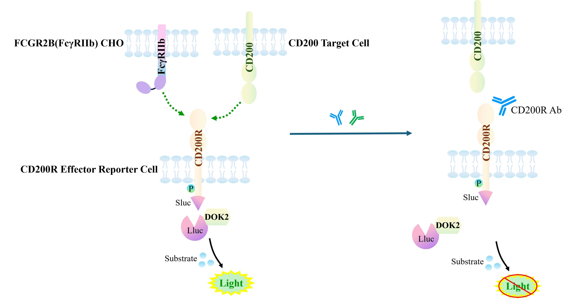
Figure 1. Schematic of the CD200 Target Cell Model.
| Classification | Co-Inhibitory |
| Family | Immunoglobulin superfamily (IgSF) |
| Gene Name | CD200 |
| Gene Aliases | MOX1;MOX2;MRC;OX-2 |
| Gene ID | 4345 |
| Accession Number | NM_005944.7 |
| UniProt Number | P41217 |
| Protein Name | OX-2 membrane glycoprotein |
| Protein Aliases | N/A |
| Target Species | Human |
| Host cell | CHO-K1 |
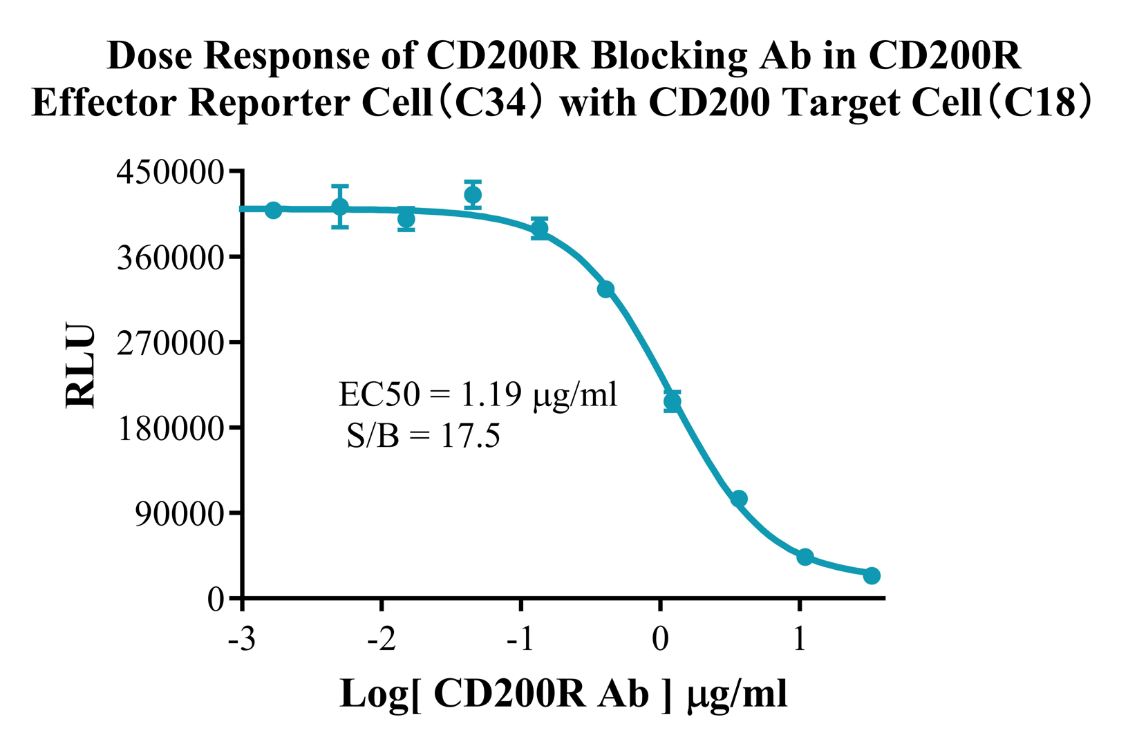
Figure 2. Dose Response of CD200R Blocking Ab in CD200R Effector Reporter Cell (C34) with CD200 Target Cell (C18).
Cell Resuscitation
1)Rapidly thaw the frozen cells in a 37 °C water bath for approximately 60 seconds. Once thawed (which may take slightly less or more than 60 seconds), immediately transfer the cell suspension from the cryovial into a 15 mL centrifuge tube containing 10 mL of pre-warmed CHO-K1Human CD200 Target Cell complete culture medium.
2)Centrifuge cells at 1000 rpm for 5 min to remove medium, then resuspend cells in 5 mL of pre-warmed complete medium.
3)Transfer the cell suspension into a T25 culture flask and incubate at 37 °C with 5% CO₂.
4)After approximately 24–36 hours, replace the medium or passage the cells to remove non-adherent dead cells.
Subculturing procedure
1)When the cell density reaches the appropriate confluency for passaging, wash the cells with PBS, then add 1 mL trypsin to detach the cells. When more than 80% of the cells detach upon gently tapping the culture flask, add complete culture medium to terminate digestion. Gently pipette to obtain a single-cell suspension, transfer to a 15 mL centrifuge tube, and centrifuge at 1000 rpm for 5 minutes.
2)Discard supernatant after centrifugation. Resuspend cells in fresh medium to a single-cell suspension and transfer to a new culture flask for continued growth.
Cell Freezing
After trypsinization and centrifugation of cells from each T75 flask or 10 cm culture dish, discard the supernatant. Add 2 mL of cryopreservation medium (90% FBS + 10% DMSO), gently resuspend thoroughly, and aliquot into two cryovials. Immediately place the cryovials into a controlled-rate freezing container (e.g., Nalgene 5100-0001), fill with isopropanol to the indicated level, and store at −80 °C. After 24 hours, transfer the cryovials to liquid nitrogen for long-term storage.
Related products
CHO-K1 Human CCR4 Cell Line
HEK293 Human NK1R CRE-Luc Cell Line
Raji-Luc-GFP
Jurkat E6.1-Luc
THP-1-GFP
THP-1-Luc
Raji-GFP
Raji-Luc
Jurkat E6.1-GFP
HEK293 Human GAL4-Luc Cell
We Are Pleased to Announce: Global Commercial Licensing Rights for Jurkat E6.1, CHO-K1, and HEK293 Cell Lines Officially Secured.
Explore