
CHO-K1 Human CCR8 β-Arrestin Cell Line
Cat. No: RQP71363
Size: 1 vial of frozen cells (>1E6 per vial in 1 mL)
Unit Price: Contact For Pricing
Description
Product Info
Biological Information
Assay Data
Cell Culture
This product is a CHO-K1 stable cell line that stably expresses tag-free human CCR8 receptor with β-Arrestin reporter system. Upon agonist binding to CCR8, β-Arrestin is recruited to the activated receptor and triggers luminescence signal. Therefore, receptor activity can be easily quantified in live cells in a dose-response manner by measuring luminescence intensity changes. This cell line is suitable for high-throughput screening (HTS) of agonists, antagonists and blocking antibodies as well as receptor functional evaluation assays.
| Cat. No | RQP71363 |
| Product Name | CHO-K1 Human CCR8 β-Arrestin Cell Line |
| Engineering method | plasmid and lentivirus |
| Culture Properties | Adherent |
| Stability | 32passages (in-house test, that not means the cell line will be instable beyond the passages we tested.) |
| Mycoplasma Status | Negative |
| Culture Medium | F12k+10%FBS+5μg/ml puromycin+5μg/ml blasticidin |
| Freeze Medium | 90% FBS+10% DMSO |
| Storage Conditions | Liquid nitrogen immediately upon delivery |
| Transducer | β-Arrestin |
| Application | Functional assays for CCR8 |
| Safety Precautions |
Wear lab coat, disposable gloves to avoid direct sample contact. For spills: apply disinfectant, allow full inactivation, then clean up. |
For research use only. Not intended for human or animal clinical trials, therapeutic or diagnostic use.
| Target Class | GPCR |
| Family | Chemokine receptors |
| Sub Family | Class A (Rhodopsin) |
| Gene Name | CCR8 |
| Gene Aliases | CD198; CCR-8; CMKBR8; TER1 |
| Gene ID | 1237 |
| Accession Number | NM_005201 |
| UniProt Number | P51685 |
| Protein Name | C-C motif chemokine receptor 8 |
| Protein Aliases | C-C chemokine receptor type 8; CCR-8; CD198 |
| Target Species | Human |
| Host cell | CHO-K1 |
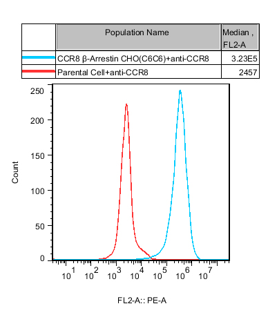
Figure 1. Recombinant CCR8 β-Arrestin/CHO-K1 stably expressing CCR8
Cells were stained with anti-CCR8 antibody. The blue curve represents CCR8 β-Arrestin/CHO-K1 cells with median fluorescence intensity (MFI) of 3.23E5; the red curve corresponds to parental CHO-K1 control cells with median fluorescence intensity of 2457. The obvious separation of two peaks demonstrates high and specific expression of CCR8 receptor on the constructed stable cell line.
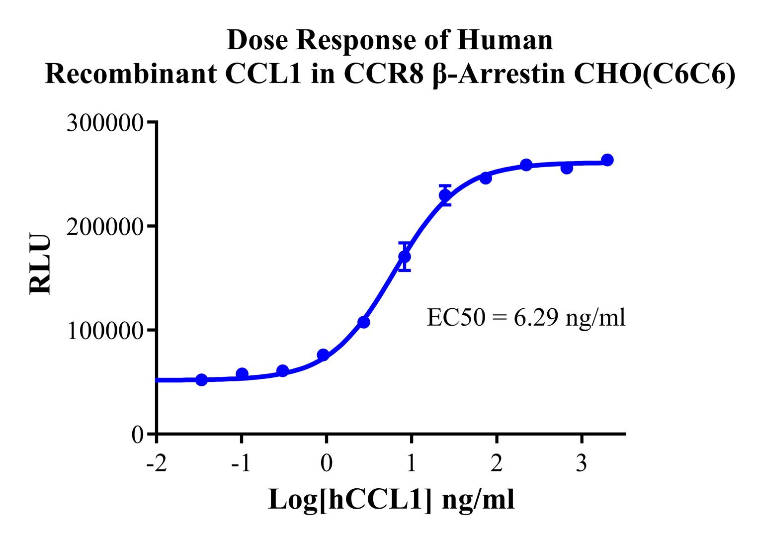
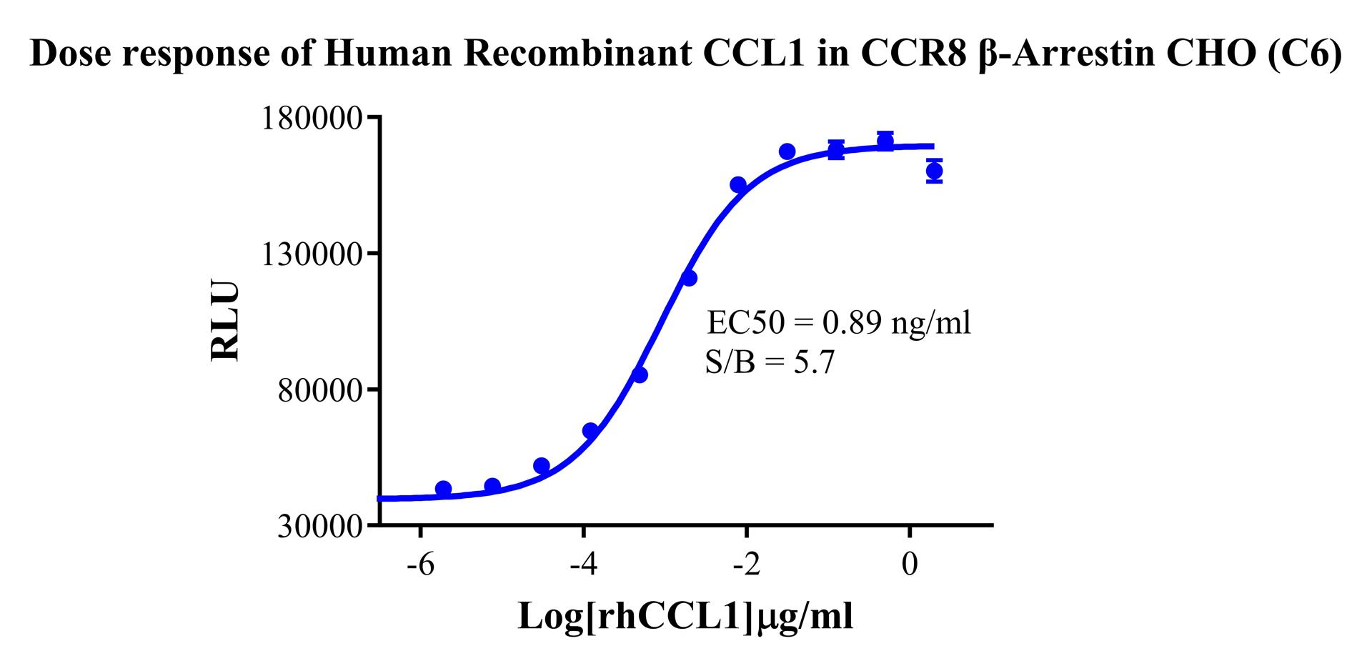
Figure 2. Dose response of Human Recombinant CCL1 in CCR8 β-Arrestin
Recombinant human CCL1 was applied as agonist to trigger receptor-mediated β-arrestin recruitment signal. The dose-response curve was plotted with RLU value against log concentration of hCCL1. The EC50 value of hCCL1 on this cell line was determined as 6.29 ng/ml, verifying that the expressed CCR8 receptor can effectively activate β-arrestin signaling pathway with good biological activity.
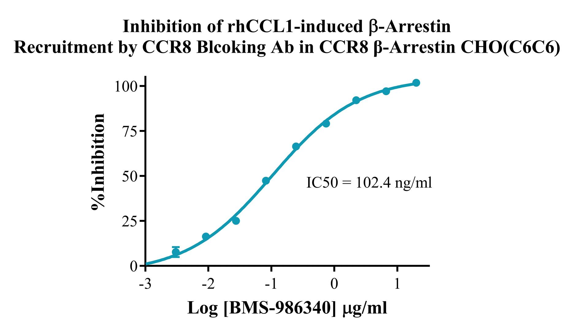
Figure 3. Inhibition of rhCCL1-induced β-Arrestin Recruitment by CCR8 Blocking Ab in CCR8 β-Arrestin CHO(C6C6)
The experiment detects the inhibitory effect of BMS-986340 on rhCCL1-triggered β-arrestin recruitment. The inhibition dose-response curve was plotted with percentage inhibition against log concentration of BMS-986340. The IC50 value was determined as 102.4 ng/ml. The result demonstrates that this cell line is suitable for antagonist and blocking antibody functional screening targeting human CCR8.
Cell Resuscitation
1)Rapidly thaw the frozen cells in a 37 °C water bath for approximately 60 seconds. Once thawed (which may take slightly less or more than 60 seconds), immediately transfer the cell suspension from the cryovial into a 15 mL centrifuge tube containing 10 mL of pre-warmed CHO-K1 Human CCR8 β-Arrestin Cell Line complete culture medium.
2)Centrifuge cells at 1000 rpm for 5 min to remove medium, then resuspend cells in 5 mL of pre-warmed complete medium.
3)Transfer the cell suspension into a T25 culture flask and incubate at 37 °C with 5% CO₂.
4)After approximately 24–36 hours, replace the medium or passage the cells to remove non-adherent dead cells.
Subculturing procedure
1)When the cell density reaches the appropriate confluency for passaging, wash the cells with PBS, then add 1 mL trypsin to detach the cells. When more than 80% of the cells detach upon gently tapping the culture flask, add complete culture medium to terminate digestion. Gently pipette to obtain a single-cell suspension, transfer to a 15 mL centrifuge tube, and centrifuge at 1000 rpm for 5 minutes.
2)Discard supernatant after centrifugation. Resuspend cells in fresh medium to a single-cell suspension and transfer to a new culture flask for continued growth.
Cell Freezing
After trypsinization and centrifugation of cells from each T75 flask or 10 cm culture dish, discard the supernatant. Add 2 mL of cryopreservation medium (90% FBS + 10% DMSO), gently resuspend thoroughly, and aliquot into two cryovials. Immediately place the cryovials into a controlled-rate freezing container (e.g., Nalgene 5100-0001), fill with isopropanol to the indicated level, and store at −80 °C. After 24 hours, transfer the cryovials to liquid nitrogen for long-term storage.
Related products
CHO-K1 Human NTSR2 Cell Line
CHO-K1 Human CCR4 Cell Line
HEK293 Human NK1R CRE-Luc Cell Line
Raji-Luc-GFP
Jurkat E6.1-Luc
THP-1-GFP
Raji-GFP
Raji-Luc
Jurkat E6.1-GFP
HEK293 Human GAL4-Luc Cell
We Are Pleased to Announce: Global Commercial Licensing Rights for Jurkat E6.1, CHO-K1, HEK293, THP-1 and RAJI Cell Lines Officially Secured.
Explore