
CHO-K1 Human CCR2 β-Arrestin Cell Line
Cat. No: RQP71394
Size: 1 vial of frozen cells (>1E6 per vial in 1 mL)
Unit Price: Contact For Pricing
Description
Product Info
Biological Information
Assay Data
Cell Culture
This product is a CHO-K1 stable cell line that stably expresses human CCR2 receptor for β-arrestin recruitment assay. Upon agonist binding to CCR2, β-arrestin is recruited to the activated receptor. The receptor activity can be quantified in live cells by measuring luminescence signal in a dose-response manner. This cell line is suitable for high-throughput screening (HTS) and receptor functional evaluation assays.
| Cat. No | RQP71394 |
| Product Name | CHO-K1 Human CCR2 β-Arrestin Cell Line |
| Engineering method | Plasmid and lentivirus |
| Culture Properties | Adherent |
| Stability | 32passages (in-house test, that not means the cell line will be instable beyond the passages we tested.) |
| Mycoplasma Status | Negative |
| Culture Medium | F12k+10%FBS+5μg/ml puromycin+5μg/ml blasticidin |
| Freeze Medium | 90% FBS+10% DMSO |
| Storage Conditions | Liquid nitrogen immediately upon delivery |
| Transducer | β-Arrestin |
| Application | Functional assay for CCR2 |
| Safety Precautions |
Wear lab coat, disposable gloves to avoid direct sample contact. For spills: apply disinfectant, allow full inactivation, then clean up. |
For research use only. Not intended for human or animal clinical trials, therapeutic or diagnostic use.
| Target Class | GPCR |
| Family | C-C motif chemokine receptors |
| Sub Family | Class A(Rhodopsin) |
| Gene Name | CCR2 |
| Gene Aliases | CC-CKR-2;CKR2;MCP-1-R;CD192;FLJ78302; |
| Gene ID | 729230 |
| Accession Number | NM_001123396 |
| UniProt Number | P41597 |
| Protein Name | C-C chemokine receptor type 2 |
| Protein Aliases | C-C CKR-2; CC-CKR-2; CCR-2; CCR2 |
| Target Species | Human |
| Host cell | CHO-K1 |
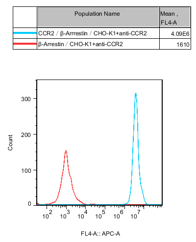
Figure 1. Recombinant CCR2/β-Arrestin/CHO-K1 constitutively expressing CCR2
Flow cytometry analysis of human CCR2 expression on CCR2/β-Arrestin CHO-K1 stable cell line. The CCR2/β-Arrestin/CHO-K1 cells (blue curve) exhibited high fluorescence intensity with Mean FL4-A = 4.09E6, while the β-Arrestin/CHO-K1 control cells (red curve) showed minimal signal (Mean FL4-A = 1610). The result verifies robust and stable membrane expression of human CCR2 receptor.
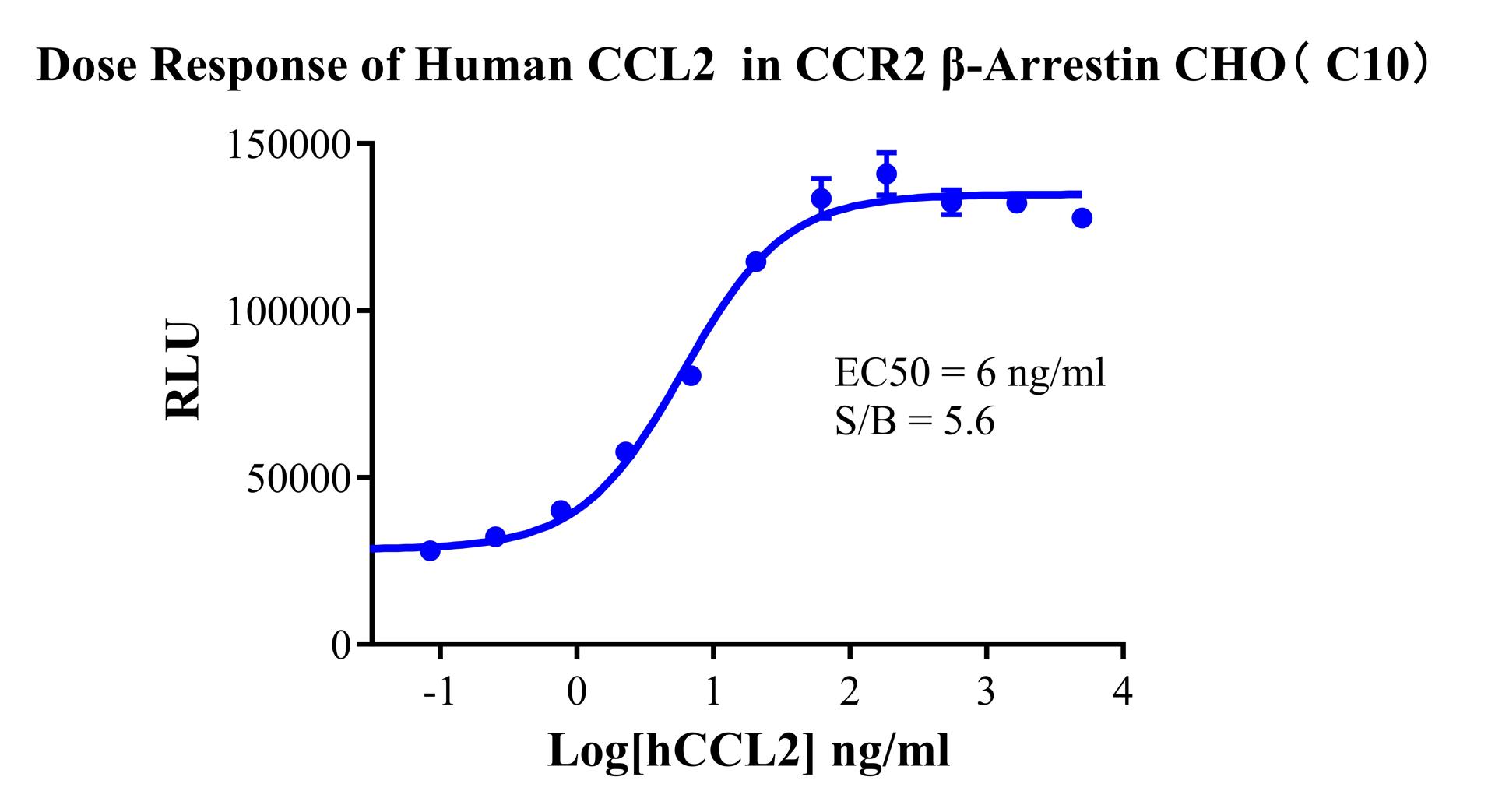
Figure 2. Dose response of Human CCL2 in CCR2 β-Arrestin CHO-K1(C10)
The CCR2 β-Arrestin/CHO-K1 (C10) stable cell line was functionally characterized by β-Arrestin recruitment luminescence assay. Human CCL2 induced a robust dose-dependent response with EC50 = 6 ng/ml and signal-to-background (S/B) ratio of 5.6, verifying specific β-Arrestin recruitment activity of human CCR2 receptor, suitable for high-throughput screening and pharmacological evaluation.
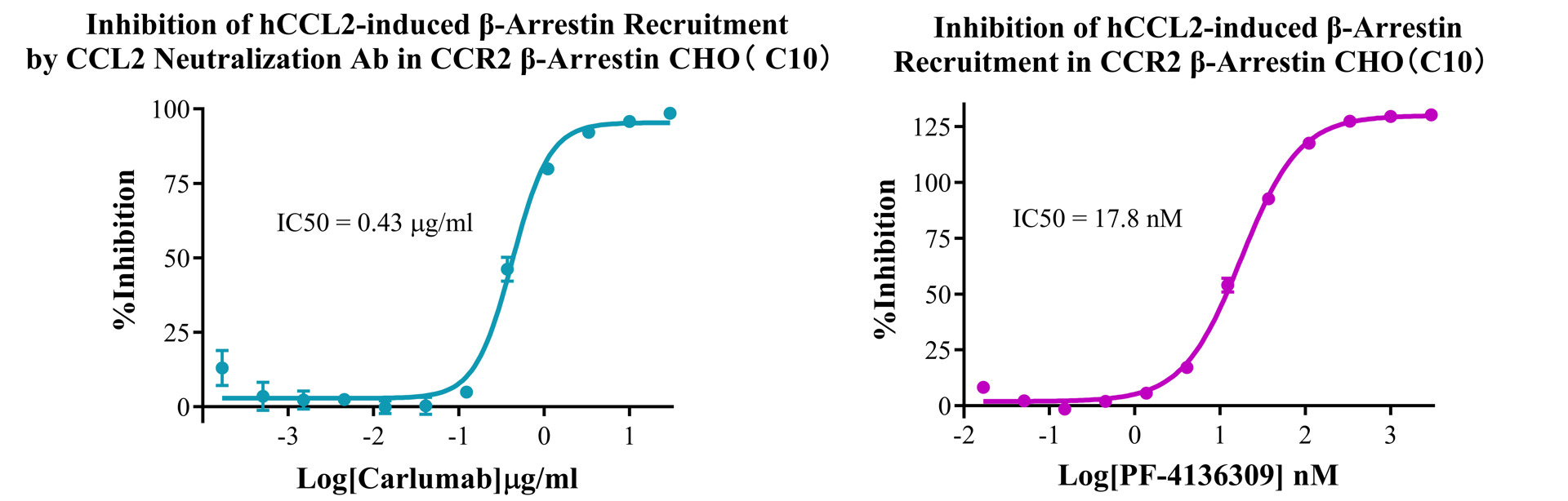
Figure 3. Dual inhibition curves of hCCL2-induced β-Arrestin recruitment in CCR2 β-Arrestin CHO-K1 (C10) cell line
Left: CCL2 neutralizing antibody (Carlumab) exhibited inhibitory activity with IC50 = 0.43 μg/ml. Right: Small molecule antagonist PF-4136309 showed potent inhibition with IC50 = 17.8 nM. This assay system is suitable for screening and potency evaluation of CCR2 antagonists and neutralizing antibodies.
Cell Resuscitation
1)Rapidly thaw the frozen cells in a 37 °C water bath for approximately 60 seconds. Once thawed (which may take slightly less or more than 60 seconds), immediately transfer the cell suspension from the cryovial into a 15 mL centrifuge tube containing 10 mL of pre-warmed CHO-K1 Human CCR2 β-Arrestin Cell Line complete culture medium.
2)Centrifuge cells at 1000 rpm for 5 min to remove medium, then resuspend cells in 5 mL of pre-warmed complete medium.
3)Transfer the cell suspension into a T25 culture flask and incubate at 37 °C with 5% CO₂.
4)After approximately 24–36 hours, replace the medium or passage the cells to remove non-adherent dead cells.
Subculturing procedure
1)When the cell density reaches the appropriate confluency for passaging, wash the cells with PBS, then add 1 mL trypsin to detach the cells. When more than 80% of the cells detach upon gently tapping the culture flask, add complete culture medium to terminate digestion. Gently pipette to obtain a single-cell suspension, transfer to a 15 mL centrifuge tube, and centrifuge at 1000 rpm for 5 minutes.
2)Discard supernatant after centrifugation. Resuspend cells in fresh medium to a single-cell suspension and transfer to a new culture flask for continued growth.
Cell Freezing
After trypsinization and centrifugation of cells from each T75 flask or 10 cm culture dish, discard the supernatant. Add 2 mL of cryopreservation medium (90% FBS + 10% DMSO), gently resuspend thoroughly, and aliquot into two cryovials. Immediately place the cryovials into a controlled-rate freezing container (e.g., Nalgene 5100-0001), fill with isopropanol to the indicated level, and store at −80 °C. After 24 hours, transfer the cryovials to liquid nitrogen for long-term storage.
Related products
CHO-K1 Human NTSR2 Cell Line
CHO-K1 Human CCR4 Cell Line
HEK293 Human NK1R CRE-Luc Cell Line
Raji-Luc-GFP
Jurkat E6.1-Luc
THP-1-GFP
Raji-GFP
Raji-Luc
Jurkat E6.1-GFP
HEK293 Human GAL4-Luc Cell
We Are Pleased to Announce: Global Commercial Licensing Rights for Jurkat E6.1, CHO-K1, HEK293, THP-1 and RAJI Cell Lines Officially Secured.
Explore