

CHO-K1 Canine PDL1 aAPC Cell
Cat. No: RQP74331
Size: 1 vial of frozen cells (>1E6 per vial in 1 mL)
Unit Price: Contact For Pricing
Product Info
Description
Biological Information
Assay Data
Cell Culture
| Cat. No | RQP74331 |
| Product Name | CHO-K1 Canine PDL1 aAPC Cell |
| Product Type | Reporter Cell |
| Culture Properties | Adherent |
| Stability | 32passages (in-house test, that not means the cell line will be instable beyond the passages we tested.) |
| Mycoplasma Status | Negative |
| Culture Medium | F12K+10%FBS+3μg/ml puromycin+600μg/ml Hygromycin B |
| Freeze Medium | 90% FBS+10% DMSO |
| Storage Conditions | Liquid nitrogen immediately upon delivery |
| Application | Functional(Report Gene) Assay |
For research use only. Not intended for human or animal clinical trials, therapeutic or diagnostic use.
Programmed Cell Death Protein 1 (PD-1), a receptor expressed on activated T cells, binds to its ligands—PD-L1 and PD-L2—to negatively regulate immune responses. PD-1 ligands are present in most cancers; the PD-1:PD-L1/2 interaction suppresses T-cell activity and enables cancer cells to evade immune surveillance. The PD-1/PD-L1 signaling pathway constitutes a critical component of tumor-induced immunosuppression, inhibiting T-lymphocyte activation and enhancing immune tolerance toward tumor cells, thereby facilitating tumor immune evasion. The binding of PD-1 to PD-L1 attenuates T-cell-mediated immune surveillance, leading to an impaired immune response or even T-cell apoptosis. PD-1/PD-L1 inhibitors can relieve the immunosuppression of anti-tumor T cells, thereby promoting T-cell proliferation, infiltration into the tumor microenvironment, and the induction of anti-tumor responses. The PD-1:PD-L1/2 pathway also plays a role in regulating autoimmune responses, making these proteins promising therapeutic targets for various cancers, as well as for conditions such as multiple sclerosis, arthritis, lupus, and Type 1 diabetes. The tyrosine phosphatase SHP2 is a key regulator of T-cell function, mediating both the activating signals downstream of the T-cell receptor (TCR) and the inhibitory signals downstream of PD-1.
The CHO-K1 Canine PDL1 aAPC Cell Model—effectively simulates the signal transduction process of PD1&PDL1 *in vivo*. The underlying principle is illustrated in the figure below.
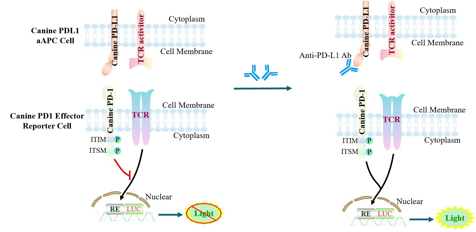
Figure 1. Schematic Diagram of the CHO-K1 Canine PDL1 aAPC Cell Model
| Classification | Co-Inhibitory |
| Family | B7 family |
| Gene Name | CD274 |
| Gene Aliases | N/A |
| Gene ID | 716043 |
| Accession Number | NM_001083889.1 |
| UniProt Number | A4GW29 |
| Protein Name | Programmed cell death 1 ligand 1 |
| Protein Aliases | B7 homolog 1 |
| Target Species | Canine |
| Host cell | CHO-K1 |
CHO cell line expressing full length PDL1. Expression is confirmed by flow cytometry.
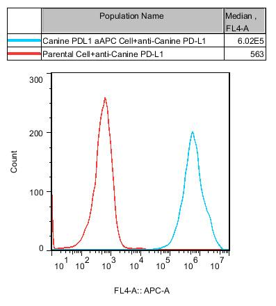
Figure 2. Recombinant Canine PDL1 aAPC Cell stably expressing Canine PDL1.
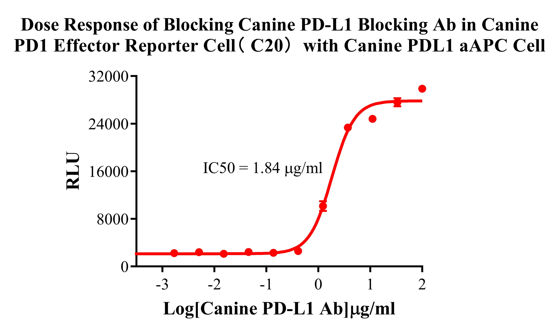
Figure 3. Dose Response of Blocking Canine PD-L1 Blocking Ab in Canine PD1 Effector Reporter Cell( C20) with Canine PDLl aAPC Cell.
Cell Resuscitation
1)Rapidly thaw the frozen cells in a 37 °C water bath for approximately 60 seconds. Once thawed (which may take slightly less or more than 60 seconds), immediately transfer the cell suspension from the cryovial into a 15 mL centrifuge tube containing 10 mL of pre-warmed CHO-K1 Canine PDL1 aAPC Cell complete culture medium.
2)Centrifuge cells at 1000 rpm for 5 min to remove medium, then resuspend cells in 5 mL of pre-warmed complete medium.
3)Transfer the cell suspension into a T25 culture flask and incubate at 37 °C with 5% CO₂.
4)After approximately 24–36 hours, replace the medium or passage the cells to remove non-adherent dead cells.
Subculturing procedure
1)When the cell density reaches the appropriate confluency for passaging, wash the cells with PBS, then add 1 mL trypsin to detach the cells. When more than 80% of the cells detach upon gently tapping the culture flask, add complete culture medium to terminate digestion. Gently pipette to obtain a single-cell suspension, transfer to a 15 mL centrifuge tube, and centrifuge at 1000 rpm for 5 minutes.
2)Discard supernatant after centrifugation. Resuspend cells in fresh medium to a single-cell suspension and transfer to a new culture flask for continued growth.
Cell Freezing
After trypsinization and centrifugation of cells from each T75 flask or 10 cm culture dish, discard the supernatant. Add 2 mL of cryopreservation medium (90% FBS + 10% DMSO), gently resuspend thoroughly, and aliquot into two cryovials. Immediately place the cryovials into a controlled-rate freezing container (e.g., Nalgene 5100-0001), fill with isopropanol to the indicated level, and store at −80 °C. After 24 hours, transfer the cryovials to liquid nitrogen for long-term storage.
Related products
CHO-K1 Human CCR4 Cell Line
HEK293 Human NK1R CRE-Luc Cell Line
Raji-Luc-GFP
Jurkat E6.1-Luc
THP-1-GFP
THP-1-Luc
Raji-GFP
Raji-Luc
Jurkat E6.1-GFP
HEK293 Human GAL4-Luc Cell
We Are Pleased to Announce: Global Commercial Licensing Rights for Jurkat E6.1, CHO-K1, and HEK293 Cell Lines Officially Secured.
Explore