

Raji Human TIM-3 Target cell
Cat. No: RQP74238
Size: 1 vial of frozen cells (>1E6 per vial in 1 mL)
Unit Price: Contact For Pricing
Product Info
Description
Biological Information
Assay Data
Cell Culture
| Cat. No | RQP74238 |
| Product Name | Raji Human TIM-3 Target Cell |
| Culture Properties | Suspension |
| Stability | 32passages (in-house test, that not means the cell line will be instable beyond the passages we tested.) |
| Mycoplasma Status | Negative |
| Culture Medium | RPMI-1640+10%FBS |
| Freeze Medium | 90% FBS+10% DMSO |
| Storage Conditions | Liquid nitrogen immediately upon delivery |
| Application | Binding Assay,immunogen, MOA research and so on |
For research use only. Not intended for human or animal clinical trials, therapeutic or diagnostic use.
T-cell immunoglobulin and mucin domain-containing protein 3 (TIM-3) is a cell-surface molecule containing both immunoglobulin and mucin domains. Originally identified as a cell-surface marker for IFN-γ-producing CD4 T helper 1 (Th1) and CD8 cytotoxic 1 (Tc1) cells, it serves as a critical immune checkpoint receptor. TIM-3 is primarily expressed on activated CD8+ T cells, exhausted T cells, Th1 cells, and innate immune cells (such as dendritic cells and macrophages). The TIM protein family consists of Type I transmembrane proteins sharing a similar structural architecture: a variable immunoglobulin (IgV) domain, a glycosylated mucin domain of varying length, and a single transmembrane domain. With the exception of TIM-4, all TIM molecules possess a C-terminal cytoplasmic tail containing conserved tyrosine-based signaling motifs.
According to the current model of TIM-3 signaling, upon T-cell activation, TIM-3 is recruited to the immunological synapse. At this site, Bat3 binds to the cytoplasmic tail of TIM-3 and recruits the catalytically active form of Lymphocyte-specific protein tyrosine kinase (Lck). However, when TIM-3 engages with its ligand, conserved tyrosine residues within its cytoplasmic tail undergo phosphorylation; this event triggers the dissociation of Bat3, thereby enabling TIM-3 to exert its inhibitory function.
The TIM-3 Target Cell serves as the target component for the TIM-3 Effector Reporter Cell system. This reporter gene-based drug target model effectively recapitulates the in vivo signaling pathway of TIM-3; the underlying principle is illustrated in the figure below.
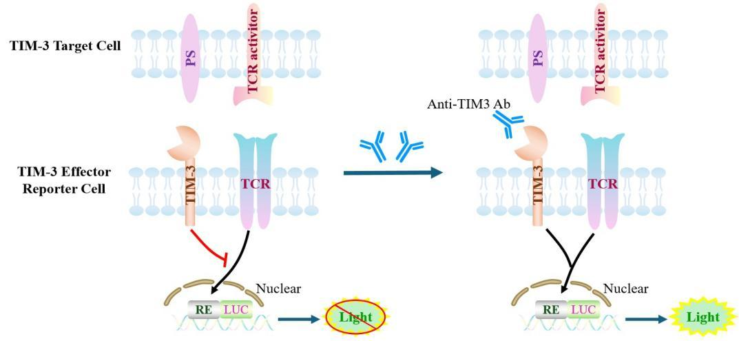
Figure 1. Schematic Diagram of the Raji Human TIM-3 Target Cell Model
| Classification | Co-Inhibitory |
| Family | immunoglobulin superfamily. TIM family |
| Gene Name | TIM-3 |
| Gene Aliases | Tim-3;TIM3;FLJ14428;TIMD3;CD366; |
| Gene ID | 84868 |
| Accession Number | NM_032782 |
| UniProt Number | Q8TDQ0 |
| Protein Name | HAVcr-2 |
| Protein Aliases | T-cell immunoglobulin and mucin domain-containing protein 3 (TIMD-3);T-cell immunoglobulin mucin receptor 3 (TIM-3);T-cell membrane protein 3 |
| Target Species | Human |
| Host cell | Raji |
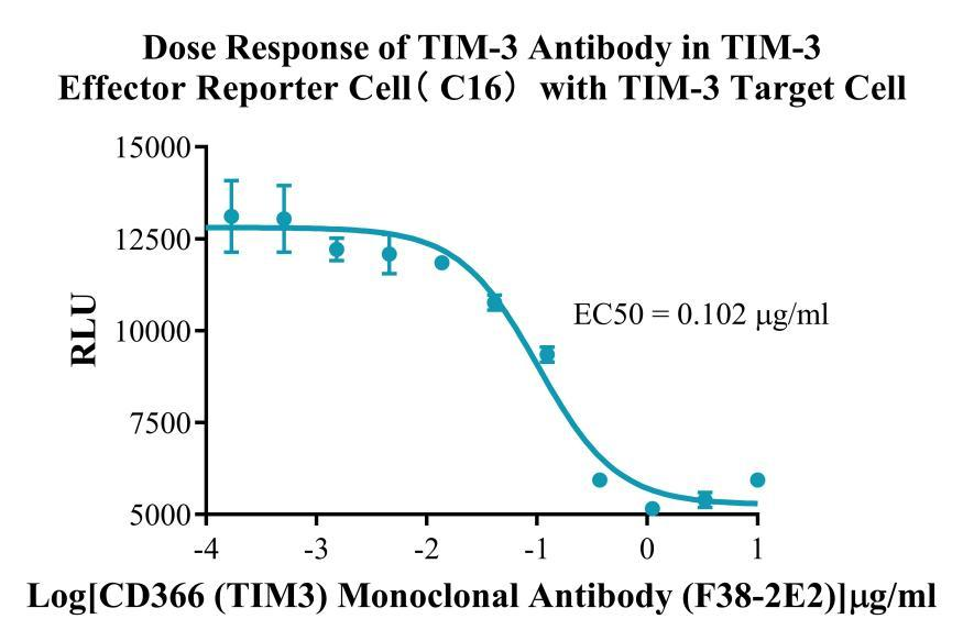
Figure 2. Dose Response of TIM-3 Antibody in TIM-3 Effector Reporter Cell(C16)with TIM-3 Target Cell.
Cell Passage Procedures
1.This cell line grows in suspension.
2.Upon receipt, cells should be thawed immediately or stored in liquid nitrogen until use.
3.Before thawing, pre-warm the water bath and culture medium to 37 °C, and prepare a small amount of dry ice.
4.Remove the cryovial from storage and transport it to the cell culture laboratory on dry ice.
5.Rapidly thaw the cells in a 37 °C water bath. Once the cells are completely thawed, spray the cryovial with 70% ethanol for disinfection and transfer it to a biosafety cabinet.
6.Add 10 mL of pre-warmed culture medium into a 15 mL centrifuge tube. Transfer the contents of the cryovial into the tube and centrifuge at 1000 rpm for 5 minutes.
7.Carefully discard the supernatant. Resuspend the cell pellet in 5 mL of pre-warmed culture medium by gentle pipetting. Immediately perform cell counting and adjust the cell density to 3–6 × 10⁵ cells/mL based on the counting results, then transfer the cells into a culture flask.
8.Count the cells every 1–2 days. When the cell density exceeds 1 × 10⁶ cells/mL, passage the cells promptly or add fresh culture medium. Maintain the cell density between 2 × 10⁵ and 1 × 10⁶ cells/mL.
Suspension Cell Cryopreservation Procedure:
1.Collect 8 × 10⁶ cells, centrifuge, and discard the supernatant.
2.Add 1 mL of cell freezing medium (90% FBS + 10% DMSO) and gently pipette to mix thoroughly. Transfer the suspension into a cryovial.
3.Immediately place the cryovial into a controlled-rate freezing container (Nalgene 5100-0001), fill with isopropanol up to the indicated level, and store at −80 °C.
4.After 24 hours, transfer the cryovial to liquid nitrogen for long-term storage.
Related products
CHO-K1 Human NTSR2 Cell Line
CHO-K1 Human CCR4 Cell Line
HEK293 Human NK1R CRE-Luc Cell Line
Raji-Luc-GFP
Jurkat E6.1-Luc
We Are Pleased to Announce: Global Commercial Licensing Rights for Jurkat E6.1, CHO-K1, HEK293, THP-1 and RAJI Cell Lines Officially Secured.
ExploreWe Are Pleased to Announce: Global Commercial Licensing Rights for Jurkat E6.1, CHO-K1, HEK293, THP-1 and RAJI Cell Lines Officially Secured.
Explore