
Jurkat E6.1 Human ADCC Bioassay Effector Cell F variant (Low Affinity)- Fcγ-NFAT Cell(CD3E KO)
Cat. No: RQP74626
Size: 1 vial of frozen cells (>1E6 per vial in 1 mL)
Unit Price: Contact For Pricing
Description
Product Info
Assay Data
Cell Culture
Antibody-dependent cell-mediated cytotoxicity (ADCC) is a process in which the Fab region of an antibody binds to epitopes on virus-infected cells or tumor cells, while its Fc region binds to Fc receptors (FcRs) on the surface of effector cells (such as NK cells and macrophages), thereby mediating the direct killing of target cells by these effector cells; this constitutes a crucial mechanism of action for therapeutic anti-tumor antibodies.
Traditional methods for assessing ADCC and ADCP (antibody-dependent cellular phagocytosis) rely primarily on the isolation and *in vitro* differentiation of relevant primary immune cells, followed by the measurement of target cell killing or phagocytosis. These methods are heavily dependent on primary cells from donors, making them costly and labor-intensive. The isolation and differentiation processes demand high technical proficiency, carry a significant risk of experimental failure, and—due to issues regarding cell purity—often result in weak, unstable, or inconsistent detection signals. Furthermore, primary cells are non-renewable; variations in donor sources across experiments can lead to poor reproducibility and batch-to-batch inconsistency. Consequently, establishing stable and reliable assay methods is challenging within the context of drug development, where rigorous quality control is essential.
The Jurkat E6.1 Human ADCC Bioassay Effector Cell F variant (Low Affinity)- Fcγ-NFAT Cell(CD3E KO) effectively simulates the in vivo ADCC signal transduction process; the underlying mechanism is illustrated in the figure below.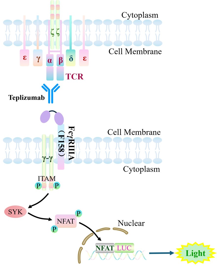
Figure 1. Schematic diagram of the Jurkat E6.1 Human ADCC Bioassay Effector Cell F variant (Low Affinity)- Fcγ-NFAT Cell(CD3E KO).
| Cat. No | RQP74626 |
| Product Name | Jurkat E6.1 Human ADCC Bioassay Effector Cell F variant (Low Affinity)- Fcγ-NFAT Cell(CD3E KO) |
| Culture Properties | suspension |
| Stability | 32passages (in-house test, that not means the cell line will be instable beyond the passages we tested.) |
| Mycoplasma Status | Negative |
| Culture Medium | RPMI-1640+10%FBS +400 μg/ml Hygromycin B+1 μg/ml Puromycin+10 μg/ml Blasticidin |
| Freeze Medium | 90% FBS+10% DMSO |
| Storage Conditions | Liquid nitrogen immediately upon delivery |
| Application | Functional(Report Gene) Assay |
For research use only. Not intended for human or animal clinical trials, therapeutic or diagnostic use.
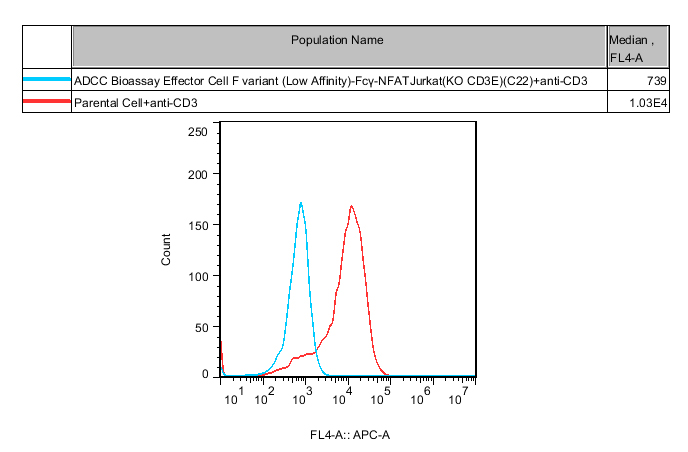
Figure 2. Flow cytometric verification of CD3 expression in ADCC Bioassay Effector Cell F variant (Low Affinity) – Fcγ-NFAT Jurkat (CD3E KO) cells.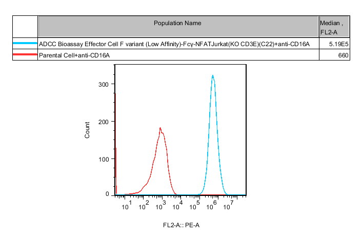
Figure 3. Flow cytometric validation of CD16A expression in ADCC Bioassay Effector Cell F variant (Low Affinity) – Fcγ-NFAT Jurkat (CD3E KO) cells.
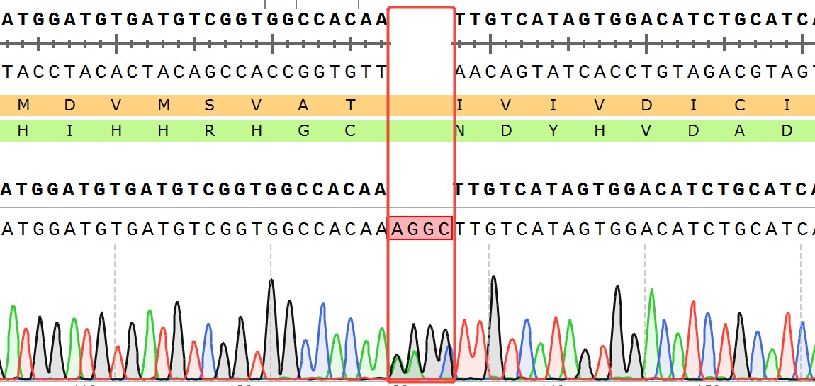
Figure 4. CD3E(NM_000733.4):c.397_398ins4bp/CD3E:p.I133Kfs
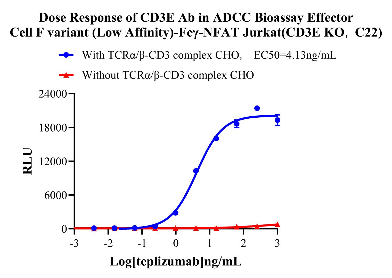
Figure 5. Dose Response of CD3E Ab in ADCC Bioassay Effector Cell F variant (Low Affinity)-Fcγ-NFAT Jurkat(CD3E KO,C22).
Cell Passage Procedures
1.This cell line grows in suspension.
2.Upon receipt, cells should be thawed immediately or stored in liquid nitrogen until use.
3.Before thawing, pre-warm the water bath and culture medium to 37 °C, and prepare a small amount of dry ice.
4.Remove the cryovial from storage and transport it to the cell culture laboratory on dry ice.
5.Rapidly thaw the cells in a 37 °C water bath. Once the cells are completely thawed, spray the cryovial with 70% ethanol for disinfection and transfer it to a biosafety cabinet.
6.Add 10 mL of pre-warmed culture medium into a 15 mL centrifuge tube. Transfer the contents of the cryovial into the tube and centrifuge at 1000 rpm for 5 minutes.
7.Carefully discard the supernatant. Resuspend the cell pellet in 5 mL of pre-warmed culture medium by gentle pipetting. Immediately perform cell counting and adjust the cell density to 3–6 × 10⁵ cells/mL based on the counting results, then transfer the cells into a culture flask.
8.Count the cells every 1–2 days. When the cell density exceeds 1 × 10⁶ cells/mL, passage the cells promptly or add fresh culture medium. Maintain the cell density between 2 × 10⁵ and 1 × 10⁶ cells/mL.
Suspension Cell Cryopreservation Procedure:
1.Collect 8 × 10⁶ cells, centrifuge, and discard the supernatant.
2.Add 1 mL of cell freezing medium (90% FBS + 10% DMSO) and gently pipette to mix thoroughly. Transfer the suspension into a cryovial.
3.Immediately place the cryovial into a controlled-rate freezing container (Nalgene 5100-0001), fill with isopropanol up to the indicated level, and store at −80 °C.
4.After 24 hours, transfer the cryovial to liquid nitrogen for long-term storage.
Related products
CHO-K1 Human NTSR2 Cell Line
CHO-K1 Human CCR4 Cell Line
HEK293 Human NK1R CRE-Luc Cell Line
Raji-Luc-GFP
Jurkat E6.1-Luc
We Are Pleased to Announce: Global Commercial Licensing Rights for Jurkat E6.1, CHO-K1, HEK293, THP-1 and RAJI Cell Lines Officially Secured.
ExploreWe Are Pleased to Announce: Global Commercial Licensing Rights for Jurkat E6.1, CHO-K1, HEK293, THP-1 and RAJI Cell Lines Officially Secured.
Explore