
CHO-K1 Human XCR1 β-Arrestin Cell Line
Cat. No: RQP71413
Size: 1 vial of frozen cells (>1E6 per vial in 1 mL)
Unit Price: Contact For Pricing
Product Info
Description
Biological Information
Assay Data
Cell Culture
| Cat. No | RQP71413 |
| Product Name | CHO-K1 Human XCR1 β-Arrestin Cell Line |
| Engineering method | plasmid and lentivirus |
| Culture Properties | Adherent |
| Stability | 32passages (in-house test, that not means the cell line will be instable beyond the passages we tested.) |
| Mycoplasma Status | Negative |
| Culture Medium | F12K+10%FBS+5μg/ml Puromycin+5μg/ml Blasticidin |
| Freeze Medium | 90% FBS+10% DMSO |
| Storage Conditions | Liquid nitrogen immediately upon delivery |
| Transducer | β-arrestin |
| Application | Functional assay for XCR1 |
For research use only. Not intended for human or animal clinical trials, therapeutic or diagnostic use.
XCR1 belongs to the C chemokine receptor family and is its sole member. Taxonomically, it belongs to the G protein-coupled receptor (GPCR) superfamily and possesses the typical seven-transmembrane helix structure of GPCRs. XCR1 exhibits high ligand specificity in ligand recognition; its only known endogenous ligands are XCL1 and XCL2 (formerly known as lymphokines), which are also the most structurally unique members of the chemokine family: containing only one disulfide bond. In terms of expression and distribution, XCR1 is the most specific lineage marker for type 1 conventional dendritic cells (cDC1) (corresponding to mouse CD8α+ DCs and human BDCA3+ DCs), and it also shows basal expression in the thymus, spleen, and placenta.
When the XCL1 ligand binds to the extracellular region of XCR1, it induces a conformational change in the receptor, preferentially coupling to Gαi proteins. This leads to the inhibition of downstream adenylate cyclase activity (reduced cAMP), while simultaneously activating phospholipase C (PLC), triggering the release of calcium ions from the endoplasmic reticulum, resulting in a rapid increase in intracellular Ca²⁺ concentration, and further cascading activation of signaling pathways such as MAPK and PI3K-AKT. Furthermore, XCR1 recruits cDC1 to T cell-rich areas via chemotaxis, and simultaneously takes up exogenous antigens through endocytosis. Under the maintenance of XCR1 signaling, it utilizes an efficient endosomal processing system to load antigens onto MHC-I molecules, thereby activating naive CD8⁺ cytotoxic T cells and natural killer (NK) cells.
The XCR1 β-Arrestin CHO reporter gene drug target model effectively simulates the in vivo XCR1/β-Arrestin signal transduction process, as shown in the figure below.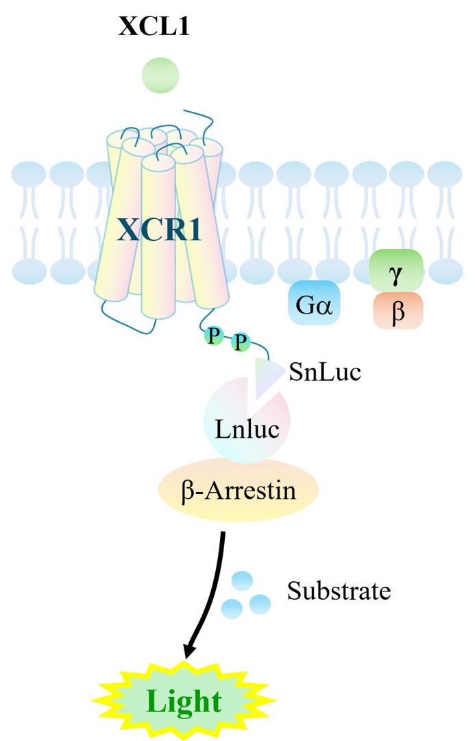
Figure 1. Schematic diagram of the XCR1 β-Arrestin CHO cell model
| Target Class | GPCR |
| Family | X-C motif chemokine receptors |
| Sub Family | Class B1(Secretin) |
| Gene Name | XCR1 |
| Gene Aliases | GPR5;CCXCR1 |
| Gene ID | 2829 |
| Accession Number | NM_001024644 |
| UniProt Number | P46094 |
| Protein Name | Chemokine XC receptor 1 |
| Protein Aliases | G-protein coupled receptor 5; Lymphotactin receptor; XC chemokine receptor 1 |
| Target Species | Human |
| Host cell | CHO-K1 |
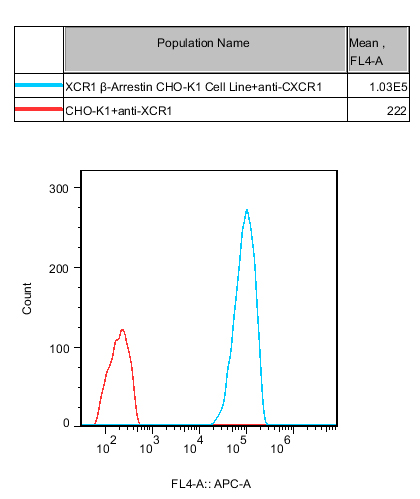
Figure 2.Recombinant XCR1/β-Arrestin/CHO-K1 constitutively expressing XCR1.
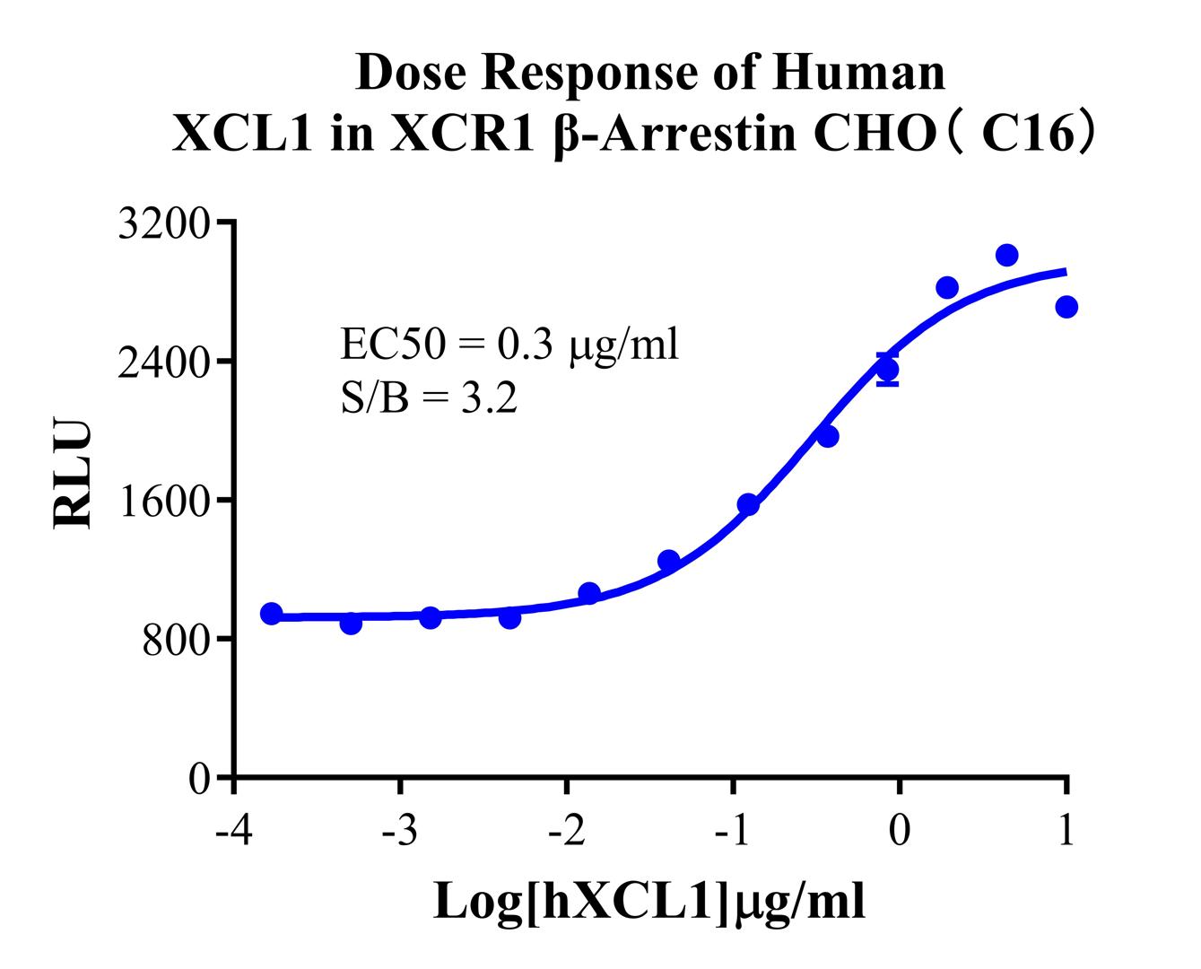
Figure 3. Dose response of Human XCL1 in XCR1 β-Arrestin CHO (C16).
Cell Resuscitation
1)Rapidly thaw the frozen cells in a 37 °C water bath for approximately 60 seconds. Once thawed (which may take slightly less or more than 60 seconds), immediately transfer the cell suspension from the cryovial into a 15 mL centrifuge tube containing 10 mL of pre-warmed CHO-K1 Human XCR1 β-Arrestin Cell Line complete culture medium.
2)Centrifuge cells at 1000 rpm for 5 min to remove medium, then resuspend cells in 5 mL of pre-warmed complete medium.
3)Transfer the cell suspension into a T25 culture flask and incubate at 37 °C with 5% CO₂.
4)After approximately 24–36 hours, replace the medium or passage the cells to remove non-adherent dead cells.
Subculturing procedure
1)When the cell density reaches the appropriate confluency for passaging, wash the cells with PBS, then add 1 mL trypsin to detach the cells. When more than 80% of the cells detach upon gently tapping the culture flask, add complete culture medium to terminate digestion. Gently pipette to obtain a single-cell suspension, transfer to a 15 mL centrifuge tube, and centrifuge at 1000 rpm for 5 minutes.
2)Discard supernatant after centrifugation. Resuspend cells in fresh medium to a single-cell suspension and transfer to a new culture flask for continued growth.
Cell Freezing
After trypsinization and centrifugation of cells from each T75 flask or 10 cm culture dish, discard the supernatant. Add 2 mL of cryopreservation medium (90% FBS + 10% DMSO), gently resuspend thoroughly, and aliquot into two cryovials. Immediately place the cryovials into a controlled-rate freezing container (e.g., Nalgene 5100-0001), fill with isopropanol to the indicated level, and store at −80 °C. After 24 hours, transfer the cryovials to liquid nitrogen for long-term storage.
Related products
CHO-K1 Human NTSR2 Cell Line
CHO-K1 Human CCR4 Cell Line
HEK293 Human NK1R CRE-Luc Cell Line
Raji-Luc-GFP
Jurkat E6.1-Luc
We Are Pleased to Announce: Global Commercial Licensing Rights for Jurkat E6.1, CHO-K1, HEK293, THP-1 and RAJI Cell Lines Officially Secured.
ExploreWe Are Pleased to Announce: Global Commercial Licensing Rights for Jurkat E6.1, CHO-K1, HEK293, THP-1 and RAJI Cell Lines Officially Secured.
Explore