

MRGPRX2: Mechanism, Therapeutics, and Cell Models
In the G protein-coupled receptor (GPCR) family, the Mas-related G protein-coupled receptors (MRGPRs) were long thought to be primarily involved in itch and pain perception. However, recent research has revealed that the MRGPR family—particularly MRGPRX2—plays a far more central role in immune regulation.
As a non-IgE-dependent activation receptor on mast cells, MRGPRX2 can be directly triggered by various cationic ligands (such as neuropeptides, antimicrobial peptides, and drug molecules). This activation induces rapid degranulation, mediating a range of allergic and inflammatory conditions, including chronic urticaria, atopic dermatitis, and drug-induced pseudo-allergies.
With pharmaceutical giants like Incyte and Novartis investing heavily in this space, MRGPRX2 has emerged as a high-profile target in the fields of autoimmunity and allergy. Leveraging our established GPCR drug screening platform, ReqBio has successfully developed a functional MRGPRX2 cell model, providing global researchers with a high-efficiency, reliable tool for early-stage drug discovery.
I. MRGPRX2: The "Rapid Response Force" for Non-IgE-Mediated Mast Cell Activation
The human MRGPR family comprises eight subtypes (MRGPRX1-X4 and MRGPRXD-XG), with MRGPRX2 standing out as the most extensively researched and clinically validated member. Structurally, it is a prototypical Class A GPCR, consisting of seven transmembrane domains, three extracellular loops, and three intracellular loops.
MRGPRX2 is primarily expressed on human skin mast cells. Its core function is to mediate an immediate activation pathway that is entirely independent of IgE. When the receptor is triggered by a diverse array of cationic ligands—including neuropeptides like Substance P, antimicrobial peptides like LL-37 and human β-defensins (hBDs), synthetic polymers such as Compound 48/80, and various drugs (e.g., Octreotide or Neuromuscular Blocking Agents)—it couples with Gαq proteins. This triggers a rapid surge in intracellular calcium levels (Ca²⁺), leading to mast cell degranulation within seconds to minutes. This process releases pre-stored mediators like histamine and tryptase, while also promoting the synthesis of new chemokines and cytokines.
While this rapid-response pathway is an evolutionary component of innate immune defense against pathogens, its overactivation or dysregulation is a direct driver of several conditions:
Chronic Spontaneous Urticaria (CSU): Wheals, hives, and intense pruritus.
Atopic Dermatitis (AD): Chronic skin inflammation and barrier dysfunction.
Drug-Induced Pseudo-Allergy: Immediate hypersensitivity reactions occurring without prior IgE sensitization.
Non-Histaminergic Pruritus: Itch sensations that do not respond to traditional antihistamines.
Allergic Contact Dermatitis: T-cell mediated inflammation exacerbated by mast cell signaling.
II. A Dual Role: Pathological Driver and Host Defender
Beyond its pathological implications, MRGPRX2 is also expressed in macrophages and plays a critical role in host defense. Small cationic antimicrobial peptides (AMPs) produced by epithelial cells, such as hBDs and LL-37, activate macrophages via MRGPRX2 to help clear cutaneous bacterial infections, promote wound healing, and prevent reinfection. Furthermore, bacterial quorum-sensing peptides have been shown to activate the murine ortholog, MRGPRB2, inhibiting bacterial growth and recruiting neutrophils to the site of infection.
This dual functionality suggests a nuanced therapeutic approach: while a complete knockout of MRGPRX2 might weaken innate immunity, selective antagonists offer the potential to alleviate allergic and inflammatory diseases without compromising essential host defense mechanisms. This balance has become the core strategic focus for drug development targeting MRGPRX2.
III. Drug Discovery Targeting MRGPRX2: Giants Compete as Clinical Validation Approaches
Currently, no MRGPRX2-targeted therapies have reached the market globally, but research momentum has intensified significantly. Between 2024 and 2025, a series of high-profile acquisitions and clinical milestones have thrust this target into the global spotlight:
|
Drug |
Developer |
Progress |
Significance |
|
EP262(Oral Small Molecule) |
Incyte (via acquisition of Escient) |
$750 million acquisition |
The first oral MRGPRX2 antagonist to enter clinical trials. |
|
KRP-M223 |
Novartis (Licensed from Kyorin) |
$55 million upfront; up to $777.5 million in milestones |
A preclinical small-molecule inhibitor; Novartis holds global rights. |
|
EVO-756 |
Evommune |
Phase 2 (Chronic Spontaneous Urticaria) |
Currently the fastest-progressing candidate; positive Phase 2 data already released. |
Table 1: Progress in MRGPRX2-Targeted Drug Development (2024–2025)
IV. Accelerating MRGPRX2 Drug Discovery: ReqBio’s Functional Cell Models and Technical Edge
In the landscape of GPCR-targeted drug discovery, stable and thoroughly validated cell models are the bedrock for identifying lead compounds and evaluating the efficacy of drug candidates. Recognizing the unique signaling profile of MRGPRX2, ReqBio has developed specialized functional cell models designed to sensitively detect downstream signals upon receptor activation.
ReqBio Recommended Cell Models for MRGPRX2:
|
Cell Line Name |
Catalog No. |
Host Cell |
Detection Mode |
Signaling Pathway |
Core Application |
|
HEK293 Human MRGPRX2 Gα15 Cell Line |
RQP71491 |
HEK293 |
Calcium Flux (FLIPR) or IP-One |
Gαq → Ca²⁺ / IP1 |
High-throughput screening (HTS) for agonists/antagonists. |
|
CHO-K1 Human MRGPRX2 β-Arrestin Receptor Cell |
RQP71585 |
CHO-K1 |
β-Arrestin Recruitment |
β-Arrestin Pathway |
Biased ligand screening; distinguishing between G-protein and β-arrestin pathways. |
Table 2: ReqBio MRGPRX2 Target Cell Models
Validation Data & Advantage Analysis
1. Calcium Flux Assay (FLIPR) Validation
HEK293 Human MRGPRX2 Gα15 Cell Line(RQP71491)
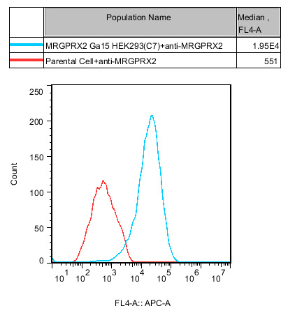
Figure 3. Recombinant MRGPRX2 Gα15 HEK293 stably expressing MRGPRX2.
Data Analysis:
This figure presents the results of a flow cytometry analysis, confirming the robust and stable expression of the MRGPRX2 receptor on the cell surface.
X-axis (FL4-A): Fluorescence intensity, representing the amount of anti-MRGPRX2 antibody bound to each cell, which serves as a direct indicator of MRGPRX2 protein expression levels.
Y-axis (Count): Number of cells.
Blue Peak: Represents the MRGPRX2 Gα15 HEK293 stable cell line. With a median fluorescence intensity (MFI) of 1.95E4, the peak shows a significant rightward shift, demonstrating that the vast majority of cells exhibit high MRGPRX2 expression.
Red Peak: Represents the Parental Cells (untransfected). The MFI is only 551, with the peak remaining on the left, indicating virtually no non-specific or endogenous signal.
Conclusion: The MRGPRX2 stable cell line was successfully established, demonstrating consistent and high-level surface expression of the MRGPRX2 protein.
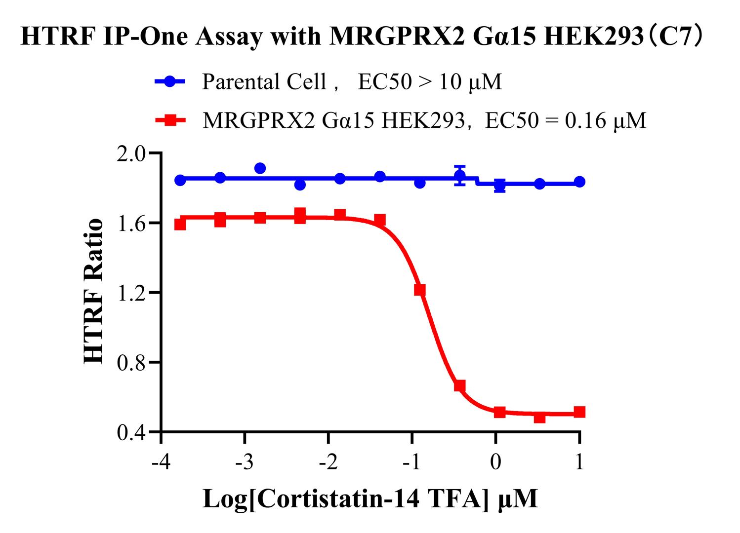
Figure 4. HTRF IP-One Assay with MRGPRX2 Gα15 HEK293(C7).
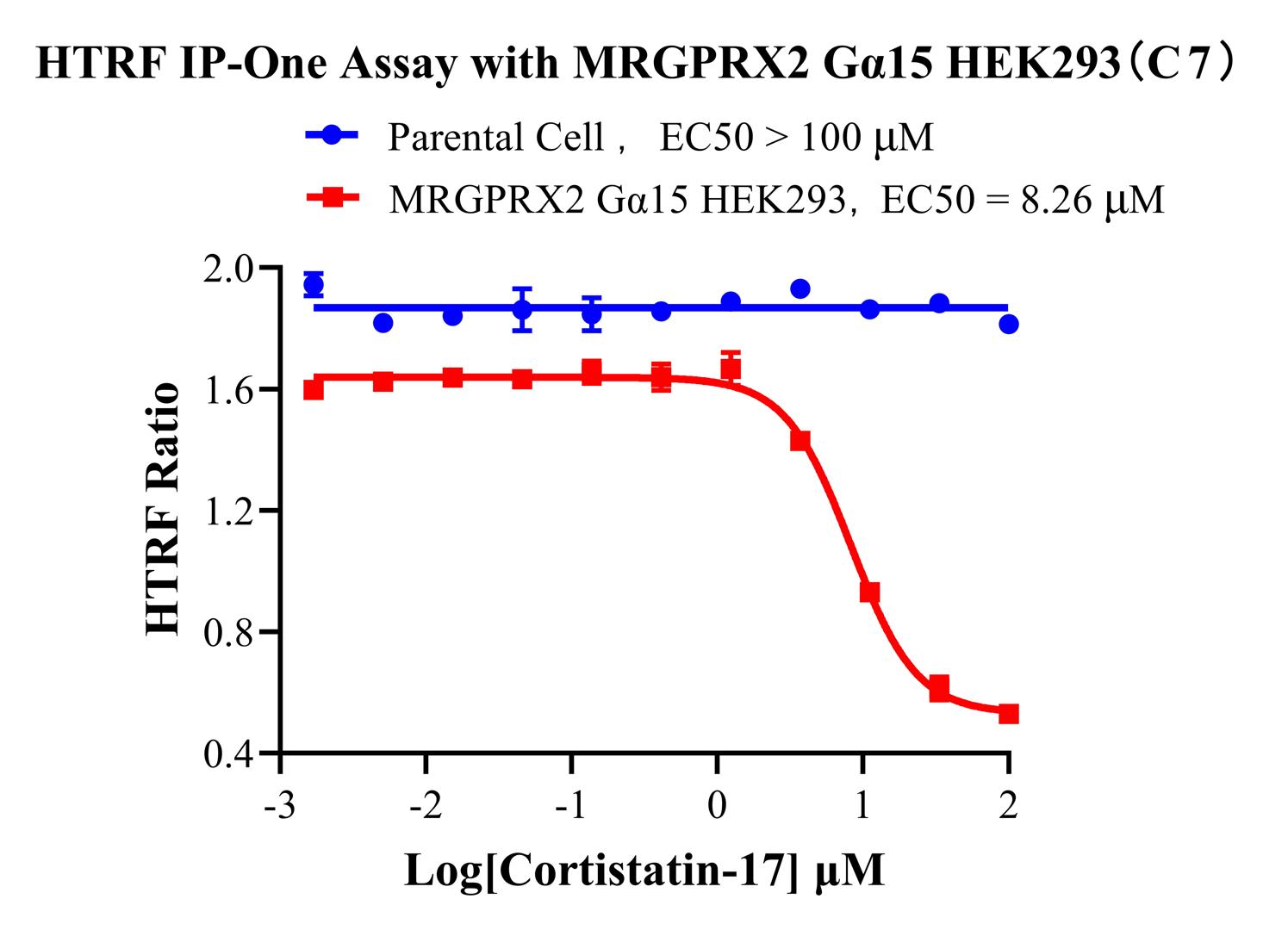
Figure 5. HTRF IP-One Assay with MRGPRX2 Gα15 HEK293(C7).
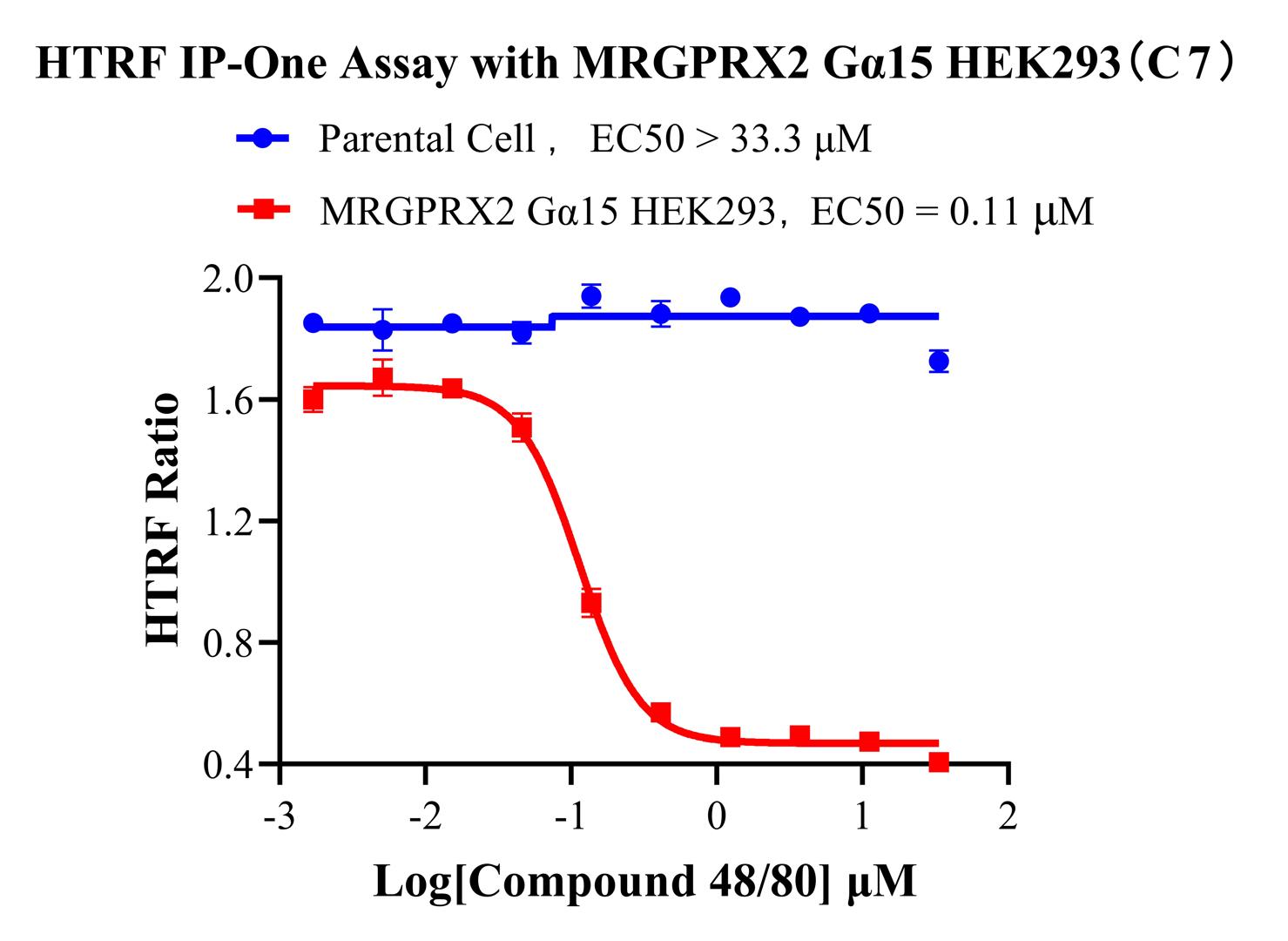
Figure 6. HTRF IP-One Assay with MRGPRX2 Gα15 HEK293(C7).
Data Analysis:
The following figures display the dose-response curves from the HTRF IP-One assay, which measures the downstream signaling (IP1 accumulation) following MRGPRX2 activation, reflecting the engagement of the Gq pathway.
X-axis: Logarithm of ligand concentration (Log [Compound], μM). Concentration increases from left to right.
Y-axis (HTRF Ratio): Measured IP1 signal. A lower ratio indicates stronger Gq pathway activation (i.e., higher IP1 accumulation).
Blue Curves (Parental Cells): Lack MRGPRX2 expression. The signal remains nearly flat despite increasing ligand concentration, confirming no non-specific activation.
Red Curves (MRGPRX2 Stable Cells): The signal decreases as ligand concentration increases, demonstrating that the response is specifically mediated through the activation of MRGPRX2.
EC50: The half-maximal effective concentration. A lower value indicates higher potency of the ligand in activating MRGPRX2.
Figure 4: Activation Curve of Cortistatin-14 TFA
MRGPRX2 Cells:EC50 = 0.16 μM
Parental Cells:EC50 > 10 μM(Inactive)
Analysis: Cortistatin-14 TFA exhibits potent agonistic activity toward MRGPRX2, achieving significant receptor activation at low concentrations.
Figure 5: Activation Curve of Cortistatin-17
MRGPRX2 Cells: EC50 = 8.26 μM
Parental Cells: EC50 > 100 μM(Inactive)
Analysis: Cortistatin-17 is capable of activating MRGPRX2, but its potency is substantially lower than that of Cortistatin-14, requiring much higher concentrations to achieve the same effect.
Figure 6: Activation Curve of Compound 48/80
MRGPRX2 Cells: EC50 = 0.11 μM
Parental Cells: EC50 > 33.3 μM(Inactive)
Analysis: Compound 48/80 is a classic, high-potency MRGPRX2 agonist. These data validate its robust activity, showing it to be the most potent activator among the three ligands tested.
2.β-Arrestin Recruitment Validation
CHO-K1 Human MRGPRX2 β-Arrestin Receptor Cell (RQP71585)
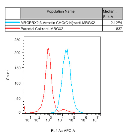
Figure 7. Recombinant MRGPRX2 β-Arrestin CHO-K1 stably expressing MRGPRX2.
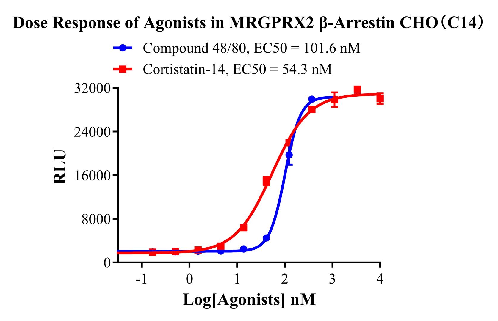
Figure 8. Dose Response of Agonists in MRGPRX2 β-Arrestin CHO-K1(C14) .
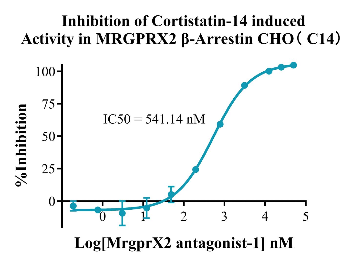
Figure 9. Inhibition of Cortistatin-14 induced Activity in MRGPRX2 β-Arrestin CHO(C14).
Product Core Advantages:
|
Advantage Dimension |
Detailed Description |
|
High-Profile Target |
MRGPRX2 has emerged as one of the most significant GPCR targets in the 2024–2025 autoimmune and allergy landscape, underscored by massive investments from industry giants like Incyte and Novartis. |
|
Precise Pathway Alignment |
By utilizing Gαq downstream detection (Calcium Flux or IP-One), our models perfectly mirror the natural signaling mechanism of MRGPRX2, ensuring that data carries high translational value for clinical applications. |
|
High Sensitivity & Wide Window |
Validation data confirms robust responses to a broad spectrum of structurally diverse ligands (cationic peptides, small molecule drugs, etc.), making it ideal for evaluating candidates across various chemical scaffolds. |
|
Flexible Assay Modalities |
We offer both Calcium Flux (real-time kinetics) and IP-One (endpoint-based) protocols to accommodate different lab equipment and varied throughput requirements, from lead optimization to HTS. |
|
Enabling Biased Signaling Discovery |
Coupled with our β-Arrestin recruitment models, researchers can differentiate between full antagonists and biased ligands, facilitating the development of next-generation therapies with improved safety profiles. |
|
Ready-to-Use (RTU) Solutions |
Each cell line undergoes rigorous monoclonal selection and functional validation. Our "plug-and-play" format allows you to initiate experiments immediately upon arrival, significantly shortening your R&D timeline. |
Table 10: Core Advantages of the Renkuan Cell Model
V. Application Scenarios: From Chronic Urticaria to Drug Hypersensitivity
ReqBio’s MRGPRX2 cell models are engineered for versatile applications across the following drug R&D scenarios:
Chronic Spontaneous Urticaria (CSU): Screening for novel oral MRGPRX2 antagonists to achieve differentiation from competitors such as EP262 and EVO-756.
Atopic Dermatitis (AD): Evaluating the ability of compounds to inhibit mast cell degranulation induced by LL-37 or human hBDs.
Drug-Induced Pseudo-Allergy: Predicting whether clinical drug candidates trigger anaphylactoid reactions via the MRGPRX2 pathway.
Biased Ligand Development: Screening for modulators that selectively block the Gαq pathway without affecting β-arrestin-mediated internalization or other signaling, thereby reducing potential side effects.
Conclusion
The rapid rise of MRGPRX2—from a relatively niche "itch receptor" to a star target in the development of drugs for allergic and inflammatory diseases—is the result of a profound convergence between fundamental immunology and clinical translational medicine. With the clinical advancement of programs like EP262 and EVO-756, the druggability of this target has already received preliminary validation.
ReqBio remains committed to focusing on cutting-edge GPCR targets. By providing high-quality functional cell models and comprehensive assay services, we aim to empower the discovery and translation of innovative medicines for patients worldwide.


news recommendation
CD3E: The T-Cell Activation Switch and Cell Models for Targeted Therapeutics
NFAT Signaling Pathway: A Critical Target for Drug R&D and Cell Models
TSHR Cell Model for Thyroid Cancer Drug Screening | GPCR Assay Platform-Reqbio
We Are Pleased to Announce: Global Commercial Licensing Rights for Jurkat E6.1, CHO-K1, and HEK293 Cell Lines Officially Secured.
Explore