

CHO-K1 Human MRGPRX2 β-Arrestin Receptor Cell
Cat. No: RQP71585
Size: 1 vial of frozen cells (>1E6 per vial in 1 mL)
Unit Price: Contact For Pricing
Product Info
Description
Biological Information
Assay Data
Cell Culture
| Cat. No | RQP71585 |
| Product Name | CHO-K1 Human MRGPRX2 β-Arrestin Receptor Cell |
| Product Type | Receptor Cell Lines |
| Product Description | CHO-K1 Human MRGPRX2 β-Arrestin Receptor Cell is a clonally stable cell line constructed using lentiviral technology,constitutively expressing the Human MRGPRX2 gene. |
| Culture Properties | Adherent |
| Stability | 32passages (in-house test, that not means the cell line will be instable beyond the passages we tested.) |
| Mycoplasma Status | Negative |
| Culture Medium | F12K+10%FBS +5 μg/ml Puromycin+5 μg/ml Blsaticidin |
| Freeze Medium | 90% FBS+10% DMSO |
| Storage Conditions | Liquid nitrogen immediately upon delivery |
| Transducer | β-Arrestin |
| Application | Functional assay for MRGPRX2 |
For research use only. Not intended for human or animal clinical trials, therapeutic or diagnostic use.
Mast cells (MCs) play a pivotal role in the pathogenesis of allergic reactions, as, upon activation, they release a diverse array of mediators from their intracellular storage granules. However, MCs are also involved in homeostasis and inflammation, innate and adaptive immunity, as well as angiogenesis across various tissues. MC degranulation is triggered by allergens, (glyco)proteins, or autoantibodies—specifically those directed against the FcεRI receptor or receptor-bound IgE antibodies—following the cross-linking and clustering of FcεRI receptors on the cell surface. MC degranulation can also occur via IgE-independent pathways, primarily mediated by various surface receptors, including Toll-like receptors (TLRs), protease-activated receptors (PARs), or Mas-related G protein-coupled receptor member X2 (MRGPRX2).
MRGPRX2 is a GPCR predominantly expressed on human MCs and is critical for mediating allergic and inflammatory responses. MRGPRX2 binds to both exogenous and endogenous ligands—such as major basic protein, eosinophil peroxidase, and neuropeptides—as well as certain pharmacological agents, including neuromuscular blocking agents, fluoroquinolones, and vancomycin. Upon activation of MRGPRX2, MCs release a cascade of chemical mediators, including histamine, tryptase, chymase, chemokines, and cytokines. These substances precipitate a range of clinical conditions—such as urticaria (hives), angioedema (swelling), chronic pruritus, and pain—and may also drive Type 2 inflammation involving the adaptive immune system.
The CHO-K1 Human MRGPRX2 β-Arrestin Receptor Cell Model—effectively simulates the signal transduction process of MRGPRX2 *in vivo*. The underlying principle is illustrated in the figure below.
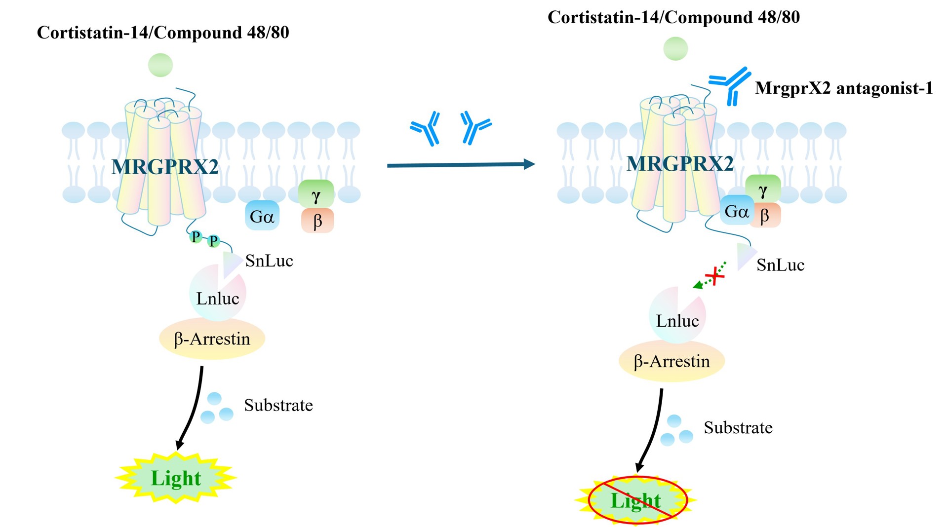
Figure 1. Schematic Diagram of the CHO-K1 Human MRGPRX2 β-Arrestin Receptor Cell Model
| Target Class | GPCR |
| Family | G protein-coupled receptors |
| Sub Family | Class A(Rhodopsin) |
| Gene Name | MRGPRX2 |
| Gene Aliases | MRGX2 |
| Gene ID | 117194 |
| Accession Number | NM_054030.4 |
| UniProt Number | Q96LB1 |
| Protein Name | Mas-related G-protein coupled receptor member X2 |
| Protein Aliases | N/A |
| Target Species | Human |
| Host cell | CHO-K1 |
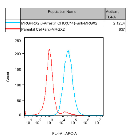
Figure 2. Recombinant MRGPRX2 β-Arrestin CHO-K1 stably expressing MRGPRX2.
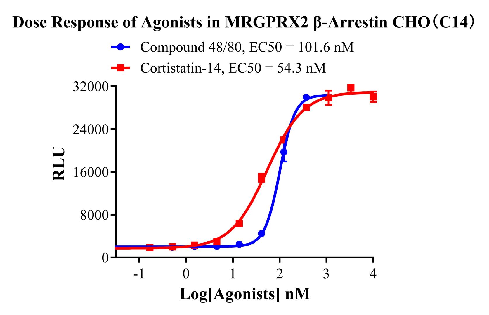
Figure 3. Dose Response of Agonists in MRGPRX2 β-Arrestin CHO-K1(C14) .
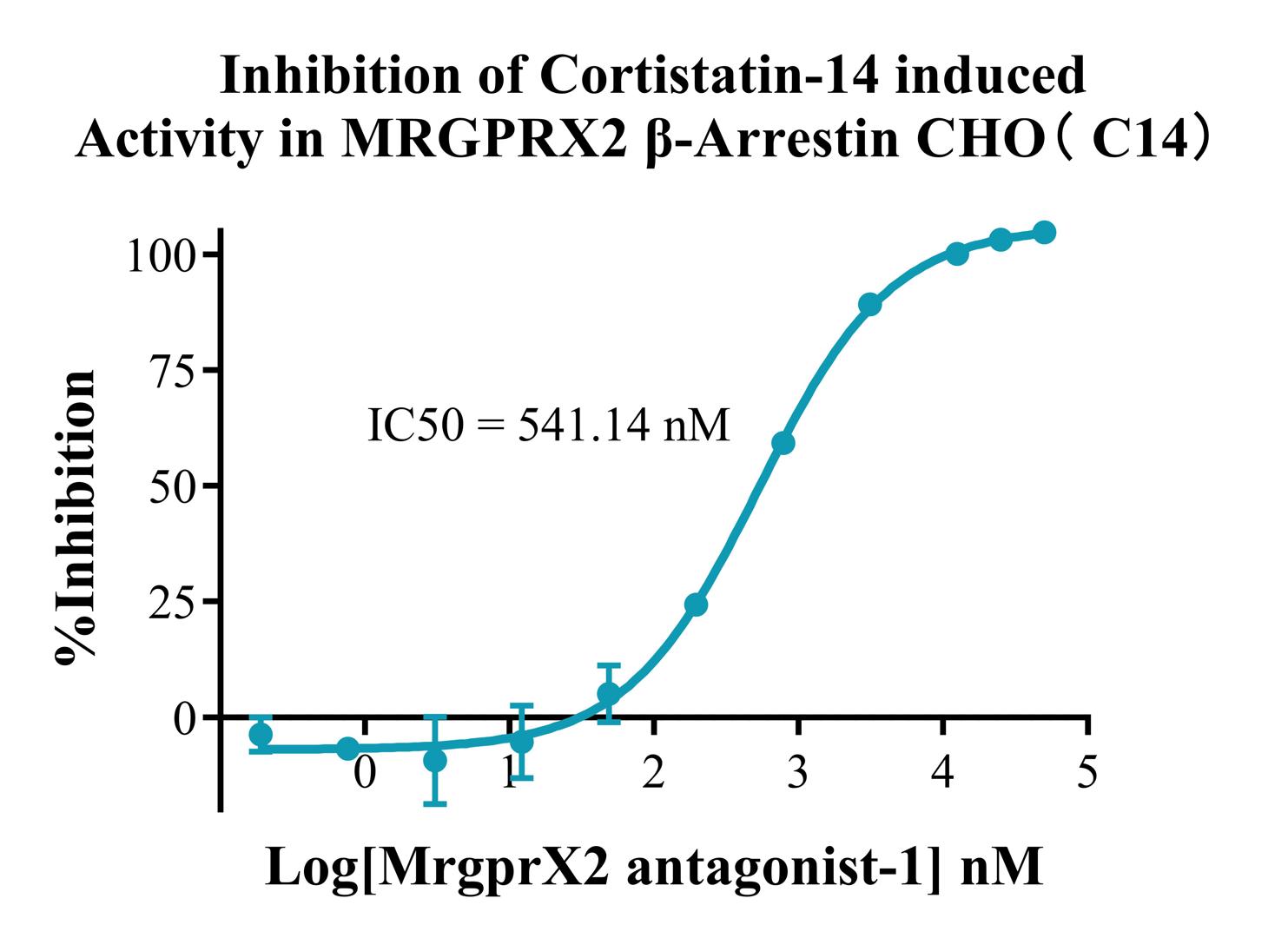
Figure 4. Inhibition of Cortistatin-14 induced Activity in MRGPRX2 β-Arrestin CHO(C14).
Cell Resuscitation
1)Rapidly thaw the frozen cells in a 37 °C water bath for approximately 60 seconds. Once thawed (which may take slightly less or more than 60 seconds), immediately transfer the cell suspension from the cryovial into a 15 mL centrifuge tube containing 10 mL of pre-warmed CHO-K1 Human MRGPRX2 β-Arrestin Receptor Cell complete culture medium.
2)Centrifuge cells at 1000 rpm for 5 min to remove medium, then resuspend cells in 5 mL of pre-warmed complete medium.
3)Transfer the cell suspension into a T25 culture flask and incubate at 37 °C with 5% CO₂.
4)After approximately 24–36 hours, replace the medium or passage the cells to remove non-adherent dead cells.
Subculturing procedure
1)When the cell density reaches the appropriate confluency for passaging, wash the cells with PBS, then add 1 mL trypsin to detach the cells. When more than 80% of the cells detach upon gently tapping the culture flask, add complete culture medium to terminate digestion. Gently pipette to obtain a single-cell suspension, transfer to a 15 mL centrifuge tube, and centrifuge at 1000 rpm for 5 minutes.
2)Discard supernatant after centrifugation. Resuspend cells in fresh medium to a single-cell suspension and transfer to a new culture flask for continued growth.
Cell Freezing
After trypsinization and centrifugation of cells from each T75 flask or 10 cm culture dish, discard the supernatant. Add 2 mL of cryopreservation medium (90% FBS + 10% DMSO), gently resuspend thoroughly, and aliquot into two cryovials. Immediately place the cryovials into a controlled-rate freezing container (e.g., Nalgene 5100-0001), fill with isopropanol to the indicated level, and store at −80 °C. After 24 hours, transfer the cryovials to liquid nitrogen for long-term storage.
Related products
CHO-K1 Human CCR4 Cell Line
HEK293 Human NK1R CRE-Luc Cell Line
Raji-Luc-GFP
Jurkat E6.1-Luc
THP-1-GFP
THP-1-Luc
Raji-GFP
Raji-Luc
Jurkat E6.1-GFP
HEK293 Human GAL4-Luc Cell
We Are Pleased to Announce: Global Commercial Licensing Rights for Jurkat E6.1, CHO-K1, and HEK293 Cell Lines Officially Secured.
Explore