

Jurkat E6.1 Human NKG2D Effector Reporter Cell
Cat. No: RQP74222
Size: 1 vial of frozen cells (>1E6 per vial in 1 mL)
Unit Price: Contact For Pricing
Product Info
Description
Biological Information
Assay Data
Cell Culture
| Cat. No | RQP74222 |
| Product Name | Jurkat E6.1 Human NKG2D Effector Reporter Cell |
| Product Type | Reporter Cell |
| Culture Properties | suspension |
| Stability | 32passages (in-house test, that not means the cell line will be instable beyond the passages we tested.) |
| Mycoplasma Status | Negative |
| Culture Medium | RPMI-1640+10%FBS+1μg/ml puromycin+800μg/ml Hygromycin B+5μg/ml blasticidin |
| Freeze Medium | 90% FBS+10% DMSO |
| Storage Conditions | Liquid nitrogen immediately upon delivery |
| Application | Functional(Report Gene) Assay |
For research use only. Not intended for human or animal clinical trials, therapeutic or diagnostic use.
NKG2D (Natural Killer Group 2D) is a C-type lectin-like activating receptor that serves as a core surveillance molecule within the innate immune system; its primary function is to recognize and eliminate infected or transformed (cancerous) cells. It is expressed on various immune cells—including NK cells, T cells, and macrophages—and participates in both innate and adaptive immune responses. NKG2D and its ligands play pivotal roles in processes such as tumorigenesis and viral infection. Structurally, NKG2D is a type II transmembrane protein; its extracellular region contains a C-type lectin domain (which binds to calcium-dependent glycosylated ligands), while its intracellular region lacks signaling motifs, necessitating signal transduction via adaptor proteins such as DAP10 or DAP12.
The ligands for NKG2D constitute a class of MHC class I-related molecules (MIC) and UL16-binding proteins (ULBP), the expression of which is induced by cellular stress signals. These ligands are barely detectable in normal cells; however, their expression is strongly induced by "danger signals"—such as DNA damage (e.g., resulting from radiotherapy), heat shock, viral infection, or oncogene activation—and they are predominantly expressed on the surfaces of tumor cells, infected cells, or damaged tissues. Upon binding to the NKG2D receptor, these ligands activate immune effector cells—such as NK cells and CD8+ T cells—thereby triggering target cell lysis and the release of inflammatory cytokines, a process that constitutes a core mechanism of immune surveillance. Conversely, soluble ligands shed within the tumor microenvironment (e.g., sMICA) can suppress this signaling pathway, thereby facilitating immune evasion.
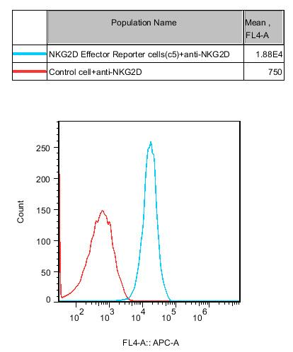
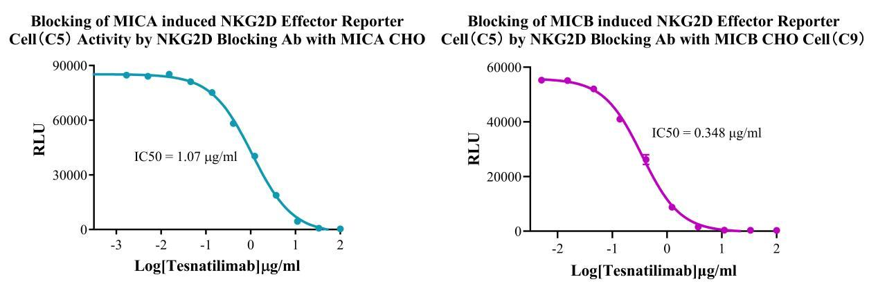
The Jurkat E6.1 Human NKG2D Effector Reporter Cell Model—effectively simulates the signal transduction process of NKG2D *in vivo*. The underlying principle is illustrated in the figure below.
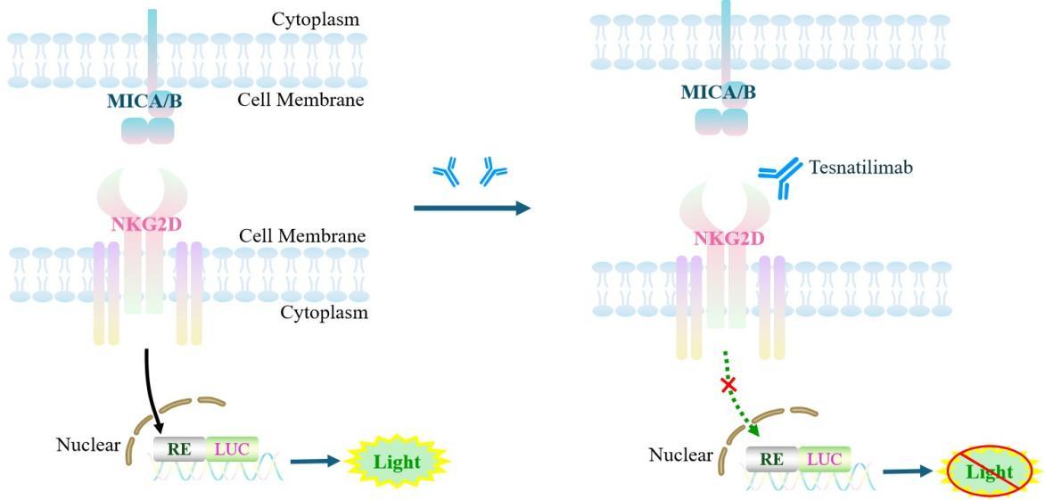
Figure 1. Schematic Diagram of the Jurkat E6.1 Human NKG2D Effector Reporter Cell Model
| Classification | Co-Stimulatory |
| Family | C‑type lectin‑like receptor (CTLR) family |
| Gene Name | KLRK1 |
| Gene Aliases | NKG2D;KLR;NKG2-D;CD314 |
| Gene ID | 22914 |
| Accession Number | NM_007360.4 |
| UniProt Number | P26718 |
| Protein Name | NKG2-D type II integral membrane protein |
| Protein Aliases | Killer cell lectin-like receptor subfamily K member 1;NK cell receptor D;NKG2-D-activating NK receptor |
| Target Species | Human |
| Host cell | Jurkat E6.1 |
Cell Passage Procedures
1.This cell line grows in suspension.
2.Upon receipt, cells should be thawed immediately or stored in liquid nitrogen until use.
3.Before thawing, pre-warm the water bath and culture medium to 37 °C, and prepare a small amount of dry ice.
4.Remove the cryovial from storage and transport it to the cell culture laboratory on dry ice.
5.Rapidly thaw the cells in a 37 °C water bath. Once the cells are completely thawed, spray the cryovial with 70% ethanol for disinfection and transfer it to a biosafety cabinet.
6.Add 10 mL of pre-warmed culture medium into a 15 mL centrifuge tube. Transfer the contents of the cryovial into the tube and centrifuge at 1000 rpm for 5 minutes.
7.Carefully discard the supernatant. Resuspend the cell pellet in 5 mL of pre-warmed culture medium by gentle pipetting. Immediately perform cell counting and adjust the cell density to 3–6 × 10⁵ cells/mL based on the counting results, then transfer the cells into a culture flask.
8.Count the cells every 1–2 days. When the cell density exceeds 1 × 10⁶ cells/mL, passage the cells promptly or add fresh culture medium. Maintain the cell density between 2 × 10⁵ and 1 × 10⁶ cells/mL.
Suspension Cell Cryopreservation Procedure:
1.Collect 8 × 10⁶ cells, centrifuge, and discard the supernatant.
2.Add 1 mL of cell freezing medium (90% FBS + 10% DMSO) and gently pipette to mix thoroughly. Transfer the suspension into a cryovial.
3.Immediately place the cryovial into a controlled-rate freezing container (Nalgene 5100-0001), fill with isopropanol up to the indicated level, and store at −80 °C.
4.After 24 hours, transfer the cryovial to liquid nitrogen for long-term storage.
Related products
CHO-K1 Human CCR4 Cell Line
HEK293 Human NK1R CRE-Luc Cell Line
Raji-Luc-GFP
Jurkat E6.1-Luc
THP-1-GFP
THP-1-Luc
Raji-GFP
Raji-Luc
Jurkat E6.1-GFP
HEK293 Human GAL4-Luc Cell
We Are Pleased to Announce: Global Commercial Licensing Rights for Jurkat E6.1, CHO-K1, and HEK293 Cell Lines Officially Secured.
Explore