

CHO-K1 Human MICB Cell
Cat. No: RQP74357
Size: 1 vial of frozen cells (>1E6 per vial in 1 mL)
Unit Price: Contact For Pricing
Product Info
Description
Biological Information
Assay Data
Cell Culture
| Cat. No | RQP74357 |
| Product Name | CHO-K1 Human MICB Cell |
| Product Type | Expression Cell line |
| Culture Properties | Adherent |
| Stability | 32passages (in-house test, that not means the cell line will be instable beyond the passages we tested.) |
| Mycoplasma Status | Negative |
| Culture Medium | F12K+10%FBS+600 μg/ml Hygromycin B |
| Freeze Medium | 90% FBS+10% DMSO |
| Storage Conditions | Liquid nitrogen immediately upon delivery |
| Application | Binding Assay,FACS |
For research use only. Not intended for human or animal clinical trials, therapeutic or diagnostic use.
NKG2D (Natural Killer Group 2D) is a C-type lectin-like activating receptor. As a core surveillance molecule of the innate immune system, its primary function is to recognize and eliminate infected or transformed (cancerous) cells. It is expressed on various immune cells—including NK cells, T cells, and macrophages—and participates in both innate and adaptive immune responses. NKG2D and its ligands play pivotal roles in processes such as tumorigenesis and viral infection. NKG2D is a type II transmembrane protein; its extracellular region contains a C-type lectin domain (which binds to calcium-dependent glycosylated ligands), while its intracellular region lacks signaling motifs, necessitating signal transduction via the adaptor proteins DAP10 or DAP12 (in mice).
The *MICB* gene (MHC class I polypeptide-related sequence B) is a non-classical MHC class I gene characterized by high polymorphism, with over 15 distinct alleles identified to date. The transmembrane glycoprotein encoded by this gene consists of three helical domains—α1, α2, and α3—and exhibits high homology with the product of the *MICA* gene (with over 90% homology in their coding regions), yet functions independently of classical MHC class I molecules. MICB protein is expressed at low levels in normal tissues but is predominantly found on the surfaces of epithelial tumor cells, gastrointestinal epithelial cells, and various immune cells. By binding to NKG2D receptors on the surfaces of NK cells, γδ T cells, and CD8+ T cells, MICB activates the cytotoxic functions and co-stimulatory signaling pathways of these immune cells, thereby playing a central role in immune surveillance.
In clinical studies, therapeutic agents targeting the MICB-NKG2D pathway—such as specific antibodies and CAR-T cell therapies—have demonstrated anti-tumor potential by inhibiting MICB shedding or enhancing NK cell activity. Furthermore, *MICB* genotyping and protein expression analysis offer novel strategies for tumor risk assessment, treatment planning, and prognostic evaluation, holding particular significance and application value within the context of combination immunotherapy regimens.
The CHO-K1 Human MICB Cell Model—effectively simulates the signal transduction process of NKG2D *in vivo*. The underlying principle is illustrated in the figure below.
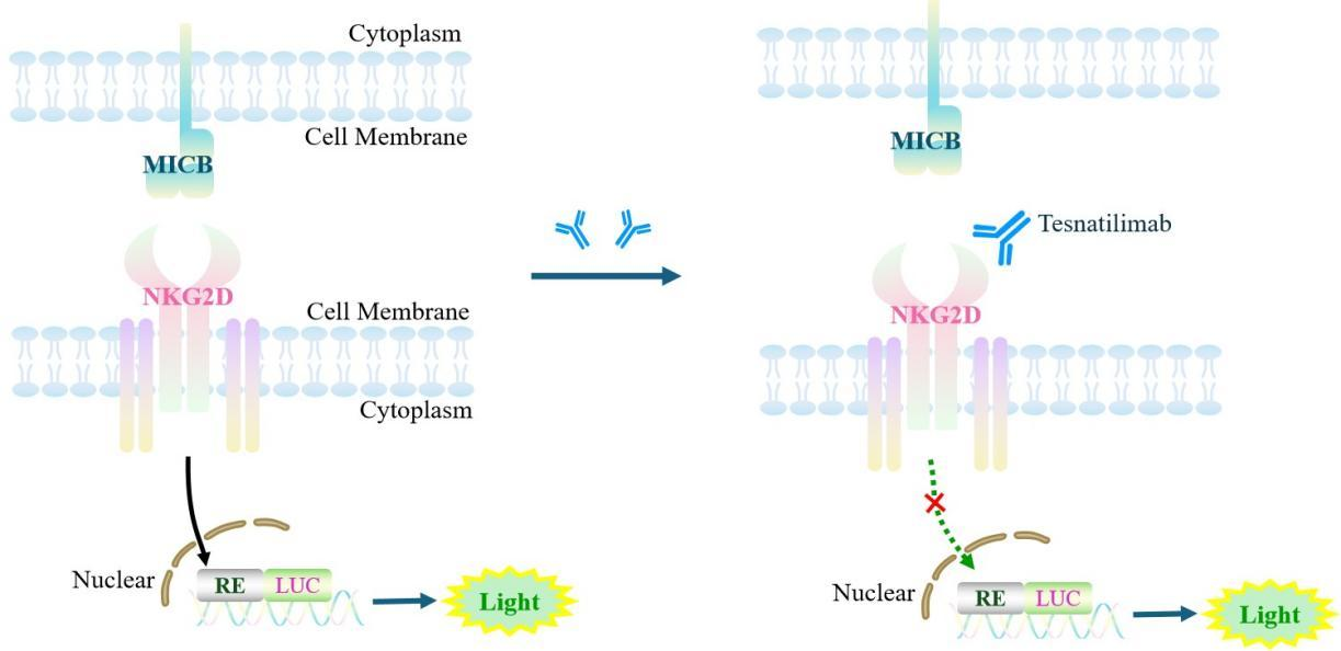
Figure 1. Schematic Diagram of the CHO-K1 Human MICB Cell Model
| Classification | Co-Stimulatory |
| Family | MHC class I family. MIC subfamily |
| Gene Name | MICB |
| Gene Aliases | PERB11.2 |
| Gene ID | 4277 |
| Accession Number | NM_005931.5 |
| UniProt Number | Q29980 |
| Protein Name | MIC-B |
| Protein Aliases | N/A |
| Target Species | Human |
| Host cell | CHO-K1 |
CHO cell line expressing full length human MICB. Expression is confirmed by flow cytometry.

Figure 2. Recombinant MICB CHO constitutively expressing MICB.
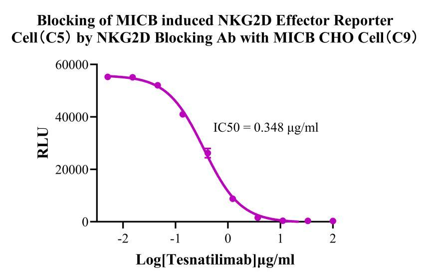
Figure 3. Blocking of MICB induced NKG2D Effector Reporter Cell(C5) by NKG2D Blocking Ab with MICB CHO Cell(C9).
Cell Resuscitation
1)Rapidly thaw the frozen cells in a 37 °C water bath for approximately 60 seconds. Once thawed (which may take slightly less or more than 60 seconds), immediately transfer the cell suspension from the cryovial into a 15 mL centrifuge tube containing 10 mL of pre-warmed CHO-K1 Human MICB Cell complete culture medium.
2)Centrifuge cells at 1000 rpm for 5 min to remove medium, then resuspend cells in 5 mL of pre-warmed complete medium.
3)Transfer the cell suspension into a T25 culture flask and incubate at 37 °C with 5% CO₂.
4)After approximately 24–36 hours, replace the medium or passage the cells to remove non-adherent dead cells.
Subculturing procedure
1)When the cell density reaches the appropriate confluency for passaging, wash the cells with PBS, then add 1 mL trypsin to detach the cells. When more than 80% of the cells detach upon gently tapping the culture flask, add complete culture medium to terminate digestion. Gently pipette to obtain a single-cell suspension, transfer to a 15 mL centrifuge tube, and centrifuge at 1000 rpm for 5 minutes.
2)Discard supernatant after centrifugation. Resuspend cells in fresh medium to a single-cell suspension and transfer to a new culture flask for continued growth.
Cell Freezing
After trypsinization and centrifugation of cells from each T75 flask or 10 cm culture dish, discard the supernatant. Add 2 mL of cryopreservation medium (90% FBS + 10% DMSO), gently resuspend thoroughly, and aliquot into two cryovials. Immediately place the cryovials into a controlled-rate freezing container (e.g., Nalgene 5100-0001), fill with isopropanol to the indicated level, and store at −80 °C. After 24 hours, transfer the cryovials to liquid nitrogen for long-term storage.
Related products
CHO-K1 Human CCR4 Cell Line
HEK293 Human NK1R CRE-Luc Cell Line
Raji-Luc-GFP
Jurkat E6.1-Luc
THP-1-GFP
THP-1-Luc
Raji-GFP
Raji-Luc
Jurkat E6.1-GFP
HEK293 Human GAL4-Luc Cell
We Are Pleased to Announce: Global Commercial Licensing Rights for Jurkat E6.1, CHO-K1, and HEK293 Cell Lines Officially Secured.
Explore