

Jurkat E6.1 Human BTLA Effector Reporter Cell
Cat. No: RQP74118
Size: 1 vial of frozen cells (>1E6 per vial in 1 mL)
Unit Price: Contact For Pricing
Product Info
Description
Biological Information
Assay Data
Cell Culture
| Cat. No | RQP74118 |
| Product Name | Jurkat E6.1 Human BTLA Effector Reporter Cell |
| Product Type | Reporter Cell |
| Culture Properties | suspension |
| Stability | 32passages (in-house test, that not means the cell line will be instable beyond the passages we tested.) |
| Mycoplasma Status | Negative |
| Culture Medium | RPMI-1640+10%FBS+1μg/ml puromycin+800μg/ml Hygromycin B |
| Freeze Medium | 90% FBS+10% DMSO |
| Storage Conditions | Liquid nitrogen immediately upon delivery |
| Application | Functional(Report Gene) Assay |
For research use only. Not intended for human or animal clinical trials, therapeutic or diagnostic use.
B and T Lymphocyte Attenuator (BTLA) is a member of the CD28 superfamily. The protein structure of BTLA is similar to Programmed Cell Death-1 (PD-1) and Cytotoxic T-Lymphocyte-Associated Antigen-4 (CTLA-4), consisting of an extracellular domain, a transmembrane domain, and a cytoplasmic domain. The cytoplasmic domain contains the Growth Factor Receptor-Bound Protein-2 (Grb-2) association motif, the Immunoreceptor Tyrosine Switch Motif (ITSM), and the Immunoreceptor Tyrosine Inhibitory Motif (ITIM). BTLA is widely expressed in lymph nodes, thymus, and spleen, but is rarely or not expressed in organs such as the heart, kidneys, brain, and liver.
Herpesvirus Entry Mediator (HVEM) belongs to the tumor necrosis factor receptor superfamily and is the only detectable ligand in human cells. HVEM can interact with BTLA in a cis or trans manner. On cells co-expressing BTLA and HVEM, BTLA interacts with HVEM in cis; whereas trans interaction occurs when BTLA and HVEM are expressed on different cells. In addition to BTLA, HVEM can also interact with CD160, lymphotoxin-α, LIGHT (TNFSF14), and Synapse-Associated Like Molecule 5 (SALM5). Notably, BTLA and CD160 compete for the same binding site within the CRD1/CRD2 region of HVEM, while LIGHT independently binds to another side of HVEM within the CRD2/CRD3 region.
The binding of HVEM to BTLA activates tyrosine phosphorylation of ITIM in BTLA, leading to the recruitment of protein tyrosine phosphatases SHP-1 and SHP-2, which contain Src Homology Domain 2 (SH2). These protein tyrosine phosphatases typically mediate immunosuppressive effects. However, binding of the Grb-2 association motif to Grb-2 results in the recruitment of the PI3K protein subunit p85 and T-cell activation.
The Jurkat E6.1 Human BTLA Effector Reporter Cell model effectively mimics the in vivo BTLA signaling transduction process. The principle is illustrated in the figure below.
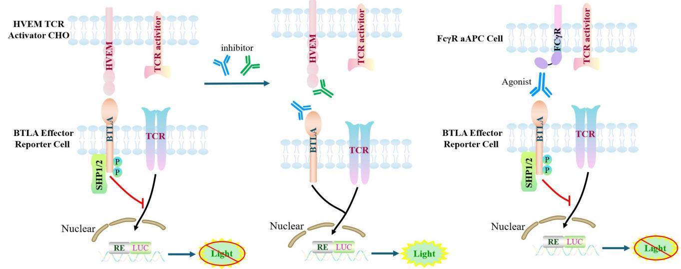
Figure 1. Schematic diagram of the Jurkat E6.1 Human BTLA Effector Reporter Cell model.
| Classification | Co-Inhibitory |
| Family | Immunoglobulin superfamily (IgSF) |
| Gene Name | BTLA |
| Gene Aliases | BTLA1;CD272; |
| Gene ID | 151888 |
| Accession Number | NM_181780.4 |
| UniProt Number | Q7Z6A9 |
| Protein Name | B- and T-lymphocyte attenuator |
| Protein Aliases | B- and T-lymphocyte-associated protein |
| Target Species | Human |
| Host cell | Jurkat E6.1 |
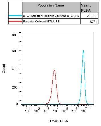
Figure 2. Recombinant BTLA Effector Reporter Cell constitutively expressing BTLA.
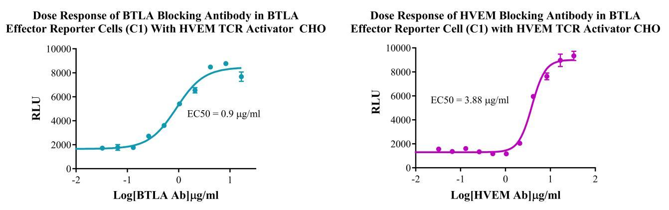
Figure 3. Dose Response of HVEM Blocking Antibody in BTLA Effector Reporter Cells (C1) with HVEM/TCR Activator CHO .
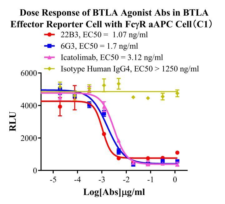
Figure 4. Dose Response of BTLA Agonist Abs in BTLA Effector Reporter Cell with FCγR aAPC Cell(C1).
Cell Passage Procedures
1.This cell line grows in suspension.
2.Upon receipt, cells should be thawed immediately or stored in liquid nitrogen until use.
3.Before thawing, pre-warm the water bath and culture medium to 37 °C, and prepare a small amount of dry ice.
4.Remove the cryovial from storage and transport it to the cell culture laboratory on dry ice.
5.Rapidly thaw the cells in a 37 °C water bath. Once the cells are completely thawed, spray the cryovial with 70% ethanol for disinfection and transfer it to a biosafety cabinet.
6.Add 10 mL of pre-warmed culture medium into a 15 mL centrifuge tube. Transfer the contents of the cryovial into the tube and centrifuge at 1000 rpm for 5 minutes.
7.Carefully discard the supernatant. Resuspend the cell pellet in 5 mL of pre-warmed culture medium by gentle pipetting. Immediately perform cell counting and adjust the cell density to 3–6 × 10⁵ cells/mL based on the counting results, then transfer the cells into a culture flask.
8.Count the cells every 1–2 days. When the cell density exceeds 1 × 10⁶ cells/mL, passage the cells promptly or add fresh culture medium. Maintain the cell density between 2 × 10⁵ and 1 × 10⁶ cells/mL.
Suspension Cell Cryopreservation Procedure:
1.Collect 8 × 10⁶ cells, centrifuge, and discard the supernatant.
2.Add 1 mL of cell freezing medium (90% FBS + 10% DMSO) and gently pipette to mix thoroughly. Transfer the suspension into a cryovial.
3.Immediately place the cryovial into a controlled-rate freezing container (Nalgene 5100-0001), fill with isopropanol up to the indicated level, and store at −80 °C.
4.After 24 hours, transfer the cryovial to liquid nitrogen for long-term storage.
Related products
CHO-K1 Human CCR4 Cell Line
HEK293 Human NK1R CRE-Luc Cell Line
Raji-Luc-GFP
Jurkat E6.1-Luc
THP-1-GFP
THP-1-Luc
Raji-GFP
Raji-Luc
Jurkat E6.1-GFP
HEK293 Human GAL4-Luc Cell
We Are Pleased to Announce: Global Commercial Licensing Rights for Jurkat E6.1, CHO-K1, and HEK293 Cell Lines Officially Secured.
Explore