
HEK293 Human TLR2 Cell
Cat. No: RQP74572
Size: 1 vial of frozen cells (>1E6 per vial in 1 mL)
Unit Price: Contact For Pricing
Product Info
Description
Biological Information
Cell Culture
| Cat. No | RQP74572 |
| Product Name | HEK293 Human TLR2 Cell |
| Product Type | Expression Cell line |
| Culture Properties | Adherent |
| Stability | 32passages (in-house test, that not means the cell line will be instable beyond the passages we tested.) |
| Mycoplasma Status | Negative |
| Culture Medium | DMEM+10%FBS+2 μg/ml Puromycin |
| Freeze Medium | 90% FBS+10% DMSO |
| Storage Conditions | Liquid nitrogen immediately upon delivery |
| Application | Binding Assay,FACS |
For research use only. Not intended for human or animal clinical trials, therapeutic or diagnostic use.
Toll-like receptors (TLRs) are highly conserved pattern recognition receptors (PRRs) that recognize various types of pathogen-associated pattern molecules (PAMPs) in microorganisms. Ten members of the Toll-like receptor family (TLR1-10) have been identified in humans, and twelve members (TLR1-9, TLR11, and TLR13) have been identified in mice. TLR1, TLR2, TLR4, TLR5, and TLR6 are located on the cell membrane surface; TLR3, TLR7, TLR8, TLR9, and TLR10 are located on the surface of cellular inclusion bodies (TLR3 recognizes dsRNA, and TLR9 recognizes dsDNA). Activation of downstream signaling of TLRs mainly depends on two classes of transcription factors: NF-κB and interfering regulatory factors (IRFs), which primarily induce the production of pro-inflammatory cytokines and type I interferon (IFN).
TLR2 is the only TLR that forms functional heterodimers with more than two other types of TLRs, dimerizing with TLR1 and TLR6, and in some cases with TLR4. TLR2 also interacts with various non-TLR molecules, enabling it to recognize multiple PAMPs from all microorganisms, including viruses, fungi, bacteria, and parasites. TLR2 can sense highly conserved lipoproteins expressed on the outer membrane of Gram-positive bacteria, as well as membrane antigens of some Gram-negative bacteria associated with its co-receptor CD14, such as lipopolysaccharide (LPS).
TLR2 primarily relies on the MyD88 and TIRAP adaptor proteins for signal transduction. The MyD88-dependent pathway involves death domain interactions that mediate intracellular signal transduction in a stepwise manner. Specifically, MyD88 recruitment is indirect and mediated by TIRAP. Activated MyD88 then sequentially triggers phosphorylation of IRAK4, IRAK1, and IRAK2. The IRAK complex binds to TRAF6, activating the complex of TAK1, TAB2, and TAB3. TAK1 phosphorylates IKKα and IKKβ, and IKK phosphorylates IκB, which is then labeled and degraded. This ultimately leads to the production of pro-inflammatory cytokines via NF-κB and AP-1, and the activation of MAPK, which regulates cell proliferation and survival.
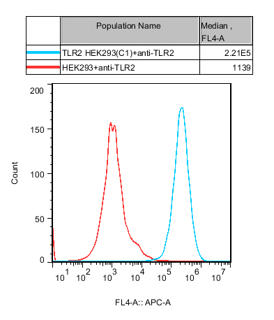
Figure 1. Recombinant TLR2 HEK293 stably expressing TLR2.
| Classification | TLR |
| Family | Toll-like receptor family |
| Gene Name | TLR2 |
| Gene Aliases | TIL4;CD282 |
| Gene ID | 7097 |
| Accession Number | NM_001318789.2 |
| UniProt Number | O60603 |
| Protein Name | Toll-like receptor 2 |
| Protein Aliases | Toll/interleukin-1 receptor-like protein 4 |
| Target Species | Human |
| Host cell | HEK293 |
Cell Resuscitation
1)Rapidly thaw the frozen cells in a 37 °C water bath for approximately 60 seconds. Once thawed (which may take slightly less or more than 60 seconds), immediately transfer the cell suspension from the cryovial into a 15 mL centrifuge tube containing 10 mL of pre-warmed HEK293 Human TLR2 Cell complete culture medium.
2)Centrifuge cells at 1000 rpm for 5 min to remove medium, then resuspend cells in 5 mL of pre-warmed complete medium.
3)Transfer the cell suspension into a T25 culture flask and incubate at 37 °C with 5% CO₂.
4)After approximately 24–36 hours, replace the medium or passage the cells to remove non-adherent dead cells.
Subculturing procedure
1)When the cell density reaches the appropriate confluency for passaging, wash the cells with PBS, then add 1 mL trypsin to detach the cells. When more than 80% of the cells detach upon gently tapping the culture flask, add complete culture medium to terminate digestion. Gently pipette to obtain a single-cell suspension, transfer to a 15 mL centrifuge tube, and centrifuge at 1000 rpm for 5 minutes.
2)Discard supernatant after centrifugation. Resuspend cells in fresh medium to a single-cell suspension and transfer to a new culture flask for continued growth.
Cell Freezing
After trypsinization and centrifugation of cells from each T75 flask or 10 cm culture dish, discard the supernatant. Add 2 mL of cryopreservation medium (90% FBS + 10% DMSO), gently resuspend thoroughly, and aliquot into two cryovials. Immediately place the cryovials into a controlled-rate freezing container (e.g., Nalgene 5100-0001), fill with isopropanol to the indicated level, and store at −80 °C. After 24 hours, transfer the cryovials to liquid nitrogen for long-term storage.