

CHO-K1 Human MC5R CRE-Luc Cell Line
Cat. No: RQP71616
Size: 1 vial of frozen cells (>1E6 per vial in 1 mL)
Unit Price: Contact For Pricing
Product Info
Description
Biological Information
Assay Data
Cell Culture
| Cat. No | RQP71616 |
| Product Name | CHO-K1 Human MC5R CRE-Luc Cell Line |
| Product Type | Receptor Cell Lines |
| Product Description | CHO-K1 Human MC5R CRE-Luc Cell Line is a clonally stable cell line constructed using lentiviral technology,constitutively expressing the Human MC5R gene. |
| Culture Properties | Adherent |
| Stability | 32passages (in-house test, that not means the cell line will be instable beyond the passages we tested.) |
| Mycoplasma Status | Negative |
| Culture Medium | F12K+10%FBS+5 μg/ml Puromycin + 600μg/ml Hygromycin B |
| Freeze Medium | 90% FBS+10% DMSO |
| Storage Conditions | Liquid nitrogen immediately upon delivery |
| Transducer | Gs |
| Application | Functional assay for MC5R |
For research use only. Not intended for human or animal clinical trials, therapeutic or diagnostic use.
Melanocortin Receptor 5 (MC5R) is the most widely distributed member of the melanocortin receptor family (MC1R–MC5R), primarily expressed in skeletal muscle, adipose tissue, sebaceous glands, immune cells, and various exocrine glands. By binding to ligands such as alpha-melanocyte-stimulating hormone (α-MSH), it participates in regulating diverse physiological processes, including energy metabolism (e.g., promoting glucose uptake and lipid mobilization), sebum secretion, immune responses, body temperature, and reproductive behaviors. Functional abnormalities of MC5R are closely associated with conditions such as obesity, diabetes, acne, dry skin, and renal diseases.
MC5R signaling is primarily mediated through two pathways: the major pathway involves activating Gs proteins to stimulate the intracellular production of the second messenger cAMP, thereby initiating a downstream signaling cascade; the minor pathway activates the MAP kinase signaling pathway (ERK1/2) via a PI3K-dependent mechanism.
The CHO-K1 Human MC5R CRE-Luc Cell Line Model—effectively simulates the signal transduction process of MC5R *in vivo*. The underlying principle is illustrated in the figure below.
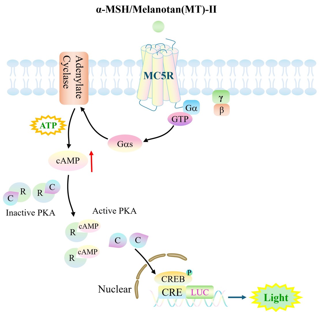
Figure 1. Schematic Diagram of the CHO-K1 Human MC5R CRE-Luc Cell Line Model
| Target Class | GPCR |
| Family | G-protein coupled receptor 1 family |
| Sub Family | Class A(Rhodopsin) |
| Gene Name | MC5R |
| Gene Aliases | N/A |
| Gene ID | 4161 |
| Accession Number | NM_005913.3 |
| UniProt Number | P33032 |
| Protein Name | MC5-R |
| Protein Aliases | MC-2 |
| Target Species | Human |
| Host cell | CHO-K1 |
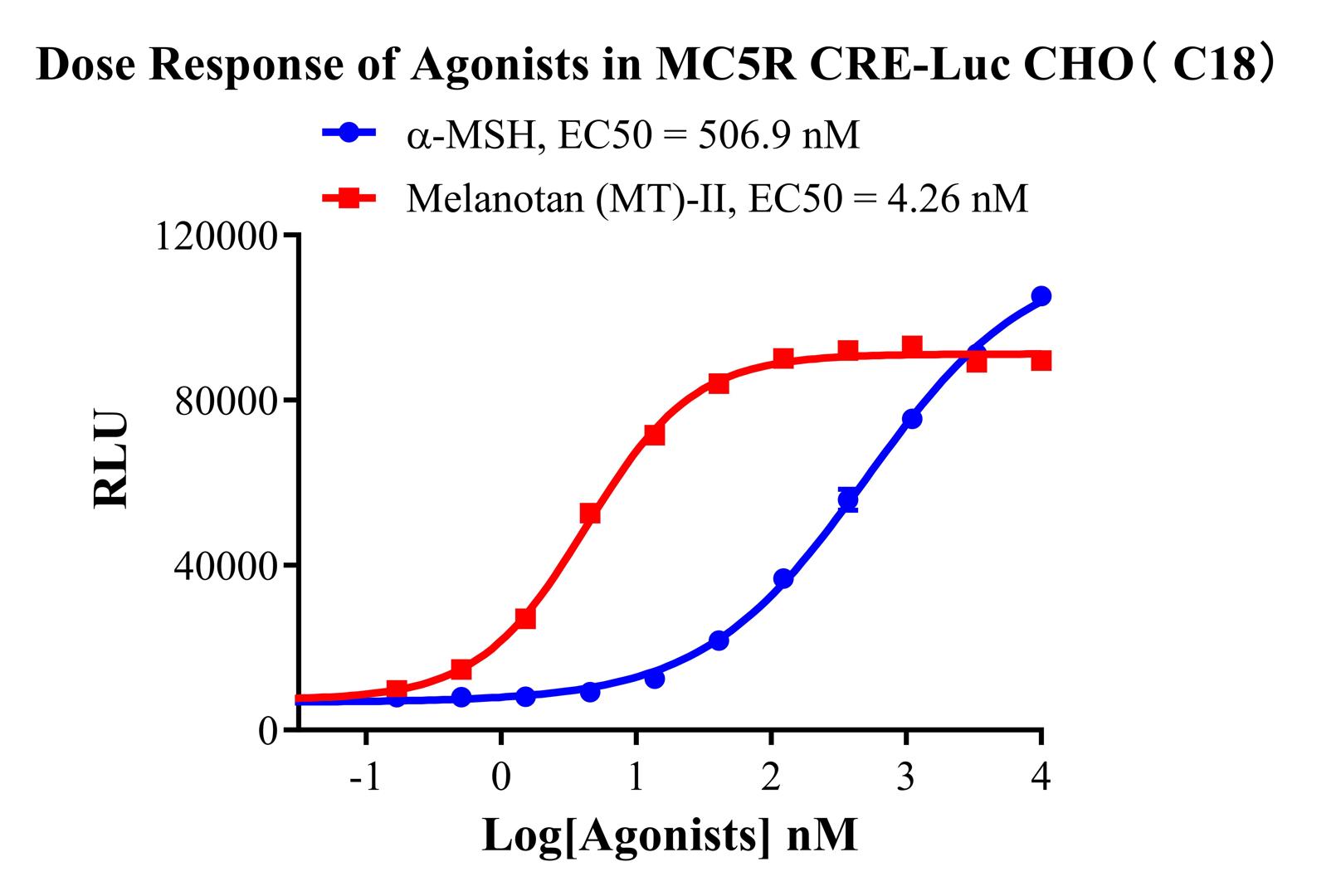
Figure 2. Dose Response of Agonists in MC5R CRE-Luc CHO( C18).
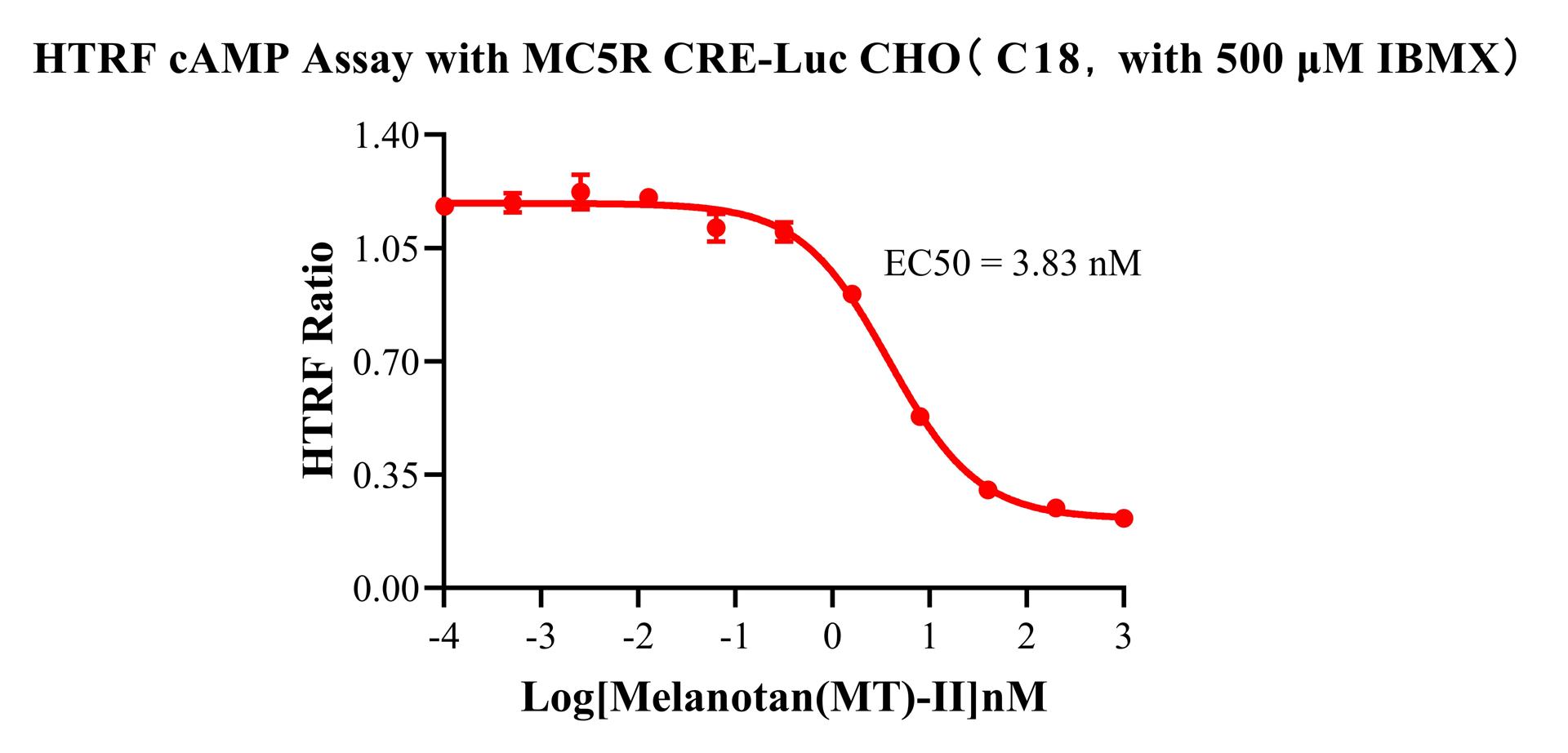
Figure 3. HTRF cAMP Assay with MC5R CRE-Luc CHO(C18, with 500 μM IBMX).
Cell Resuscitation
1)Rapidly thaw the frozen cells in a 37 °C water bath for approximately 60 seconds. Once thawed (which may take slightly less or more than 60 seconds), immediately transfer the cell suspension from the cryovial into a 15 mL centrifuge tube containing 10 mL of pre-warmed CHO-K1 Human MC5R CRE-Luc Cell Line complete culture medium.
2)Centrifuge cells at 1000 rpm for 5 min to remove medium, then resuspend cells in 5 mL of pre-warmed complete medium.
3)Transfer the cell suspension into a T25 culture flask and incubate at 37 °C with 5% CO₂.
4)After approximately 24–36 hours, replace the medium or passage the cells to remove non-adherent dead cells.
Subculturing procedure
1)When the cell density reaches the appropriate confluency for passaging, wash the cells with PBS, then add 1 mL trypsin to detach the cells. When more than 80% of the cells detach upon gently tapping the culture flask, add complete culture medium to terminate digestion. Gently pipette to obtain a single-cell suspension, transfer to a 15 mL centrifuge tube, and centrifuge at 1000 rpm for 5 minutes.
2)Discard supernatant after centrifugation. Resuspend cells in fresh medium to a single-cell suspension and transfer to a new culture flask for continued growth.
Cell Freezing
After trypsinization and centrifugation of cells from each T75 flask or 10 cm culture dish, discard the supernatant. Add 2 mL of cryopreservation medium (90% FBS + 10% DMSO), gently resuspend thoroughly, and aliquot into two cryovials. Immediately place the cryovials into a controlled-rate freezing container (e.g., Nalgene 5100-0001), fill with isopropanol to the indicated level, and store at −80 °C. After 24 hours, transfer the cryovials to liquid nitrogen for long-term storage.
Related products
CHO-K1 Human CCR4 Cell Line
HEK293 Human NK1R CRE-Luc Cell Line
Raji-Luc-GFP
Jurkat E6.1-Luc
THP-1-GFP
THP-1-Luc
Raji-GFP
Raji-Luc
Jurkat E6.1-GFP
HEK293 Human GAL4-Luc Cell
We Are Pleased to Announce: Global Commercial Licensing Rights for Jurkat E6.1, CHO-K1, and HEK293 Cell Lines Officially Secured.
Explore