

Home>
Resources>
Learning>
Articles>
LTBR: From Tertiary Lymphoid Structure Formation to Cell-Based Models for Solid Tumor Immunotherapy
LTBR: From Tertiary Lymphoid Structure Formation to Cell-Based Models for Solid Tumor Immunotherapy
Among the members of the tumor necrosis factor receptor (TNFR) superfamily, lymphotoxin beta receptor (LTBR, also designated TNFRSF3) is a long-underappreciated immune regulatory molecule. During embryogenesis, LTBR orchestrates the development of secondary lymphoid organs (SLOs) such as the spleen and lymph nodes; in adult tissues, it drives the formation of tertiary lymphoid structures (TLS), which are closely associated with the efficacy of immune checkpoint blockade (ICB) and the immune activity of the tumor microenvironment. As the role of TLS in solid tumor therapy gains wider recognition, LTBR is transitioning from fundamental immunology toward the clinical translational frontier, emerging as a target for agonistic antibodies, bispecific antibodies, and combination therapies. Leveraging its mature cell engineering platform, Reqbio has successfully developed both an LTBR effector reporter cell and an LTBR CHO overexpression cell, providing researchers worldwide with efficient, reliable tools for LTBR-targeted drug screening and bioactivity characterization.
I. LTBR: A Central Hub Linking Immune Organ Development and Tertiary Lymphoid Structure Formation
LTBR expression is highly cell type-specific, with predominant distribution in lymphoid tissue fibroblasts, endothelial cells, epithelial cells, and myeloid cells. It fulfills three core roles in maintaining immune homeostasis:
Secondary lymphoid organ (SLO) development: LTBR orchestrates the embryonic formation of the spleen, lymph nodes, and Peyer’s patches. LTBR gene deficiency results in lymph node aplasia and impaired B cell differentiation, predisposing affected individuals to recurrent infections.
Tertiary lymphoid structure (TLS) induction: LTBR induces TLS formation in non-lymphoid tissues by promoting the secretion of chemokines (CXCL13, CCL19), which recruit T and B cell infiltration and thereby enhance both anti-infective and anti-tumor immunity.
Tissue microenvironment homeostasis: LTBR participates in hepatic regeneration and lipid metabolism regulation, contributing to tissue homeostasis.
LTBR signaling exhibits a "tumor-suppressive/tumor-promoting" duality: sustained LTBR activation can drive the polarization of immunosuppressive macrophages and promote tumor progression, whereas LTBR-induced TLS can simultaneously enhance the efficacy of ICB and improve patient outcomes. Furthermore, biallelic loss-of-function mutations in LTBR can cause primary immunodeficiency. Consequently, the central challenge in drug development is precise modulation of LTBR signaling — rather than simple activation or inhibition.
II. LTBR Structure and Signal Transduction: Canonical and Non-Canonical NF-κB Pathways
LTBR is a type I single-pass transmembrane glycoprotein whose natural ligands include the membrane-bound form lymphotoxin α1β2 (LTα1β2) and the soluble ligand LIGHT (TNFSF14). Its structure comprises three functional domains:
- Extracellular domain: Contains four cysteine-rich domains (CRDs) that mediate ligand binding with specificity.
- Transmembrane domain: A single-pass α-helix that mediates receptor dimerization and multimerization.
- Intracellular domain: Proline-rich region that directly recruits TRAF (TRAF2/3) proteins, serving as the central hub of signal transduction.
Upon activation, LTBR primarily engages two NF-κB pathways:
|
Pathway |
Key Molecules |
Downstream Effects |
Biological Function |
|
Non-canonical NF-κB |
NIK → IKKα → p100 → p52/RelB |
Chemokines CXCL13, CCL19; adhesion molecules |
Drives TLS formation, immune cell recruitment, anti-tumor immunity |
|
Canonical NF-κB |
IKKα/β → IκBα degradation → RelA/p50 |
Pro-inflammatory cytokines IL-6, TNF-α; cell proliferation genes |
Mediates inflammatory responses, cell survival and proliferation |
Of these, the non-canonical NF-κB pathway is the core mechanism through which LTBR drives TLS formation and provides the theoretical basis for developing LTBR agonists in solid tumor therapy.
III. Therapeutic Targeting of LTBR: Focus on Agonists and Bispecific Antibodies
Building on LTBR’s central role in TLS formation and immune activation, global pharmaceutical companies are accelerating their LTBR-targeted drug programs. Current development is primarily focused on agonistic bispecific antibodies that co-target LTBR and tumor-associated antigens such as FAP, enabling tumor microenvironment-selective immune activation.
|
Drug |
Developer |
Clinical Stage |
Drug Type |
Targets |
Indication |
|
RO-7567132 |
Roche |
Phase 1 |
Bispecific antibody |
FAP × TNFRSF3 (LTBR) |
Solid tumors |
|
MST-0312 |
Mestag Therapeutics |
Phase 1 |
Bispecific antibody |
FAP × TNFRSF3 (LTBR) |
Solid tumors |
In addition, agonistic LTBR monoclonal antibodies and combination regimens with PD-1/PD-L1 inhibitors are under preclinical investigation. Because LTBR activation depends on receptor clustering (ligand-induced or antibody cross-linking), the bispecific antibody format has emerged as the preferred strategy for achieving tumor-localized activation while avoiding systemic inflammation.
IV. Accelerating LTBR Drug Discovery: Reqbio’s Cell Model Portfolio and Analytical Advantages
Functional drug screening and bioactivity assays targeting LTBR require cell-based models capable of sensitively and specifically detecting downstream signaling events following LTBR activation — particularly through the non-canonical NF-κB pathway. Reqbio has developed two complementary cell models for the LTBR target, covering both reporter gene-based screening and binding specificity validation.
Reqbio LTBR Cell Model:
|
Cell Line |
Cat. No. |
Cell Features |
Detection Mode |
Key Applications |
|
Jurkat E6.1 Human LTBR Effector Reporter Cell |
RQP74259 |
Stably expresses LTBR and an NF-κB reporter gene (non-canonical pathway-responsive) |
Luciferase reporter assay |
High-throughput screening and bioactivity assays for LTBR agonists/antagonists |
|
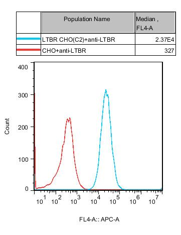
CHO-K1 Human LTBR Cell |
RQP74447 |
Stably overexpresses human LTBR (see Figure 5) |
Flow cytometry/FACS |
Antibody binding specificity assessment, affinity determination, CAR-T target cell validation |
1.Jurkat E6.1 Human LTBR Effector Reporter Cell(RQP74259)
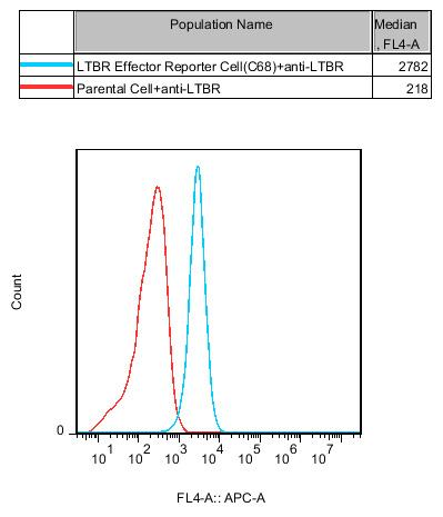
Figure 1 Expression Confirmation: Flow cytometry or western blotting confirms stable LTBR surface expression on the reporter cell. This cell line also carries a luciferase reporter driven by an NF-κB response element, enabling direct quantification of downstream signaling intensity following LTBR activation.
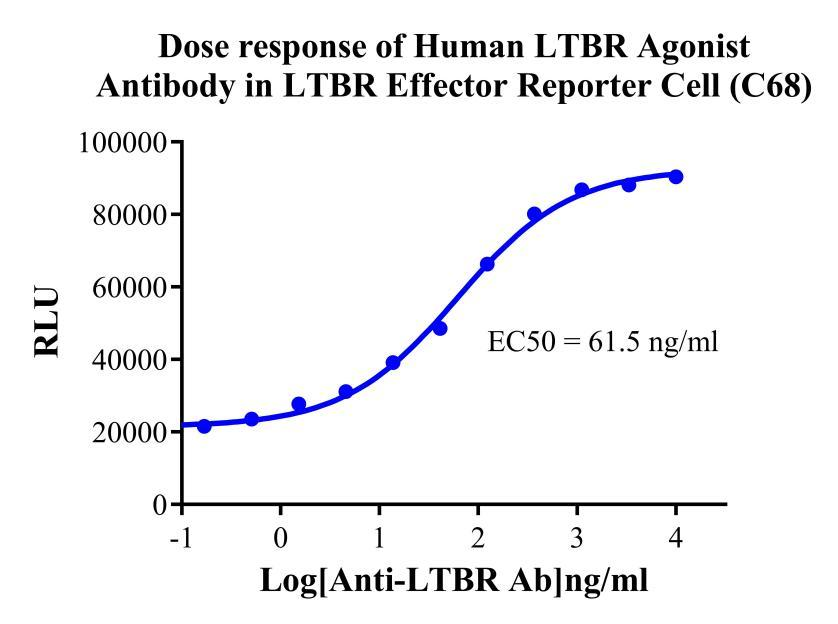
Figure 2 Functional Validation :
Detection principle: Upon binding of an LTBR agonist antibody (or bispecific antibody) to cell-surface LTBR, the non-canonical NF-κB pathway is activated, driving luciferase expression.
Data interpretation: The human LTBR agonist antibody produces a classic sigmoidal dose-response curve with a well-defined EC₅₀ and a wide signal window (typically >10-fold). Background in the no-antibody control is minimal, confirming assay specificity.
Analytical Advantages:
- Pathway-precise targeting: The reporter cell responds primarily to LTBR-mediated non-canonical NF-κB signaling — the pathway that drives TLS formation — making it more target-relevant than generic NF-κB reporters.
- High sensitivity and wide dynamic range: The luciferase assay is simple, rapid, and quantitative, well-suited for high-throughput screening in 384-well plate format.
- Multi-modality compatibility: Supports detection of LTBR agonistic monoclonal antibodies, bispecific antibodies (with cross-linking or co-culture of target cells), and fusion proteins.
- Lot-release and stability testing: The model has been validated against standards analogous to those applied by the NIFDC and is suitable for bioactivity assays and quality control of antibody drug products.
2.CHO-K1 Human LTBR Cell(RQP74447)
Key Applications:
- Antibody binding specificity: Flow cytometric detection of candidate antibody binding to LTBR-CHO cells, with exclusion of cross-reactivity against endogenous CHO antigens.
- Affinity determination: Compatible with ForteBio, SPR, or quantitative flow cytometry (Scatchard analysis).
- CAR-T and bispecific antibody target cell: Serves as a positive target cell to validate the binding and killing function of immune effector molecules.
Analytical Advantages:
- High expression and purity: Following single-cell cloning and flow cytometric validation, inter-lot consistency is excellent.
- No endogenous interference: CHO cells do not express endogenous LTBR ligands, eliminating the risk of auto-activation.
- Ready-to-use: Cryopreserved cells are functional immediately upon thaw, saving the time required to generate stable cell lines in-house.
Product Advantage Summary
|
Advantage |
Details |
|
Emerging Target |
LTBR is a next-generation target for TLS formation and solid tumor immunotherapy. Roche and Mestag have already entered the clinic, leaving the competitive landscape relatively uncrowded and the R&D value high. |
|
Complete Model Portfolio |
The combination of an effector reporter cell and an overexpression cell covers the full screening cascade from functional activity to binding specificity. |
|
Pathway Specificity |
The reporter cell is purpose-built for LTBR-mediated non-canonical NF-κB signaling — the pathway directly responsible for TLS induction — giving data that are more physiologically relevant and translationally meaningful than generic NF-κB reporters. |
|
High Sensitivity & Wide Window |
Figure 4 demonstrates a well-defined dose-response curve for agonist antibodies with a large signal window, suitable for compound screening and antibody lot-release testing. |
|
Broad Applicability |
Compatible with agonistic monoclonal antibodies, bispecific antibodies (e.g., FAP×LTBR), fusion proteins, and small-molecule agonists. |
|
Regulatory Compliance |
Reporter gene-based bioactivity assays are widely accepted by the NIFDC and by pharmaceutical companies, and are suitable for IND submissions and lot-release testing. |
|
Ready-to-Use |
All cell models have been functionally validated and quality-controlled for cryopreservation. Cells are ready for experiments immediately upon thaw, shortening development timelines. |
V. Representative Application Scenarios
- LTBR agonistic bispecific antibody screening: Use LTBR effector reporter cells in the presence of FAP-positive target cells (providing cross-linking) to evaluate the LTBR agonist activity of bispecific antibodies.
- LTBR antagonist development: In the presence of ligand (LTα1β2 or LIGHT), assess the capacity of candidate antibodies or small molecules to block LTBR signaling.
- Antibody binding specificity validation: Use LTBR-CHO cells alongside wild-type CHO cells to select high-selectivity antibodies by flow cytometry.
- CAR-T functional validation: Use LTBR-CHO cells as target cells to evaluate CAR-T cell binding and cytotoxic activity.
Conclusion
As a pivotal molecule at the intersection of lymphoid organ development, tertiary lymphoid structure formation, and tumor immunotherapy, LTBR is advancing from basic research toward clinical translation. With bispecific antibodies from Roche and Mestag now in clinical trials, LTBR agonist therapy is poised to become an important strategy in next-generation solid tumor immunotherapy. Reqbio’s LTBR effector reporter cell and LTBR CHO cell model — characterized by precise pathway design and rigorous functional validation — provide global drug developers with efficient, reliable evaluation tools to accelerate the advancement of LTBR-targeted therapeutics.


news recommendation
CD3E: The T-Cell Activation Switch and Cell Models for Targeted Therapeutics
NFAT Signaling Pathway: A Critical Target for Drug R&D and Cell Models
TSHR Cell Model for Thyroid Cancer Drug Screening | GPCR Assay Platform-Reqbio
We Are Pleased to Announce: Global Commercial Licensing Rights for Jurkat E6.1, CHO-K1, and HEK293 Cell Lines Officially Secured.
Explore